. 2014 Jun;88(11):6128-36.
doi: 10.1128/JVI.00333-14. Epub 2014 Mar 19.

Alpha/beta interferon receptor signaling amplifies early proinflammatory cytokine production in the lung during respiratory syncytial virus infection

Affiliations

Alpha/beta interferon receptor signaling amplifies early proinflammatory cytokine production in the lung during respiratory syncytial virus infection

Michelle Goritzka et al. J Virol. 2014 Jun.

Abstract

Type I interferons (IFNs) are produced early upon virus infection and signal through the alpha/beta interferon (IFN-α/β) receptor (IFNAR) to induce genes that encode proteins important for limiting viral replication and directing immune responses. To investigate the extent to which type I IFNs play a role in the local regulation of inflammation in the airways, we examined their importance in early lung responses to infection with respiratory syncytial virus (RSV). IFNAR1-deficient (IFNAR1(-/-)) mice displayed increased lung viral load and weight loss during RSV infection. As expected, expression of IFN-inducible genes was markedly reduced in the lungs of IFNAR1(-/-) mice. Surprisingly, we found that the levels of proinflammatory cytokines and chemokines in the lungs of RSV-infected mice were also greatly reduced in the absence of IFNAR signaling. Furthermore, low levels of proinflammatory cytokines were also detected in the lungs of IFNAR1(-/-) mice challenged with noninfectious innate immune stimuli such as selected Toll-like receptor (TLR) agonists. Finally, recombinant IFN-α was sufficient to potentiate the production of inflammatory mediators in the lungs of wild-type mice challenged with innate immune stimuli. Thus, in addition to its well-known role in antiviral resistance, type I IFN receptor signaling acts as a central driver of early proinflammatory responses in the lung. Inhibiting the effects of type I IFNs may therefore be useful in dampening inflammation in lung diseases characterized by enhanced inflammatory cytokine production.

Importance: The initial response to viral infection is characterized by the production of interferons (IFNs). One group of IFNs, the type I IFNs, are produced early upon virus infection and signal through the IFN-α/β receptor (IFNAR) to induce proteins important for limiting viral replication and directing immune responses. Here we examined the importance of type I IFNs in early responses to respiratory syncytial virus (RSV). Our data suggest that type I IFN production and IFNAR receptor signaling not only induce an antiviral state but also serve to amplify proinflammatory responses in the respiratory tract. We also confirm this conclusion in another model of acute inflammation induced by noninfectious stimuli. Our findings are of relevance to human disease, as RSV is a major cause of infant bronchiolitis and polymorphisms in the IFN system are known to impact disease severity.


Figures

FIG 1
FIG 1
Viral load and weight loss after RSV infection. C57BL/6 wt and IFNAR1−/− mice were infected intranasally with 2 × 10⁶ FFU of RSV. RNA was isolated from lung tissue, and after conversion into cDNA, copy numbers of RSV L gene RNA were determined using quantitative PCR (qPCR). (A) Levels of RSV L gene RNA in the lung tissue of wt and IFNAR1−/− mice at different time points after RSV infection. Copy numbers were determined using a plasmid standard. Data shown are pooled data from 3 individual experiments with 3 to 5 mice per group in each experiment. The dotted line represents the detection limit. ND, not detectable. (B) Weights of infected and noninfected mice were monitored daily and plotted as a percentage of weight on the day of infection (day 0). The data shown are representative of at least 3 experiments with 3 to 5 mice per group in each experiment. Error bars indicate the SEM. ***, P ≤ 0.001; **, P ≤ 0.01; *, P ≤ 0.05 comparing RSV-infected wt with RSV-infected IFNAR1−/− mice. ##, P ≤ 0.01 comparing RSV-infected wt with naive wt mice.
FIG 2
FIG 2
Interferon responses after RSV infection of IFNAR1−/− and wt mice. C57BL/6 wt and IFNAR1−/− mice were intranasally infected with 2 × 10⁶ FFU of RSV. (A) The level of IFN-α was determined in bronchoalveolar lavage (BAL) fluid by ELISA. (B) RNA was isolated from lungs, and gene expression levels of IFN-β were determined by qPCR. (C to E) IFN-γ (C), IFN-λ (D), and interferon-stimulated gene (ISG) CXCL10 (E) gene expression levels in lung tissue and protein production in BAL fluid were quantified. (F and G) RIG-I (Ddx58) (F) and Mx-1 (G) mRNA was determined by qPCR. Gene expression relative to GAPDH was calculated for CXCL10, RIG-I, and Mx-1. For IFN-β, IFN-λ, and IFN-γ, copy numbers were determined using a plasmid standard. The detection limit for all assays was 200 copies and is represented by the dotted line. Data shown are representative of at least 2 experiments with 4 or 5 mice per group. Error bars indicate the SEM. Significance when comparing RSV-infected wt with RSV-infected IFNAR1−/− mice: **, P ≤ 0.01; *, P ≤ 0.05.
FIG 3
FIG 3
Diminished induction of proinflammatory cytokine mRNA in IFNAR1−/− mice during RSV infection. C57BL/6 wt and IFNAR1−/− mice were intranasally infected with 2 × 10⁶ FFU of RSV. (A) RNA was isolated from lungs, and gene expression levels of IL-6, IL-1-β, and TNF-α were determined by qPCR. (B) Percentage of neutrophils in the BAL fluid at indicated days postinfection. Data shown are pooled data from 3 individual experiments with 3 to 5 mice per group in each experiment. (C) CXCL1 (KC) gene expression levels in lung tissue and protein production in BAL fluid were quantified. Gene expression relative to GAPDH was calculated for IL-6, IL-1β, and CXCL1. For TNF-α, copy numbers were determined using a plasmid standard. Dotted lines represent the detection limit. Data shown are representative of at least 2 experiments with 4 or 5 mice per group, or for CXCL1 data shown are pooled data from 2 individual experiments with 3 to 5 mice per group in each experiment. Error bars indicate the SEM. Significance when comparing RSV-infected wt with RSV-infected IFNAR1−/− mice: ***, P ≤ 0.001; **, P ≤ 0.01; *, P ≤ 0.05.
FIG 4
FIG 4
Reduced levels of proinflammatory cytokines in IFNAR1−/− mice during RSV infection. C57BL/6 wt and IFNAR1−/− mice were intranasally infected with 2 × 10⁶ FFU of RSV. At different time points postinfection protein levels of IL-6, IL-1β, TNF-α, IL-1α, GM-CSF, IL-5, IL-12p40, CXCL9, and CCL3 production were determined in BAL fluid by Luminex. Data shown are representative of at least 2 experiments with 4 or 5 mice per group. Error bars indicate the SEM. Significance when comparing RSV-infected wt with RSV-infected IFNAR1−/− mice: **, P ≤ 0.01; *, P ≤ 0.05.
FIG 5
FIG 5
Induction of cytokines after intranasal challenge of IFNAR1−/− and wt mice with innate stimuli. C57BL/6 wt and IFNAR1−/− mice were intranasally challenged with TLR agonists [CpG, 1.25 μg/g body weight; poly(I·C), 3.5 μg/g body weight; LPS, 500 ng/g body weight]. Lungs and BAL fluid were collected 24 h postchallenge. (A) RNA was isolated from lungs, and gene expression levels of IFN-λ, CXCL10, IL-6, IL-1β, and TNF-α were determined by qPCR. Gene expression relative to GAPDH was calculated for IL-1β, IL-6, and CXCL10. For IFN-λ and TNF-α, copy numbers were determined using a plasmid standard. (B) IFN-α and IL-6 detected in the BAL fluid using ELISA. Data shown are pooled data from 2 individual experiments with 4 or 5 mice per group in each experiment. Error bars indicate the SEM. Significance when comparing RSV-infected wt with RSV-infected IFNAR1−/− mice: ***, P ≤ 0.001; **, P ≤ 0.01; *, P ≤ 0.05.
FIG 6
FIG 6
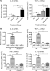
Recombinant IFN-α can amplify proinflammatory cytokine induction. (A) C57BL/6 wt mice were intranasally challenged with a suboptimal dose of LPS (50 ng/g body weight) with or without recombinant IFN-α11 (rIFN-α11) (500 ng/mouse), and lungs were collected after 24 h. (B) C57BL/6 wt and IFNAR1−/− mice were intranasally challenged with rIFN-α (500 ng/mouse), and lungs were collected after 12 h. RNA was isolated from lungs, and gene expression levels of IL-6 and TNF-α (A) and IL-6, TNF-α, IFN-γ, and IL-1β (B) were determined by qPCR. Gene expression relative to GAPDH was calculated for IL-6 and IL-1β, and for TNF-α and IFN-γ copy numbers were determined using a plasmid standard. Data shown are pooled data from 2 individual experiments with 4 or 5 mice per group in each experiment. Error bars indicate the SEM. Significance when comparing RSV-infected wt with RSV-infected IFNAR1−/− mice: ***, P ≤ 0.001; **, P ≤ 0.01; *, P ≤ 0.05.

Similar articles

Cited by

References

    1. Randall RE, Goodbourn S. 2008. Interferons and viruses: an interplay between induction, signalling, antiviral responses and virus countermeasures. J. Gen. Virol. 89:1–47. 10.1099/vir.0.83391-0 - DOI - PubMed
    1. Durbin RK, Kotenko SV, Durbin JE. 2013. Interferon induction and function at the mucosal surface. Immunol. Rev. 255:25–39. 10.1111/imr.12101 - DOI - PMC - PubMed
    1. González-Navajas JM, Lee J, David M, Raz E. 2012. Immunomodulatory functions of type I interferons. Nat. Rev. Immunol. 12:125–135. 10.1038/nri3133 - DOI - PMC - PubMed
    1. Takeuchi O, Akira S. 2009. Innate immunity to virus infection. Immunol. Rev. 227:75–86. 10.1111/j.1600-065X.2008.00737.x - DOI - PMC - PubMed
    1. Kumagai Y, Takeuchi O, Kato H, Kumar H, Matsui K, Morii E, Aozasa K, Kawai T, Akira S. 2007. Alveolar macrophages are the primary interferon-alpha producer in pulmonary infection with RNA viruses. Immunity 27:240–252. 10.1016/j.immuni.2007.07.013 - DOI - PubMed

Publication types