. 2014 May;25(10):1641-52.
doi: 10.1091/mbc.E13-12-0717. Epub 2014 Mar 19.

Nucleophosmin modulates stability, activity, and nucleolar accumulation of base excision repair proteins

Affiliations

Nucleophosmin modulates stability, activity, and nucleolar accumulation of base excision repair proteins

Mattia Poletto et al. Mol Biol Cell. 2014 May.

Abstract

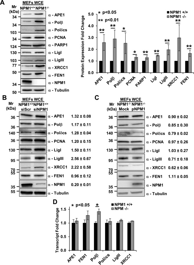
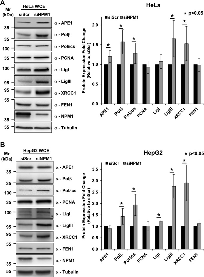
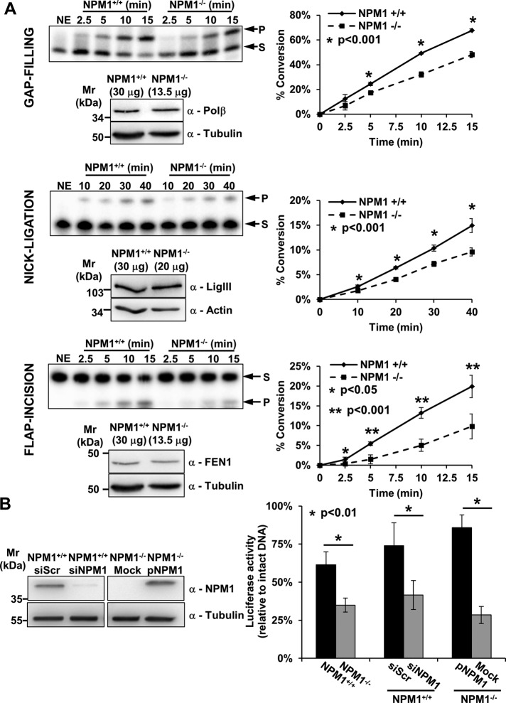
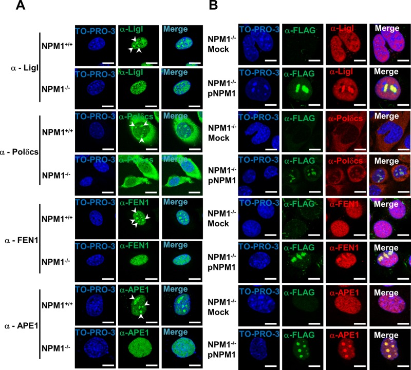
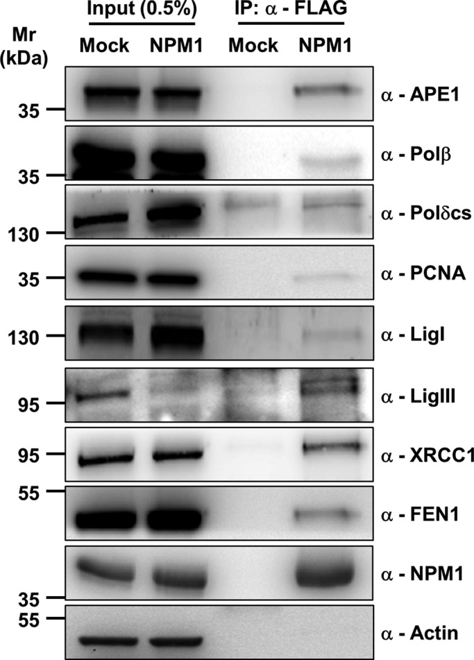
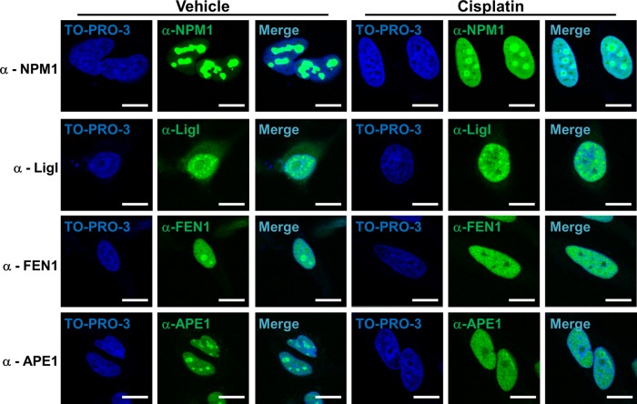
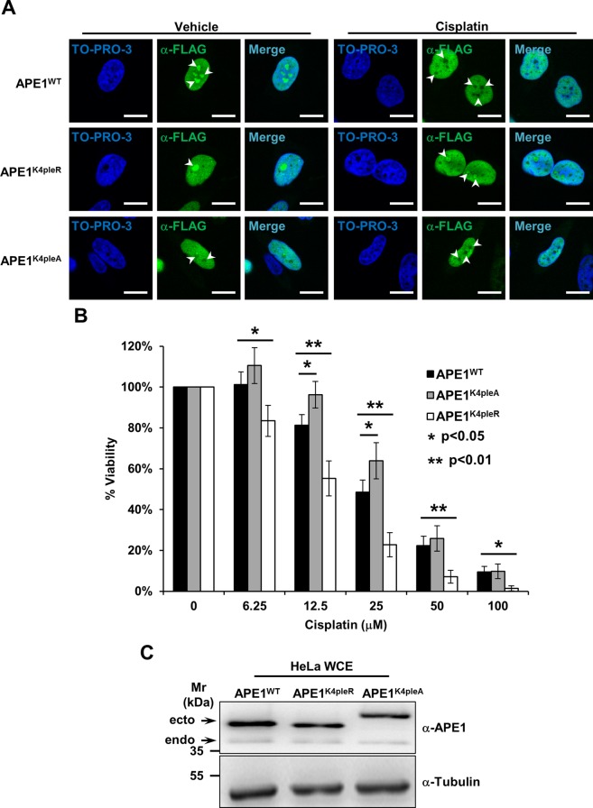
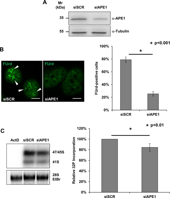
Nucleophosmin (NPM1) is a multifunctional protein that controls cell growth and genome stability via a mechanism that involves nucleolar-cytoplasmic shuttling. It is clear that NPM1 also contributes to the DNA damage response, yet its exact function is poorly understood. We recently linked NPM1 expression to the functional activation of the major abasic endonuclease in mammalian base excision repair (BER), apurinic/apyrimidinic endonuclease 1 (APE1). Here we unveil a novel role for NPM1 as a modulator of the whole BER pathway by 1) controlling BER protein levels, 2) regulating total BER capacity, and 3) modulating the nucleolar localization of several BER enzymes. We find that cell treatment with the genotoxin cisplatin leads to concurrent relocalization of NPM1 and BER components from nucleoli to the nucleoplasm, and cellular experiments targeting APE1 suggest a role for the redistribution of nucleolar BER factors in determining cisplatin toxicity. Finally, based on the use of APE1 as a representative protein of the BER pathway, our data suggest a function for BER proteins in the regulation of ribogenesis.


Figures

FIGURE 1:
FIGURE 1:
Depletion of NPM1 positively modulates BER protein amounts. (A) Representative Western blotting analysis on NPM1+/+ and NPM1/ whole-cell extracts (60 μg), showing the higher levels of BER proteins in the absence of NPM1 expression (left). Antibodies used are indicated on the right-hand side. Right, histogram reporting densitometric quantification of Western blotting signals from at least three independent experiments. Protein amounts are expressed as mean ± SD of the signal, considering NPM1+/+ as reference sample. Either actin or tubulin was used as loading control. (B, C). Representative Western blotting analysis on whole-cell extracts (50 μg) from scrambled siRNA (siScr), NPM1 siRNA (siNPM1)-treated NPM1+/+ cells (B), and mock- or NPM1-reconstituted (pNPM1) NPM1/ cells (C). Change in BER protein levels upon NPM1 down-regulation or reexpression are indicated on the right-hand side. siScr cells or mock-transfected cells were used as reference, respectively. Values indicated are mean ± SD from at least three independent replicates. Antibodies used are indicated on the right-hand side; tubulin was used as loading control. (D) Real-time PCR analysis comparing the relative amount of transcript for the indicated BER factors between NPM1+/+ and NPM1/ cells. Among the genes tested, only Polβ showed a statistically significant increment. Values reported are expressed as mean ± SD of the expression level in three independent replicates, considering NPM1+/+ as reference sample.
FIGURE 2:
FIGURE 2:
NPM1 knockdown positively modulates BER proteins independently of p53 status. (A) Representative Western blotting analysis on HeLa whole-cell extracts (50 μg) from scrambled siRNA (siScr)– or NPM1 siRNA (siNPM1)–treated cells (left). An increased amount of a subset of BER proteins is visible upon NPM1 down-regulation. Antibodies used are indicated on the right-hand side. Right, histogram reporting the quantitative increment of different BER proteins in HeLa cells upon NPM1 down-regulation. Values reported are expressed as mean ± SD of the fold of induction relative to siScr-treated cells in at least three independent replicates. (B) Representative Western blotting analysis on HepG2 whole-cell extracts (50 μg) obtained from siScr or siNPM1 cells (left). NPM1 depletion leads to accumulation of different BER proteins. Antibodies used are indicated on the right-hand side. *Unspecific band. Right, histogram reporting the increase of BER protein levels in HepG2 cells upon NPM1 down-regulation. Values reported are expressed as mean ± SD of the fold of induction relative to siScr-treated cells in at least three independent replicates.
FIGURE 3:
FIGURE 3:
NPM1 contributes to the tuning of BER capacity. (A) In vitro BER assays using NPM1+/+ and NPM1/ whole-cell extracts at equal amounts of DNA Polβ, DNA ligase III (LigIII), or FEN1 reveal impaired gap-filling, nick-ligation, and flap-incision activities, respectively, in the absence of NPM1. For each enzymatic activity we report a representative denaturing gel analysis, along with a Western blot indicating the amount of cell extract used to obtain comparable quantities of each BER factor (left). Graphs (right) describe the percentage of substrate (S) converted to product (P) as a function of time. Values reported are mean ± SD of at least three independent experimental replicates. (B) In vivo BER capacity assessment using a reporter-based system as indicated in Materials and Methods (left). The assays highlight a positive role for NPM1 in the modulation of BER capacity. Histograms show mean ± SD of at least three independent replicates. Luciferase activity for the depurinated plasmid relative to that of undamaged DNA is reported. A statistically significant impairment in BER capacity is observed when comparing NPM1+/+ and NPM1/ cells. A similar defect in BER-mediated repair is also visible when comparing siScr- and siNPM1-treated NPM1+/+ cells. Reconstitution of NPM1/ cells with human recombinant NPM1 (pNPM1) instead restored the BER defect. A representative Western blotting analysis (right) shows NPM1 levels in the siScr- and siNPM1-treated NPM1+/+ cells and in the mock- and NPM1-reconstituted (pNPM1) NPM1/ cells used in the in vivo BER assays. Tubulin was used as loading control.
FIGURE 4:
FIGURE 4:
NPM1 promotes the accumulation of BER proteins within nucleoli. (A) Representative immunofluorescence images showing the differential subcellular localization of diverse BER factors in NPM1+/+ and NPM1/ cells. Each protein analyzed (left) shows nucleolar accumulation (white arrowheads) only in the presence of NPM1. TO-PRO-3 counterstaining was used to highlight nuclei; bars, 16 μm. (B) Representative immunofluorescence analysis on mock- and NPM1-reconstituted (pNPM1) NPM1/ cells. NPM1 staining (α-FLAG, green) was used to localize cells positively transfected with FLAG-tagged human NPM1; NPM1 reexpression restores the accumulation of BER proteins (red) within nucleoli. Nuclei are counterstained with TO-PRO-3; bars, 16 μm.
FIGURE 5:
FIGURE 5:
NPM1 is present in complexes containing the majority of the BER proteins. Western blot analysis on coimmunoprecipitated material obtained from HeLa cells either mock-transfected or transfected with FLAG-tagged human NPM1. Immunoprecipitation was performed with an anti-FLAG antibody, and the Western blot analysis was carried out with the indicated antibodies (right). Material coimmunoprecipitated with NPM1 shows enrichment in BER components. Actin was used as loading control for the input fractions.
FIGURE 6:
FIGURE 6:
NPM1 modulates BER protein localization during nucleolar stress. Representative immunofluorescence analysis on the indicated proteins underlines the dramatic change in subcellular localization upon nucleolar stress induced by cisplatin treatment (100 μM for 6 h) in HeLa cells. BER proteins, as well as NPM1, relocalize from nucleoli to the nucleoplasm; for some BER factors (APE1, FEN1, LigI) nucleolar emptying is also evident. TO-PRO-3 counterstaining was used to highlight nuclei; bars, 16 μm.
FIGURE 7:
FIGURE 7:
APE1 redistribution upon cisplatin requires active modification of lysine residues and protects cells from cisplatin cytotoxicity. (A) Representative immunofluorescence analysis on HeLa cells expressing different FLAG-tagged APE1 forms. On cisplatin treatment (100 μM for 6 h) only APE1WT redistributes from nucleoli (white arrowheads), whereas the nonacetylatable APE1K4pleR does not display major relocalization. The APE1K4pleA form fails to accumulate within nucleoli, regardless of the cisplatin treatment. TO-PRO-3 counterstaining was used to highlight nuclei; bars, 16 μm. (B) Viability assay on HeLa cell clones expressing the indicated APE1 forms. Cells were treated with doxycycline to induce endogenous APE1 knockdown (Lirussi et al., 2012) and challenged with increasing amounts of cisplatin for 24 h. The histogram reports the mean ± SD of at least three independent experimental replicates. The APE1K4pleR-expressing clone is hypersensitive to cisplatin, indicating that modification on critical APE1 lysine residues and redistribution of the protein from nucleoli are important to cell survival. The APE1K4pleA-expressing cells, conversely, show a protective phenotype linked to a decreased cell proliferation rate. (C) Representative Western blot analysis on HeLa cell clones used for the experiments in B. HeLa whole-cell extracts (20 μg) from the indicated clones were probed with an anti-APE1 antibody. The analysis shows comparable expression of the ectopic APE1 forms (ecto) and minimum residual of the endogenous protein (endo). Tubulin was used as loading control.
FIGURE 8:
FIGURE 8:
Depletion of APE1 leads to nucleolar impairment. (A) Representative Western blot analysis on HeLa cells treated with either a scrambled siRNA (siScr) or an APE1-targeting siRNA (siAPE1) shows the efficiency of APE1 knockdown. Tubulin was used as loading control. (B) FUrd labeling of siScr- and siAPE1-treated HeLa cells. Left, representative immunofluorescence showing the preferential accumulation of FUrd within transcriptionally active nucleoli only in APE1-proficient cells (white arrowheads). Bars, 8 μm. Right, histogram reporting the average FUrd nucleolar incorporation efficiency in siScr and siAPE1 cells. Values reported express the average amount of FUrd-positive cells ± SD from at least three independent experiments. (C) [32P]phosphate metabolic labeling of nascent rRNA transcripts in HeLa cells treated with siScr, siAPE1, or actinomycin D (as positive control) reveals the nucleolar impairment in APE1-depleted cells. Cells were transfected with siRNAs and 48 h later pulsed for 1 h with [32P]orthophosphate as described in Materials and Methods. Left panel, total RNA was extracted and the amount of labeled rRNA precursors was measured through autoradiography. Ethidium bromide staining was used as loading control. Right, histogram reporting the average [32P]phosphate incorporation into the 47S precursor in siScr and siAPE1 cells. Values reported express the mean incorporation ± SD from at least three independent experiments. siScr was used as reference sample.

References

    1. Ali MM, Frei E, Straub J, Breuer A, Wiessler M. Induction of metallothionein by zinc protects from daunorubicin toxicity in rats. Toxicology. 2002;179:85–93. - PubMed
    1. Antoniali G, Lirussi L, Poletto M, Tell G. Emerging roles of the nucleolus in regulating the DNA damage response: the non-canonical DNA repair enzyme APE1/Ref-1 as a paradigmatical example. Antioxid Redox Signal. 2014;20:621–639. - PMC - PubMed
    1. Apicelli AJ, et al. A non-tumor suppressor role for basal p19ARF in maintaining nucleolar structure and function. Mol Cell Biol. 2008;28:1068–1080. - PMC - PubMed
    1. Becherel OJ, Gueven N, Birrell GW, Schreiber V, Suraweera A, Jakob B, Taucher-Scholz G, Lavin MF. Nucleolar localization of aprataxin is dependent on interaction with nucleolin and on active ribosomal DNA transcription. Hum Mol Genet. 2006;15:2239–2249. - PubMed
    1. Burger K, et al. Chemotherapeutic drugs inhibit ribosome biogenesis at various levels. J Biol Chem. 2010;16:12416–12425. - PMC - PubMed

Publication types

MeSH terms

LinkOut - more resources