

Effects of exposure to bisphenol A during pregnancy and lactation on the testicular morphology and caspase-3 protein expression of ICR pups
- PMID: 24648961
- PMCID: PMC3917723
- DOI: 10.3892/br.2013.79
Effects of exposure to bisphenol A during pregnancy and lactation on the testicular morphology and caspase-3 protein expression of ICR pups
Abstract
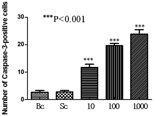
Bisphenol A (BPA), a xenoestrogen and endocrine-disrupting chemical, is a cause for concern due to its being a potential human carcinogen. The aim of this study was to investigate the effects of continued maternal exposure to BPA on the testicular structures and expression of caspase-3 protein in male ICR offspring during pregnancy and lactation and explore its possible mechanism. Pregnant ICR mice were divided into two control groups, which were either given or not given the solvent dimethyl sulfoxide (DMSO) and three treatment groups, which were gavaged with water-soluble BPA dissolved in DMSO at three different concentrations from gestational day 0 to weaning on postnatal day (PND) 21. The number of mice pups and ratios of males to females were recorded. On PND 21, male offspring were sacrificed to measure their wet weights and testicular coefficients. Electron microscopy was used to observe testicular morphological changes, Hoechst 33258 staining to detect cell apoptosis and immunohistochemistry to measure caspase-3 expression. Although there was no significant difference between offspring of the control group and the treatment group in litter size and male-female ratio (P>0.05), the testicular viscera coefficient in the latter decreased (P<0.01). Specifically, compared with offspring of the control group, in addition to increased cell apoptosis, those of the treatment groups were found to have changes in mitochondrial and endoplasmic reticulum in their spermatogenous, Sertoli, Leydig and peritubular myoid cells, which were concomitant with an elevated expression of caspase-3 in the cytoplasm (P<0.01). In conclusion, exposure of pregnant mice to BPA during pregnancy and lactation has some toxic effects on the testes of male ICR offspring and these may originate from increased apoptosis.
Keywords: bisphenol A; caspase-3; offspring mice.
Figures


References
-
- Takahashi A, Higashino F, Aoyagi M, et al. Bisphenol A from dental polycarbonate crown upregulates the expression of hTERT. J Biomed Mater Res B Appl Biomater. 2004;71:214–221. - PubMed
-
- Besaratinia A, Pfeifer GP. A review of mechanisms of acrylamide carcinogenicity. Carcinogenesis. 2007;28:519–528. - PubMed
-
- World Health Organization . FAO/WHO Consultation on the Health Implications of Acrylamide in Food-Summary Report. Geneva, Switzerland: WHO; 2002. pp. 1–12.
LinkOut - more resources
Full Text Sources
Other Literature Sources
Research Materials