. 2013 May;1(3):444-452.
doi: 10.3892/mco.2013.76. Epub 2013 Feb 15.

Marine- and plant-derived ω-3 fatty acids differentially regulate prostate cancer cell proliferation

Affiliations

Marine- and plant-derived ω-3 fatty acids differentially regulate prostate cancer cell proliferation

Pinar O Eser et al. Mol Clin Oncol. 2013 May.

Abstract

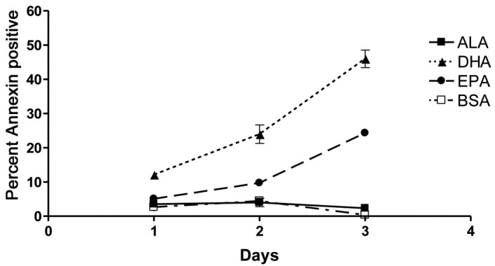
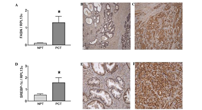
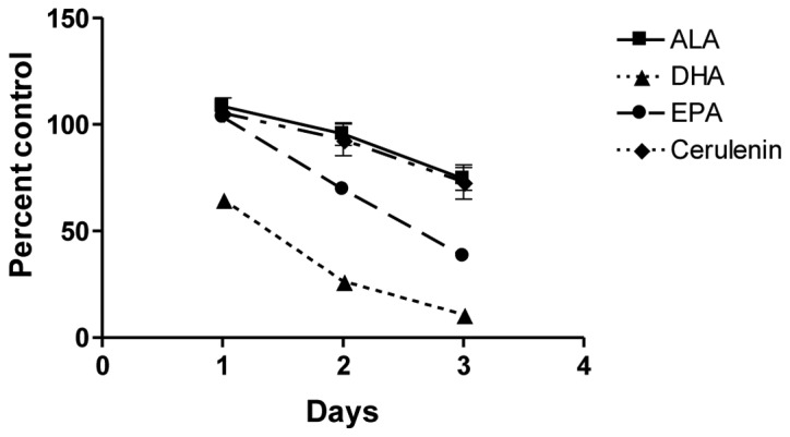
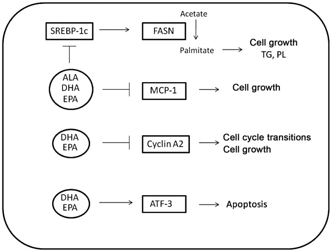
Fish oil contains the marine ω-3 polyunsaturated fatty acids (ω-3 PUFAs) docosahexaenoic (DHA) and eicosapentaenoic acid (EPA). The consumption of diets rich in these fatty acids is associated with a decreased incidence of prostate cancer. However, there is limited knowledge regarding the non-marine ω-3 PUFA α-linolenic acid (ALA). To study which ω-3 PUFAs are more effective in prostate cancer prevention, and whether the mechanisms of action are conserved between them, we investigated the effect of DHA, EPA and ALA on the human prostate cancer cell lines PC-3 and LNCaP. Different trends of inhibition of PC-3 cell proliferation were observed for the three ω-3 PUFA, with DHA having the most pronounced effects on cell proliferation, while ALA had the minimum effects of the three ω-3 PUFAs. All the ω-3 PUFAs decreased fatty acid synthase (FASN) mRNA. Concerning genes involved in inflammation, cell cycle and apoptosis, DHA regulated the most genes in all categories, followed by EPA and then ALA. In addition, DHA and EPA increased the gene expression of the pro-apoptotic protein activating transcription factor 3 mRNA. Moreover, these two fatty acids significantly induced apoptosis. In conclusion, while some mechanisms of cancer cell inhibition are conserved among ω-3 PUFA, the extent, magnitude, and duration of transcriptional changes vary for each individual fatty acid.

Keywords: docosahexaenoic acid; eicosapentaenoic acid; prostate cancer; α-linolenic acid; ω-3.


Figures

Figure 1
Figure 1
ω-3 fatty acids differentially inhibit androgen-independent and -dependent prostate cancer cell line growth. (A) PC-3 and (B) LNCaP cells were treated with 100 μM α-linolenic (ALA), docosahexaenoic (DHA) or eicosapentaenoic (EPA) acid conjugated to BSA following overnight serum starvation. Cell proliferation was measured using the CellTiter-Glo® Luminescent Cell Viability assay. Three trends of inhibition emerged in PC-3 and LNCaP cells treated with the ω-3 PUFAs. DHA inhibited the two cell lines rapidly and to the greatest extent. EPA also effectively inhibited cancer cell growth, albeit more gradually compared to DHA. PC-3 cells treated with EPA reached approximately the same endpoint compared to DHA-treated cells, ultimately exhibiting a similar magnitude of inhibition. ALA inhibited cells more slowly and to a lesser extent compared to DHA and EPA. While the same inhibitory trends were observed in androgen independent and dependent cell lines, the more protracted response in LNCaP cells may be attributed to their slower rate of cell division. Values are shown as the mean of replicates from four independent experiments with error bars indicating standard error.
Figure 2
Figure 2
Fish oil ω-3 fatty acids induce cell apoptosis. PC-3 cells were treated with 100 μM α-linolenic (ALA), docosahexaenoic (DHA) or eicosapentaenoic (EPA) acid conjugated to bovine serum albumin (BSA) following overnight serum starvation. Cells and supernatants were collected every 24 h for 3 days and analyzed for apoptosis (annexin staining) and cell death (7-AAD staining) by flow cytometry. DHA significantly induced apoptosis within 24 h, demonstrating the greatest overall induction. EPA also significantly induced apoptosis, but at a later stage compared to DHA and not to the same extent. ALA did not induce apoptosis above BSA control levels. Significance was assessed using one-way analysis of variance (ANOVA). Values are shown as the mean of replicates from three independent experiments with error bars indicating standard error.
Figure 3
Figure 3
Fatty acid synthase (FASN) and its regulator sterol response element binding protein-1c (SREBP-1c) are upregulated in clinical prostate cancer samples. Normal prostate (NPT) and prostate cancer tissues (PCT) isolated from patients at prostatectomy were analyzed and compared by real-time PCR gene expression and immunohistochemistry. RNA was isolated from the clinical samples or PC-3 cells treated with ω-3 PUFAs and gene expression was assessed using quantitative real-time PCR. Clinical samples were demonstrated to have elevated transcript levels of (A) FASN and (D) its regulator SREBP-1c when compared to normal prostate tissue. These trends were immunohistochemically confirmed. Normal prostate tissue was demonstrated to have low levels of (B) FASN and (E) SREBP-1c expression (magnification, ×200), compared to Gleason grade 5 tumor showing (C) FASN in the cytoplasm and (F) nuclear staining of SREBP-1c (magnification, ×400). (A and D) For the clinical samples, statistical significance was assessed using the paired Student’s t-test; *P<0.05.
Figure 4
Figure 4
α-linolenic acid (ALA) inhibits PC-3 cell proliferation in an identical pattern as FASN inhibition. PC-3 cells were treated with 100 μM of BSA-conjugated ω-3 fatty acids or 2 μM of the FASN inhibitor cerulenin following overnight serum starvation. Cell proliferation was measured using the CellTiter-Glo® Luminescent Cell Viability assay. In a time course experiment, ALA produced an identical viability curve to cerulenin, while DHA and EPA inhibited PC-3 cell growth more rapidly and effectively, suggesting the activation of additional anti-proliferative pathways by the fish oils. Values are shown as the mean of replicates with error bars representing standard error. ALA, α-linolenic acid; DHA, docosahexaenoic acid; EPA, eicosapentaenoic acid.
Figure 5
Figure 5
Cellular pathways involved in fatty acid metabolism, inflammation, cell cycle regulation and apoptosis are differentially modulated by ω-3 fatty acids. The expression of all the genes was quantified in PC-3 cells using quantitative real-time PCR. ALA, DHA and EPA inhibited fatty acid synthase (FASN) and its regulator sterol response element binding protein-1c to 50% of control levels. All three ω-3 PUFAs also inhibited the inflammatory protein and autocrine prostate cancer growth factor MCP-1, although the extent of inhibition varied. DHA caused the most effective inhibition, with MCP-1 levels decreasing to 10% of control levels in DHA-treated PC-3 cells, while EPA inhibited MCP-1 transcript levels by 30%, and ALA caused a decrease to 50% of control. DHA and EPA reduced the transcript levels of cyclin A2 to 20% of control levels to reduce cell proliferation. Only DHA activated ATF-3, leading to the inhibition of anti-apoptotic factors downstream of NF-κB, thereby inducing apoptosis in PC-3 cells.

References

    1. Rambeaud JJ. Intermittent complete androgen blockade in meta-static prostate cancer. Eur Urol. 1999;35(Suppl 1):32–36. - PubMed
    1. Harris KA, Reese DM. Treatment options in hormone-refractory prostate cancer: current and future approaches. Drugs. 2001;61:2177–2192. - PubMed
    1. Chan JM, Gann PH, Giovannucci EL. Role of diet in prostate cancer development and progression. J Clin Oncol. 2005;23:8152–8160. - PubMed
    1. King IB, Kristal AR, Schaffer S, Thornquist M, Goodman GE. Serum trans-fatty acids are associated with risk of prostate cancer in beta-Carotene and Retinol Efficacy Trial. Cancer Epidemiol Biomarkers Prev. 2005;14:988–992. - PubMed
    1. Haaland CM, Heaphy CM, Butler KS, Fischer EG, Griffith JK, Bisoffi M. Differential gene expression in tumor adjacent histologically normal prostatic tissue indicates field cancerization. Int J Oncol. 2009;35:537–546. - PubMed

LinkOut - more resources