Review
. 2014 Apr;87(1036):20130677.
doi: 10.1259/bjr.20130677. Epub 2014 Feb 3.

Imaging in head and neck squamous cell carcinoma: the potential role of PET/MRI

Affiliations
Review

Imaging in head and neck squamous cell carcinoma: the potential role of PET/MRI

Minerva Becker et al. Br J Radiol. 2014 Apr.

Abstract

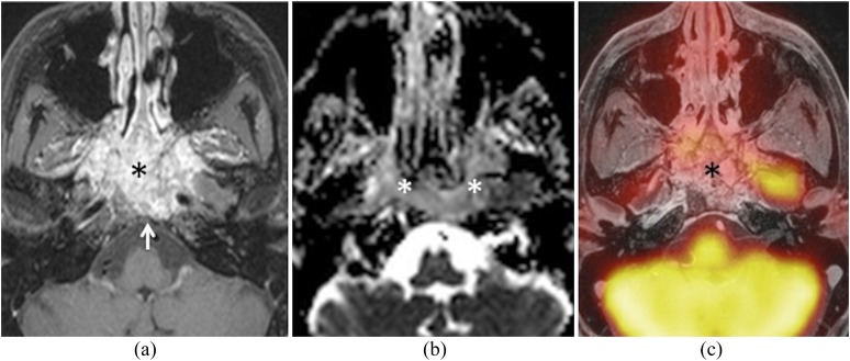
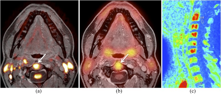
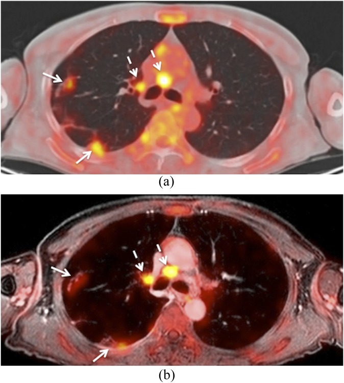
In head and neck oncology, the information provided by positron emission tomography (PET)/CT and MRI is often complementary because both the methods are based on different biophysical foundations. Therefore, combining diagnostic information from both modalities can provide additional diagnostic gain. Debates about integrated PET/MRI systems have become fashionable during the past few years, since the introduction and wide adoption of software-based multimodality image registration and fusion and the hardware implementation of integrated hybrid PET/MRI systems in pre-clinical and clinical settings. However, combining PET with MRI has proven to be technically and clinically more challenging than initially expected and, as such, research into the potential clinical role of PET/MRI in comparison with PET/CT, diffusion-weighted MRI (DW MRI) or the combination thereof is still ongoing. This review focuses on the clinical applications of PET/MRI in head and neck squamous cell carcinoma (HNSCC). We first discuss current evidence about the use of combined PET/CT and DW MRI, and, then, we explain the rationale and principles of PET/MR image fusion before summarizing the state-of-the-art knowledge regarding the diagnostic performance of PET/MRI in HNSCC. Feasibility and quantification issues, diagnostic pitfalls and challenges in clinical settings as well as ongoing research and potential future applications are also discussed.


Figures

Figure 1.
Figure 1.
This patient was a follow-up case of a squamous cell carcinoma of the floor of the mouth. (a) Axial positron emission tomography (PET)/CT image shows streak artefacts from bilateral dental implants hampering interpretation. Tumour recurrence could not be excluded on the basis of PET/CT. (b) Corresponding hybrid PET/MRI (PET fused with axial gadolinium-enhanced water-only Dixon image) shows the absence of tumour recurrence. In this patient, PET/MRI is less affected by dental artefacts (arrows) than PET/CT.
Figure 2.
Figure 2.
Hybrid positron emission tomography (PET)/MRI obtained for primary staging of advanced laryngeal squamous cell carcinoma. (a) Fused T2 and b 1000 image (colour overlay) illustrate restricted diffusivity in the right Level VI region (arrow), suggesting metastatic lymph nodes. Note geometric distortion of the overlaid b 1000 image in comparison with T2. Position of the spinal cord (long dashed arrow) and T1 nerve roots (arrowheads) on b 1000. Position of the spinal cord on T2 (short dashed arrow). (b) Corresponding fused T2 and PET show hypermetabolic thyroid nodule (arrow) and absent Level VI metastatic nodes. Ultrasonography with fine-needle aspiration cytology and surgery revealed a benign thyroid nodule and absent Level VI metastases, respectively.
Figure 3.
Figure 3.
Positron emission tomography (PET)/MRI and PET/CT obtained for primary staging of squamous cell carcinoma of the hypopharynx. (a) Axial PET/CT image shows a hypermetabolic tumour located in the posterior hypopharyngeal wall (arrow). (b) Corresponding hybrid PET/MRI (fused PET and T2) shows poor data fusion due to patient motion. Note anterior displacement of the PET image in comparison with T2, suggesting hypermetabolic base of the tongue–vallecula tumour (dashed arrow). True location of the tumour in the hypopharynx (arrow).
Figure 4.
Figure 4.
Hybrid positron emission tomography (PET)/MRI obtained 6 months after radiotherapy of laryngeal squamous cell carcinoma. Clinically, recurrence was suspected. (a) Axial fat-saturated T2 shows diffuse oedema with posterior commissure involvement (arrow). No evidence of recurrence. (b) Fused T2 and b 1000 illustrate absent restriction of diffusivity (arrow). Normal high signal of the spinal cord and nerve roots on b 1000. No major geometric distortion. (c) Fused PET and gadolinium-enhanced T1 show increased 18-fludeoxyglucose uptake (mean standardized uptake value = 3.8; maximum standardized uptake value = 5.2) in the posterior commissure (arrow) suggesting recurrence. Surgical biopsy and follow-up revealed scar tissue.
Figure 5.
Figure 5.
Hybrid positron emission tomography (PET)/MRI obtained 6 months after proton therapy and chemotherapy for undifferentiated sinonasal carcinoma. Recurrence in the nasopharynx was suspected clinically. (a) Axial fat-saturated gadolinium-enhanced T1 shows a large nasopharyngeal mass (asterisk) with extensive destruction of the clivus (arrow) and central skull base, suggesting recurrence vs radiation-induced inflammation. (b) Apparent diffusion coefficient (ADC) map reveals restricted diffusivity (ADCmean = 0.98) suggesting recurrence (asterisks). (c) Corresponding fused PET and gadolinium-enhanced fat-saturated T1 reveal absent 18-fludeoxyglucose uptake (asterisk) suggesting inflammation. Surgical biopsy and follow-up revealed inflammatory tissue.
Figure 6.
Figure 6.
Images obtained from the same hybrid positron emission tomography (PET)/MRI examination as shown in Figure 5. (a) Fused b 1000 and gadolinium-enhanced water-only Dixon image show restricted diffusivity in the C2 vertebral body (arrow). (b) Fused PET and gadolinium-enhanced water-only Dixon image illustrate increased 18-fludeoxyglucose uptake (mean standardized uptake value = 4.4; maximum standardized uptake value = 6) in the C2 vertebral body (arrow). Similar findings were present in the vertebral bodies of C3–C6 (not shown). The vertebral bodies had been included in the radiation portal. Nevertheless, bone metastases were suspected. (c) Sagittal maximum enhancement perfusion map obtained by dynamic gadolinium-enhanced MRI shows increased vascularization in the vertebral bodies of C2–C6 (in red) supporting the diagnosis of bone metastases. Bone biopsy and follow-up confirmed metastases.
Figure 7.
Figure 7.
This patient was a follow-up case of a squamous cell carcinoma of the oropharynx. (a) Axial positron emission tomography (PET)/CT shows metastatic mediastinal lymph nodes (dashed arrows) and metastatic lung nodules (arrows). (b) Corresponding hybrid PET/MRI (fused PET and gadolinium-enhanced water-only Dixon image and slice thickness of 2 mm) shows similar findings. Metastatic mediastinal nodes (dashed arrows). Lung metastases (arrows). Note that lung nodule conspicuity is slightly better on PET/CT than on PET/MRI.

References

    1. Argiris A, Karamouzis MV, Raben D, Ferris RL. Head and neck cancer. Lancet 2008; 371: 1695–709. doi: 10.1016/S0140-6736(08)60728-X - DOI - PMC - PubMed
    1. Calabrese L, Ostuni A, Ansarin M, Giugliano G, Maffini F, Alterio D, et al. Future challenges in head and neck cancer: from the bench to the bedside? Crit Rev Oncol Hematol 2012; 84(Suppl. 1): e90–6. doi: 10.1016/j.critrevonc.2010.11.001 - DOI - PubMed
    1. Richards PS, Peacock TE. The role of ultrasound in the detection of cervical lymph node metastases in clinically N0 squamous cell carcinoma of the head and neck. Cancer Imaging. 2007; 7: 167–78. doi: 10.1102/1470-7330.2007.0024 - DOI - PMC - PubMed
    1. Becker M, Zbären P, Casselman JW, Kohler R, Dulguerov P, Becker CD. Neoplastic invasion of laryngeal cartilage: reassessment of criteria for diagnosis at MR imaging. Radiology 2008; 249: 551–9. doi: 10.1148/radiol.2492072183 - DOI - PubMed
    1. Becker M, Burkhardt K, Dulguerov P, Allal A. Imaging of the larynx and hypopharynx. Eur J Radiol 2008; 66: 460–79. doi: 10.1016/j.ejrad.2008.03.027 - DOI - PubMed

Publication types

MeSH terms