. 2014 Jun 1;390(1):1-13.
doi: 10.1016/j.ydbio.2014.03.004. Epub 2014 Mar 18.

Foxi transcription factors promote pharyngeal arch development by regulating formation of FGF signaling centers

Affiliations

Foxi transcription factors promote pharyngeal arch development by regulating formation of FGF signaling centers

Renée K Edlund et al. Dev Biol. .

Abstract

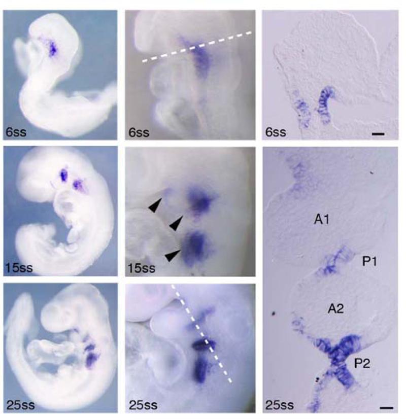
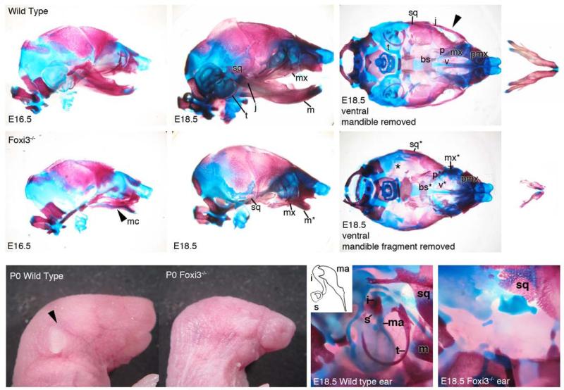
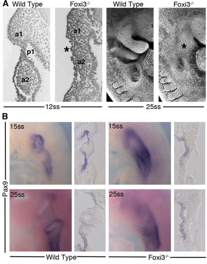
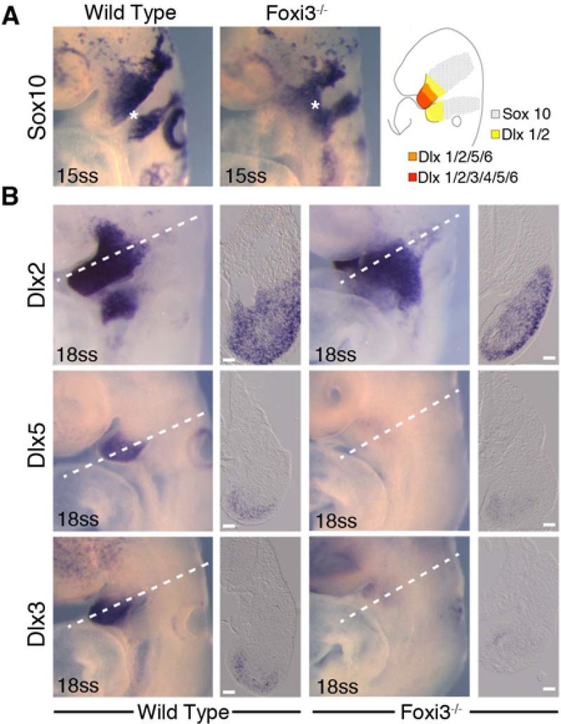
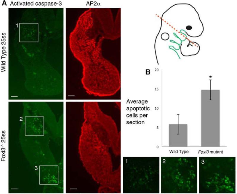
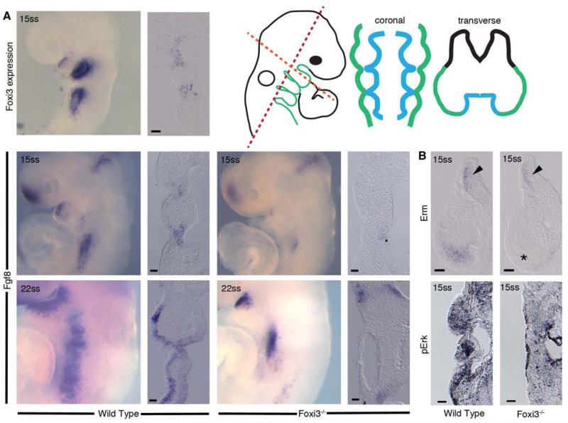
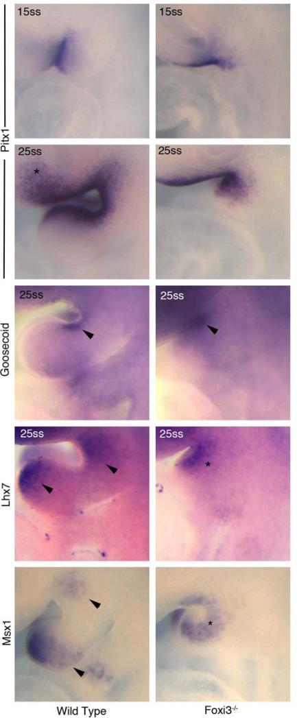
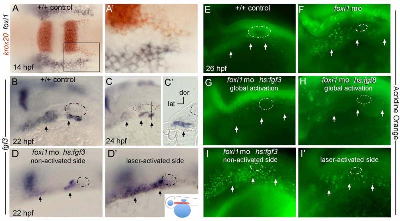
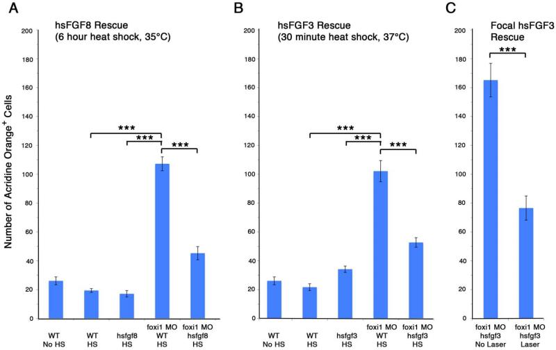
The bones of the vertebrate face develop from transient embryonic branchial arches that are populated by cranial neural crest cells. We have characterized a mouse mutant for the Forkhead family transcription factor Foxi3, which is expressed in branchial ectoderm and endoderm. Foxi3 mutant mice are not viable and display severe branchial arch-derived facial skeleton defects, including absence of all but the most distal tip of the mandible and complete absence of the inner, middle and external ear structures. Although cranial neural crest cells of Foxi3 mutants are able to migrate, populate the branchial arches, and display some elements of correct proximo-distal patterning, they succumb to apoptosis from embryonic day 9.75 onwards. We show this cell death correlates with a delay in expression of Fgf8 in branchial arch ectoderm and a failure of neural crest cells in the arches to express FGF-responsive genes. Zebrafish foxi1 is also expressed in branchial arch ectoderm and endoderm, and morpholino knock-down of foxi1 also causes apoptosis of neural crest in the branchial arches. We show that heat shock induction of fgf3 in zebrafish arch tissue can rescue cell death in foxi1 morphants. Our results suggest that Foxi3 may play a role in the establishment of signaling centers in the branchial arches that are required for neural crest survival, patterning and the subsequent development of branchial arch derivatives.

Keywords: Craniofacial development; FGF; Neural Crest; Pharyngeal arch.


Figures

Figure 1
Figure 1. Foxi3 is expressed in pharyngeal ectoderm and endoderm
Prior to branchial arch outgrowth, Foxi3 is expressed broadly in the pharyngeal region in both ectoderm and endoderm. As arch development progresses, Foxi3 expression is refined to the clefts between the arches (arrowheads). Foxi3 is expressed in both ectoderm and endoderm, but is not expressed in the mesoderm or neural crest cells that make up the mesenchyme of the arches. A1 – branchial arch 1, A2 – branchial arch 2, P1 – pharyngeal pouch 1, P2 – pharyngeal pouch 2, ss – somite stage of development. Scale bars are 100μm.
Figure 2
Figure 2. Generation of Foxi3 mutant mice
A: Diagram of the mouse Foxi3 locus, composed of two coding exons. A targeting vector was generated containing a loxP site upstream of exon 2 and an Frt-PGKNeo-Frt-LoxP sequence downstream of exon. B: The successfully targeted allele introduced an extra NheI and EcoRV site into the locus that could be detected by Southern blotting with 5’ and 3’ probes. Crossing successfully targeted offspring with CMV-Cre mice recombined the loxP sites to generate the Foxi3-del allele,
Figure 3
Figure 3. Neural crest derived bones of the head are malformed or absent in Foxi3 mutants
In Foxi3 mutants at E16.5, Meckel's cartilage (mc) is misshapen and a distinct mandible is absent. At E18.5 the full range of Foxi3 mutant skeletal defects are apparent. A truncated mandible (m*) is fused to the maxilla (mx*). The maxilla is malformed and the jugal (j) is absent (missing structures indicated by arrowheads), and the squamosal bone (sq*) and palatines (p*) are misshapen. The middle and inner ears are absent, indicated by an asterisk (*). Removed from the maxilla, the fused portion of mandible is small, truncated, and asymmetrical. Foxi3 mutant pups lack an external ear and the lower half of the face is covered in continuous ectoderm. In a magnified view of the Foxi3 mutant ear all ear structures are absent, including tympanic ring (t), malleus (ma), incus (i), and stapes (s). Basisphenoid – bs; premaxilla – pmx; vomer – v.
Figure 4
Figure 4. Foxi3 mutant mice do not form pharyngeal pouches
(A) Coronal sections through 12ss embryos stained with DAPI reveal absence of a distinct pharyngeal pouch 1 (p1) between the first two pharyngeal arches (a1, a2) in the Foxi3 mutant (asterisk). At 25 somites, confocal imaging of DAPI-stained embryos shows that Foxi3 mutant embryos have clearly failed to form distinct arches (asterisk). (B) Whole mount embryos at 15 somites and 25 somites show less distinct patterns of Pax9 expression around the presumptive pharyngeal pouches. Coronal sections through these embryos reveal continuous expression of Pax9 along the entire extent of pharyngeal endoderm at both ages in Foxi3 mutants, whereas Pax9 is restricted to pouches in wild type embryos.
Figure 5
Figure 5. Neural crest cells migrate, populate and pattern the Foxi3 mutant pharyngeal region
A: In 15 somite staged embryos, neural crest cells expressing Sox10 migrate out of the neural tube and into the pharyngeal region (asterisks), but do not divide into separate arch populations (asterisk in wild type embryo). After entering the arches, neural crest cells establish a proximal-distal axis through nested and combinatorial expression of Dlx transcription factors (illustration in A). B: In 18 somite staged Foxi3 mutants, neural crest cells adopt the nested expression pattern (A), but the population of cells expressing the intermediate and distal factors Dlx5 and Dlx3 are smaller in the mutant first arch. Scale bars are 100μm.
Figure 6
Figure 6. Neural crest cells undergo apoptosis in Foxi3 mutant arches
(A) Neural crest cells, stained with AP2α, undergo apoptosis, indicated by active caspase-3 in the distal tip of Foxi3 mutant arches (box 3). Some apoptosis in the proximal region of wild type and Foxi3 mutant arches (boxes 1 and 2). We excluded these cells from neural crest cell death quantification because they exist in both populations and are AP2α-negative. Higher magnification images of the regions highlighted with white boxes are shown below. Scale bars are 100μm. (B) The increase in apoptosis in Foxi3 mutant arches is statistically significant (p=0.04). Foxi3 mutants n=5; wild type n=6 embryos. Error bars represent the standard error of the mean.
Figure 7
Figure 7. Fgf8 expression is delayed in Foxi3 mutant pharyngeal ectoderm
The expression pattern of Fgf8 at 15 somites is similar to the Foxi3 expression pattern at the same stage. In 15 somite staged Foxi3 mutants, Fgf8 expression is substantially reduced and is present only in pharyngeal endoderm but not pharyngeal ectoderm. Expression of downstream transcription factor Erm is reduced in Foxi3 mutants to a few cells adjacent to the endoderm. MAPK signaling as indicated by di-phosphorylated Erk (pErk) is detectable in many fewer mesenchymal cells in Foxi3 mutant embryo arches. The level of staining in Foxi3 mutants was similar to that seen in wild type embryos incubated for 30 minutes in the MAPK inhibitor U0126 (not shown). Expression of Fgf8 partially recovers in pharyngeal ectoderm by 22 somites, but is still not expressed as broadly as in wild type embryos of corresponding ages. Scale bars are 100μm.
Figure 8
Figure 8. Patterning of truncated arches is minimally altered in Foxi3 mutants
At both 15 somites and 25 somites, Pitx1 expression around BA1 is unaltered. In the Foxi3 mutant there is a reduction in Pitx1-positive cells anterior to BA1 (asterisk in Pitx1 25ss). Gsc expression remains unchanged in Foxi3 mutants (arrowheads in Goosecoid). At 25 somites, Lhx7 is found in two distinct domains in wild type embryos (arrowheads in Lhx7). Only one domain is found in Foxi3 mutant embryos at the same age (asterisk in Lhx7). Similarly, at 25 somites in wild type embryos, Msx1 is found in two domains, whereas only one domain is present in Foxi3 mutants. This pattern may represent a loss of the distal Lhx7 and Msx1 domains, or a fusion of the two domains for each of these factors.
Figure 9
Figure 9. Fgf mis-expression in zebrafish rescues the cell death phenotype in foxi1 morphants
A, A’: At 14 hpf (10 somites), krox20 (red) marks streams of nascent neural crest migrating from the hindbrain whereas foxi1 (black) marks non-neural ectoderm abutting the hindbrain. No cells co-express both markers. The boxed area in A is magnified in A’. Images show a dorsal view with anterior to the left. B-I’: Embryos at later stages are oriented with anterior to the left and dorsal up, with the otic vesicle circled with a dashed line and pharyngeal pouches marked with arrows. B-C’: Expression of fgf3 in wild-type control embryos marks pharyngeal pouch endoderm at 22 hpf (B) and 24 hpf (C, C’). The vertical line in C marks the plane of section in C’. D, D’: A hs:fgf3 transgenic embryo injected with foxi1-MO shows a reduced level of fgf3 expression on the non-activated side at 22 hpf (D) but shows dramatically elevated expression on the laser-activated side (D’). Note the horizontal axis in D is inverted to facilitate comparison. Laser-activation was performed at 20 hpf, focusing on the pharyngeal region, shown in the inset to panel D’. E-I’: Live embryos were incubated with acridine orange at 26 hpf to label cells undergoing apoptosis. A wild-type control embryo shows very few apoptotic cells (E) whereas a foxi1 morphant shows a marked increase in apoptosis (F). The cell death phenotype normally seen in foxi1 morphants was strongly suppressed by global low-level activation of hs:fgf3 (37°C for 30 minutes beginning at 20 hpf) (G) or hs:fgf8 (35°C for 6 hours beginning at 20 hpf) (H). A hs:fgf3 transgenic embryo injected with foxi1-MO showed elevated apoptosis on the non-activated side (I, horizontal axis inverted for easier comparison), but apoptosis was strongly suppressed on the laser-irradiated side (I’).
Figure 10
Figure 10. Over-expression of fgf8 or fgf3 in zebrafish foxi1 morphants significantly reduces numbers of apoptotic cells in the arches
Foxi1 morphants have a significant increase in apoptosis in the arch mesenchyme over wild type zebrafish. The apoptosis phenotype can be partially rescued by global heat shock to over-express fgf8 (A) or fgf3 (B), both under the control of heat shock promoters. Fgf induction by heat shock in the absence of foxi1 knock-down does not increase cell death in the arches (A and B). The rescue effect is specific to ectodermal fgf. Induction of fgf3 in the arch ectoderm using a low energy laser significantly rescues cell death in the arches of foxi1 morphants (C). For all graphs, error bars represent the standard error of the mean, and tests of statistical significance are shown by brackets. *** = p<0.05.

References

    1. Abu-Issa R, Smyth G, Smoak I, Yamamura K, Meyers EN. Fgf8 is required for pharyngeal arch and cardiovascular development in the mouse. Development. 2002;129:4613–4625. - PubMed
    1. Adams DJ, Quail MA, Cox T, van der Weyden L, Gorick BD, Su Q, Chan WI, Davies R, Bonfield JK, Law F, Humphray S, Plumb B, Liu P, Rogers J, Bradley A. A genome-wide, end-sequenced 129Sv BAC library resource for targeting vector construction. Genomics. 2005;86:753–758. - PubMed
    1. Beermann F, Kaloulis K, Hofmann D, Murisier F, Bucher P, Trumpp A. Identification of evolutionarily conserved regulatory elements in the mouse Fgf8 locus. Genesis. 2006;44:1–6. - PubMed
    1. Brewer S, Jiang X, Donaldson S, Williams T, Sucov HM. Requirement for AP-2alpha in cardiac outflow tract morphogenesis. Mechanisms of development. 2002;110:139–149. - PubMed
    1. Brito JM, Teillet MA, Le Douarin NM. An early role for sonic hedgehog from foregut endoderm in jaw development: ensuring neural crest cell survival. Proceedings of the National Academy of Sciences of the United States of America. 2006;103:11607–11612. - PMC - PubMed

Publication types

MeSH terms