. 2014 Mar 20;10(3):e1004217.
doi: 10.1371/journal.pgen.1004217. eCollection 2014 Mar.

The Kil peptide of bacteriophage λ blocks Escherichia coli cytokinesis via ZipA-dependent inhibition of FtsZ assembly

Affiliations

The Kil peptide of bacteriophage λ blocks Escherichia coli cytokinesis via ZipA-dependent inhibition of FtsZ assembly

Daniel P Haeusser et al. PLoS Genet. .

Abstract

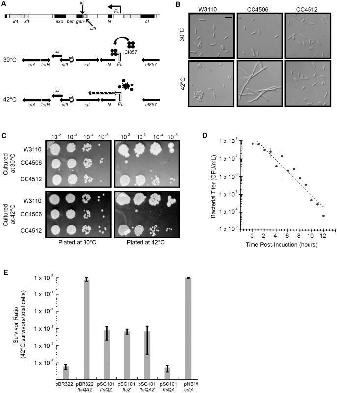
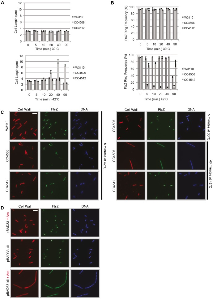
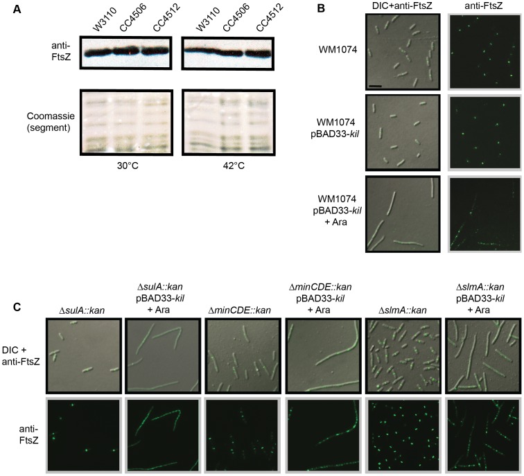
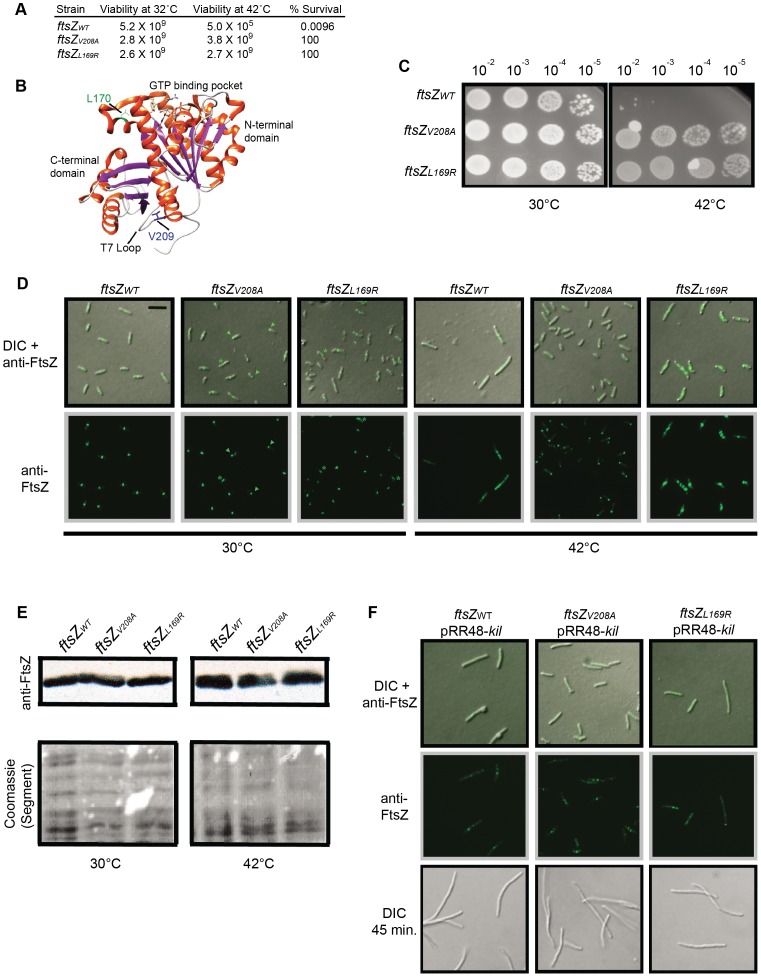
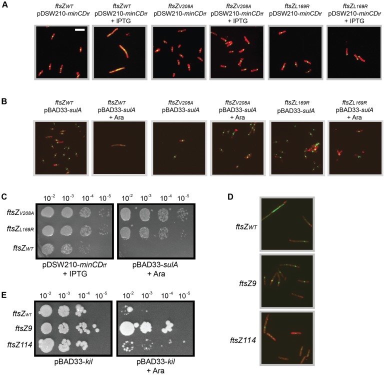
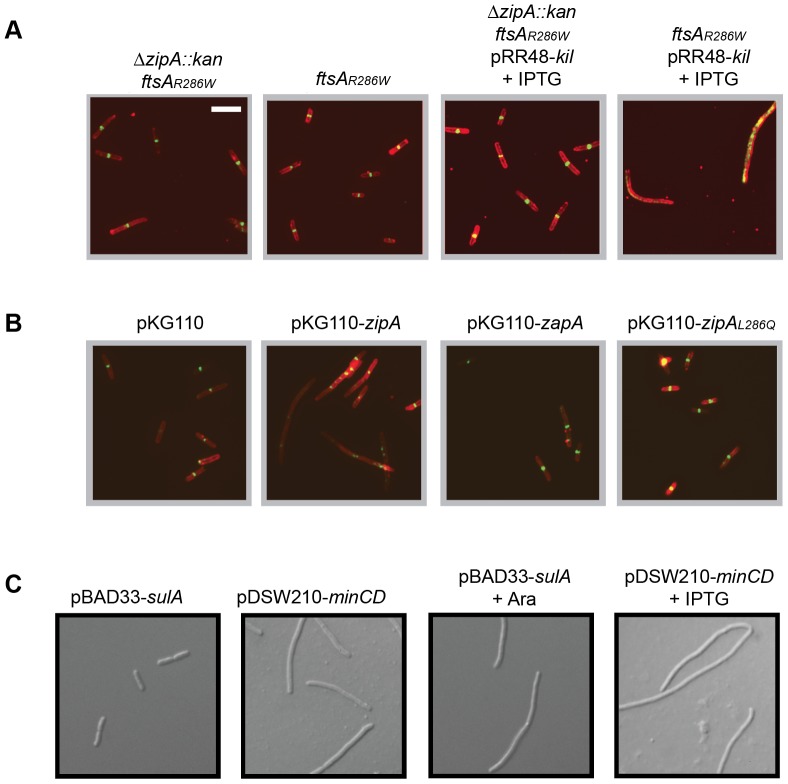
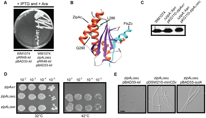
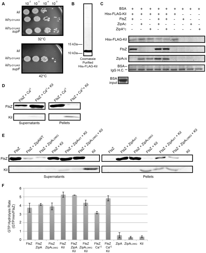
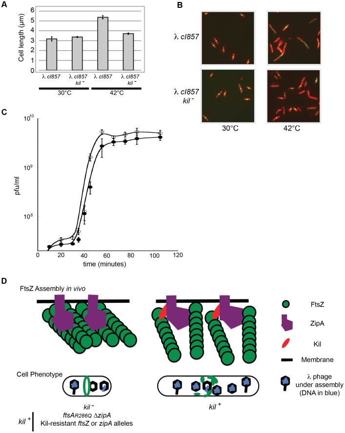
Assembly of the essential, tubulin-like FtsZ protein into a ring-shaped structure at the nascent division site determines the timing and position of cytokinesis in most bacteria and serves as a scaffold for recruitment of the cell division machinery. Here we report that expression of bacteriophage λ kil, either from a resident phage or from a plasmid, induces filamentation of Escherichia coli cells by rapid inhibition of FtsZ ring formation. Mutant alleles of ftsZ resistant to the Kil protein map to the FtsZ polymer subunit interface, stabilize FtsZ ring assembly, and confer increased resistance to endogenous FtsZ inhibitors, consistent with Kil inhibiting FtsZ assembly. Cells with the normally essential cell division gene zipA deleted (in a modified background) display normal FtsZ rings after kil expression, suggesting that ZipA is required for Kil-mediated inhibition of FtsZ rings in vivo. In support of this model, point mutations in the C-terminal FtsZ-interaction domain of ZipA abrogate Kil activity without discernibly altering FtsZ-ZipA interactions. An affinity-tagged-Kil derivative interacts with both FtsZ and ZipA, and inhibits sedimentation of FtsZ filament bundles in vitro. Together, these data inspire a model in which Kil interacts with FtsZ and ZipA in the cell to prevent FtsZ assembly into a coherent, division-competent ring structure. Phage growth assays show that kil+ phage lyse ∼30% later than kil mutant phage, suggesting that Kil delays lysis, perhaps via its interaction with FtsZ and ZipA.


Conflict of interest statement

The authors have declared that no competing interests exist.

Figures

Figure 1
Figure 1. Induction of λ kil interferes with E. coli cell division, but is suppressed by ftsZ overexpression.
Strains: W3110 = wild type, CC4506 = kil+, CC4512 = kil. (A) Location of kil in the λ genome and the defective prophage used for this study. The λ kil gene overlaps cIII and gam as part of the operon under PL control (top schematic). PL expression of kil in the defective prophage (CC4506) is under control of the temperature-sensitive cI857 repressor, allowing repression at 30°C (middle schematic) and expression at 42°C (bottom schematic). (B) Induction of kil causes cell filamentation. Differential interference contrast (DIC) microscopy of E. coli strains back-diluted from a mid-log 30°C culture to low optical density (OD600∼0.025) and grown to late-log in LB at 30°C (top panels) or 42°C (bottom panels). Scale bar = 5 µm. (C) Induction of kil leads to a loss in cell viability. Serial spot dilutions of cells grown in LB at 30°C (top panels) or 42°C for 90 minutes (bottom panels) and then plated onto LB agar incubated at 30°C (left panels) or 42°C (right panels) overnight. (D) Prolonged induction of kil leads to an irreversible loss of viability. Bacterial titers of CC4506, expressed as colony-forming units (cfu) per mL, after indicated time of kil induction. Time points with error bars represent the average and standard deviation between three replicate experiments. (E) Over-expression of the E. coli cell division gene ftsZ suppresses the loss of cell viability caused by kil induction. Plating efficiencies expressed as survivor ratios (cfu/mL surviving at 42°C divided by total cells) for CC4506 with indicated plasmids containing different combinations of the genes in the ftsQAZ operon. Error bars represent the standard deviation between three replicate experiments.
Figure 2
Figure 2. Induction of λ kil rapidly blocks FtsZ ring formation.
(A) Induction of kil causes cell filamentation. Average cell lengths (µm) at various time points (min) following temperature shift to 42°C (bottom panel) or continued incubation (control) at 30°C (top panel), measured from IFM micrographs. Error bars represent standard deviation between average length measurements from three separate images. (B & C) Midcell FtsZ ring localization is rapidly lost upon kil induction. (B) FtsZ ring frequency (%) at various time points (min) following temperature shift to 42°C (bottom panel) or continued incubation (control) at 30°C (top panel), counted from IFM micrographs. Error bars represent standard deviation between FtsZ ring frequency counts from three separate images. Cell length and ring frequency measurements at each point were obtained using a minimum number of 100 cells. (C) Representative IFM micrographs of indicated strains at indicated times and temperature from images used for measurements/counts in (A) & (B). Cell wall signal is from rhodamine-conjugated wheat-germ agglutinin, FtsZ signal is from AlexaFluor 488-conjugated, goat α–rabbit recognition of rabbit α-FtsZ, and DNA signal is from 4′,6-diamidino-2-phenylindole (DAPI). Scale bar = 5 µm. (D) Induction of kil from pBAD33 in W3110 (lacking λ phage) blocks FtsZ ring formation and leads to cell filamentation. Shown are representative IFM micrographs of W3110 cells containing indicated plasmid under indicated induction conditions. Signals and scale bar are as in (C).
Figure 3
Figure 3. Kil does not alter FtsZ levels or act through SulA, the Min system, or SlmA.
(A) FtsZ levels are unchanged by kil expression. Shown is an immunoblot against FtsZ (upper panels) from whole cell extracts of indicated strains harvested after 40 minutes of growth at 30°C (left panels) or 42°C (right panels). Relative levels of total protein are shown as a segment of Coomassie stained lanes (bottom panels). (B) Expression of kil in an MG1655-derived WM1074 background also blocks FtsZ ring formation and causes cell filamentation. DIC (left panels) and α-FtsZ IFM (left & right panels) micrographs are shown from mid-log cultures (40 minute induction) of indicated strains. (C) Kil-dependent filamentation remains in cells lacking SulA, the Min system, or SlmA (WM1074 background). DIC (top panels) and α-FtsZ IFM (top & bottom panels) micrographs from fixed mid-log cultures (40 min induction) of indicated strains are shown. Scale for micrographs in (B)–(C) is indicated by 5 µm scale bar in (B).
Figure 4
Figure 4. Characterization of Kil-resistant ftsZ alleles.
(A) The ftsZV208A and ftsZL169R mutants are Kil-resistant alleles. Shown are cfu at 32°C and 42°C and resulting survivor frequencies (cfu/mL at 42°C divided by cfu/mL at 32°C) for kil-expressing strains with ftsZWT, ftsZV208A, or ftsZL169R. (B) Location of L170 and V209 in the crystal structure of Pseudomonas aeruginosa FtsZ , corresponding to L169 and V208 of E. coli FtsZ, generated by Chimera (http://www.cgl.ucsf.edu/chimera/). α-helices are red and β-sheets are purple. The L170 sidechain, adjacent to the GTP-binding pocket, is indicated in green and the V209 sidechain, within the T7 loop, is indicated in blue. (C) Placement of isolated ftsZ alleles in a fresh background confers resistance to kil expression. Spot dilutions of indicated ftsZ alleles in a CC4506 background strain under conditions where kil is not expressed (30°C, left) and expressed (42°C, right). (D) Expression of kil in strains harboring ftsZV208A or ftsZL169R does not result in cell filamentation and permits FtsZ ring formation, although mutant FtsZ ring localization is not completely normal upon kil expression at 42°C. Representative DIC (top) or α-FtsZ (top & bottom) micrographs of fixed cells are shown with indicated ftsZ alleles in the CC4506 background at 30°C (left) and after 40 minutes of kil expression at 42°C (right). α-FtsZ signal is as described in Figure 2. Scale bar = 5 µm. For ftszV208A cells, arrowheads mark rings or puncta at cell poles and asterisks mark ring doublets. For ftsZL169R cells, asterisks mark aberrant FtsZ structures. (E) FtsZV208A and FtsZL169R are synthesized at comparable levels to FtsZWT. Shown are immunoblots against FtsZ (upper panels) from whole cell extracts of indicated strains harvested after 40 minutes of growth at 30°C (left panels) or 42°C (right panels). Relative levels of total protein are shown as a cropped segment of Coomassie-stained lanes (bottom panels). (F) The amount of Kil produced from a plasmid overcomes the resistance conferred by ftsZV208A or ftsZL169R. Shown are representative DIC (top and bottom) or α-FtsZ (top & middle) IFM of fixed cells with indicated ftsZ alleles in the CC4506 background containing pRR48-kil at 30°C, with 1 mM IPTG to induce kil from the plasmid for 15 (top and middle) or 45 minutes (bottom). Signal and scale are as in (D).
Figure 5
Figure 5. General resistance of isolated ftsZ alleles to assembly inhibition.
(A) Cells with ftsZV208A or ftsZL169R form FtsZ rings and do not filament despite overexpression of minCD. Representative IFM micrographs are shown of cell wall (red) and α-FtsZ (green) signal from fixed cells of the CC4506 background strain with pDSW210-his-minCD (minC-minD gene fusion) and the indicated ftsZ alleles grown at 30°C (no kil induction) harvested after 40 minutes of minCD induction (+IPTG) or non-inducing conditions. Cell wall and α-FtsZ signal are shown as in Figure 2. Scale bar = 5 µm. (B) Cells with ftsZV208A or ftsZL169R form FtsZ rings and do not filament despite sulA overexpression. Shown are representative IFM micrographs of cell wall (red) or α-FtsZ (green) signal from fixed cells of the CC4506 background strain with pBAD33-sulA and the indicated ftsZ alleles grown at 30°C (no kil induction) harvested after 40 minutes of sulA induction (+Ara) or non-inducing conditions. Signal and scale are as in (A). (C) The ftsZ mutations confer resistance to minCD- or sulA-induced lethality. Spot dilutions are shown of the CC4506 background strain with pDSW210-his-minCD or pBAD33-sulA and the indicated ftsZ alleles plated at 30°C (no kil expression) on LB agar with 1 mM IPTG (+IPTG) or 0.2% arabinose (+Ara). (D) Cells with the characterized ftsZ9 and ftsZ114 alleles partially suppress inhibition of FtsZ ring formation by Kil. Shown are representative IFM micrographs of cell wall (red) or α-FtsZ (green) signal from fixed cells containing the indicated ftsZ allele and pBAD33-kil grown after 40 minutes of kil induction (+Ara). Signal and scale as in (A). (E) Cells with the ftsZ9 allele completely suppress, and those with the ftsZ114 allele partially suppress the loss of viability upon kil expression. Shown are spot dilutions of cells containing the indicated ftsZ allele and pBAD33-kil plated on LB agar with (right) or without (left) 0.2% arabinose at the indicated dilutions.
Figure 6
Figure 6. Kil activity in vivo requires ZipA.
(A) Cells harboring ftsAR286W to allow bypass of zipAzipA::aph) are resistant to kil expression, but are not resistant when containing ftsAR286W alone. Shown are representative IFM micrographs of cell wall (red) or α-FtsZ (green) signal from fixed cells of the indicated genotypes in a WM1074 pRR48-kil background with (+IPTG) or without kil induction for 40 minutes. Cell wall and α-FtsZ signal are shown as in Figure 2. Scale bar = 5 µm. (B) Complementation of ftsAR286W ΔzipA::aph with zipAWT in trans from pKG110 specifically restores Kil-mediated filamentation. Shown are representative IFM micrographs of cell wall (red) or α-FtsZ (green) signal from fixed cells grown under kil-inducing conditions for 40 minutes (1 mM IPTG) of the indicated genotypes in a WM1074 ftsAR286W ΔzipA::aph pRR48-kil background. Signal and scale as in (A). (C) SulA and MinCD activity do not require zipA. Representative DIC micrographs of live cells with indicated plasmids in a WM1074 ftsAR286W ΔzipA::aph background grown with (bottom panels) or without (top panels) the indicated inducers for 40 min. Scale as in (A).
Figure 7
Figure 7. Isolation and characterization of Kil-resistant zipA alleles.
(A) The zipAL286Q allele is a spontaneously isolated mutant resistant to loss of viability caused by kil expression from dual plasmids. Shown is an image of an LB plate with 1 mM IPTG and 0.2% arabinose to induce kil from pRR48 and pBAD33 simultaneously, with the original strain (left) and the kil-resistant isolate (right). Targeted sequencing of the zipA locus identified the resistant mutation as zipAL286Q. (B) Location of L286 and Q290 of ZipA in the co-crystal structure of the E. coli ZipA C-terminal domain (ZipAC) with an E. coli C-terminal FtsZ peptide (FtsZC) , as generated by Chimera (http://www.cgl.ucsf.edu/chimera/). For ZipAC, α-helices are colored red and β-sheets are colored purple. The L286 sidechain is indicated in green and the Q290 sidechain in blue. FtsZC is colored cyan. (C) ZipAWT and ZipAL286Q expressed from pKG110 are made at levels comparable to native ZipA. Shown is an immunoblot against ZipA (upper panels) from whole cell extracts of indicated strains harvested during mid-log growth in the presence of 0.5 µM sodium salicylate. (D) Replacement of the native zipA gene with zipAL286Q or zipAL286R by recombineering confers Kil resistance. Spot dilutions of indicated zipA alleles in a CC4506 background strain are shown under conditions where kil is not expressed (32°C, left) and expressed (42°C, right). (E) Cells harboring zipAL286Q are resistant to Kil specifically, and not to MinCD or SulA. Representative DIC micrographs of WM1074 cells with the indicated plasmids are shown under inducing conditions (0.2% arabinose for pBAD33; 1 mM IPTG for pDSW210). Scale bar = 5 µm.
Figure 8
Figure 8. Kil protein copurifies with FtsZ and ZipA and inhibits FtsZ bundling in vitro.
(A) The product of the kil gene is a protein. Spot dilutions incubated at 32 or 42°C of cells containing, under PL control, a wild type copy of kil (top row), a copy of kil where the thirty-first codon is replaced with an amber stop codon (middle row), or cells of an isogenic strain containing the supF gene that allows translation through the amber stop codon (bottom row). (B) His6-FLAG-Kil purifies as a single, intense band from BL21(DE3) ftsAR286W ΔzipA::aph pBS58 cells. Shown is a Coomassie-stained 20% acrylamide SDS gel loaded with 15 µL purified His6-FLAG-Kil, with location of indicated size makers noted. (C) His6-FLAG-Kil interacts with FtsZ, ZipAC, and ZipA*C in pull down assays. Various protein samples as indicated were incubated with α-FLAG resin and extensively washed, followed by SDS-PAGE and Coomassie staining. Proteins associated with His6-FLAG-Kil should be strongly enriched in the gels. Bovine serum albumin (BSA) was added to all input samples at the level shown and migrates immediately above the IgG heavy chain (IgG H.C.) in gels after pull downs (bottom strip). (D) Kil inhibits in vitro FtsZ assembly as assayed by sedimentation of calcium-bundled FtsZ polymer bundles. FtsZ and His6-FLAG-Kil bands are shown from Coomassie-stained gels of supernatant and pellet samples, following sedimentation of FtsZ assembly reactions in the presence of 1 mM GTP and other indicated components. (E) Kil inhibits FtsZ bundling by purified ZipAWT or ZipAL286Q C-terminal domains (ZipAC or ZipA*C). Shown are FtsZ and His6-FLAG-Kil bands from Coomassie-stained gels of supernatant and pellet samples as in (D). (F) Kil slightly increases the GTPase activity of FtsZ. Rates of GTP hydrolysis are expressed as average GTP/min/FtsZ for the indicated combination of purified components in the presence of 1 mM GTP as in the sedimentation reactions in (D). Kil refers to His6-FLAG-Kil and ZipA or ZipAL286Q refer to His-tagged C-terminal domains. Error bars represent the range of values between two replicate experiments.
Figure 9
Figure 9. λ lysogen induction inhibits host cell division in a kil-dependent manner.
(A) Induction of a λ lysogen results in a kil-dependent increase in E. coli cell length prior to host cell lysis. The average fixed-cell lengths of indicated kil+ and kil strain populations 40 minutes post λ lysogen induction (42°C), or control non-inducing conditions (30°C) are plotted. Error bars represent standard deviation between three separate population measurements. (B) Induction of a λ lysogen results in a kil-dependent, near-total ablation of FtsZ rings prior to host lysis. Representative IFM micrographs stained for cell wall (red) and α-FtsZ (green) are shown from the same fixed cells used for (A); the indicated genotypes and growth temperatures are listed. Cell wall and α-FtsZ signals are as described in Figure 2. Scale bar = 5 µm. (C) A one-step growth curve of kil+ phage (solid circles) and kil phage (open circles) showing plaque forming units (pfu) per mL over time in MG1655 at 39°C. (D) A model (with key to shapes on right) for Kil activity on FtsZ ring formation (top cartoons) is depicted for the indicated strain genotypes, and the resulting effects on cellular phenotype (lower cartoons). The top left cartoon illustrates the kil condition alone, not all Kil-resistant conditions. The recruitment of Kil to midcell inhibits FtsZ assembly by completely blocking normal FtsZ ring assembly. FtsZ blobs and spirals seen by IFM suggest that FtsZ assembles aberrantly in the presence of Kil in vivo, unable to resolve into a closed ring structure. See discussion for the potential role of ZipA. Cell filamentation may increase lysis time in an un-compartmentalized host.

References

    1. Weisberg RA, Gallant JA (1967) Dual function of the lambda prophage repressor. J Mol Biol 25: 537–544. - PubMed
    1. Court D, Oppenheim AB (1983) Phage lambda's accessory genes. In: Hendrix RW, Roberts JW, Stahl FW, Weisberg RA, editors. Lambda II. Cold Spring Harbor, N.Y.: Cold Spring Harbor Laboratory. 251–278.
    1. Greer H (1975) The kil gene of bacteriophage lambda. Virology 66: 589–604. - PubMed
    1. Daniels D, Schroeder J, Szybalski W, Sanger F, Coulson A, et al... (1983) Complete annotated lambda sequence. In: Hendrix RW, Roberts JW, Stahl FW, Weisberg RA, editors. Lambda II. Cold Spring Harbor, N.Y.: Cold Spring Harbor Laboratory. 519–676.
    1. Bahl H, Echols H, Straus DB, Court D, Crowl R, et al. (1987) Induction of the heat shock response of E. coli through stabilization of sigma 32 by the phage lambda CIII protein. Genes Dev 1: 57–64. - PubMed

Publication types

MeSH terms