. 2014 Mar 20;9(3):e91859.
doi: 10.1371/journal.pone.0091859. eCollection 2014.

Relaxed rotational and scrunching changes in P266L mutant of T7 RNA polymerase reduce short abortive RNAs while delaying transition into elongation

Affiliations

Relaxed rotational and scrunching changes in P266L mutant of T7 RNA polymerase reduce short abortive RNAs while delaying transition into elongation

Guo-Qing Tang et al. PLoS One. .

Abstract

Abortive cycling is a universal feature of transcription initiation catalyzed by DNA-dependent RNA polymerases (RNAP). In bacteriophage T7 RNAP, mutation of proline 266 to leucine (P266L) in the C-linker region connecting the N-terminal promoter binding domain with the C-terminal catalytic domain drastically reduces short abortive products (4-7 nt) while marginally increasing long abortives (9-11 nt). Here we have investigated the transcription initiation pathway of P266L with the goal of understanding the mechanistic basis for short and long abortive synthesis. We show that the P266L mutation does not alter the affinity for the promoter, mildly affects promoter opening, and increases the +1/+2 GTP K(d) by 2-fold. However, unlike wild-type T7 RNAP that undergoes stepwise rotation of the promoter binding domain and DNA scrunching during initial transcription, the P266L mutant does not undergo coupled rotational/scrunching movements until 7 nt RNA synthesis. The lack of rotation/scrunching correlates with greater stabilities of the initiation complexes of the P266L and decreased short abortive products. The results indicate that the increased flexibility in the C-linker due to P266L mutation enables T7 RNAP to absorb the stress from the growing RNA:DNA hybrid thereby decreasing short abortive products. Increased C-linker flexibility, however, has an adverse effect of delaying the transition into elongation by 1-2 nt, which gives rise to long abortive products. However, a mutation in the upstream promoter region greatly decreases long abortive products in P266L reactions, rendering the combination of P266L and A-15C promoter a desirable pair for efficient in vitro transcription for RNA production. We conclude that the conformational rigidity in the C-linker region conferred by the proline at position 266 is responsible for the undesirable short abortive products, but the rigidity is critical for efficient promoter clearance and transition into elongation.


Conflict of interest statement

Competing Interests: The authors have declared that no competing interests exist.

Figures

Figure 1
Figure 1. C-linker region of T7 RNAP.
(A) The C-linker region (residues 251 to 296 in cartoon format) adopts different conformations in the initiation state (yellow, 1QLN, IC3), the elongation state (Pink, 1MSW, EC), and with 7 nt transcript bound (blue, 3E2E, IC7). The P266 and L266 residue is shown in stick format. The amino acids from 255 to 263 are disordered in the P266L structure (3E2E) and shown as dashed line. The direction of rotation of the linker near the hinge region is marked with arrows. The C-terminal domains (residues 300–883) of the three structures were aligned using Pymol (Molecular graphics systems). (B) Conservation of proline residue in the linker region between N-terminal domain and C-terminal domain at positions 266 and 270 in single-subunit RNAPs of phage, bacterium, and eukaryotic mitochondria. The N-terminal 1–300 amino acid sequence of T7 RNAP was used as a query in a BLAST amino acid search of the NCBI database for sequence alignment.
Figure 2
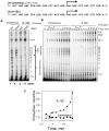
Figure 2. P266L mutation reduces short abortive without affecting the initial steps of transcription.
(A) Transcription by WT or P266L T7 RNAP (5 µM) on the T7 φ9 consensus or φ9(A-15C) promoter (10 µM) was carried out at 25°C for 1 min and products were resolved on a 23% sequencing gel and visualized by [γ-32P]GTP labeling. (B) Time course of transcription by P266L(15 µM) on consensus and A-15C promoter (10 µM). (C) The productive (runoff) to abortive ratio is higher when the P266L T7 RNAP is used in combination with the A-15C promoter mutant than with the consensus promoter. The productive to abortive ratio was obtained from data in (B).
Figure 3
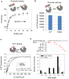
Figure 3. Promoter binding, opening, GTP binding and RNA turnover.
(A) TAMRA fluorophore labeled promoter DNA (20 nM) was titrated with increasing P266L T7 RNAP (0 to 100 nM) and increase in fluorescence anisotropy was fit to the quadratic equation with K d of 3.6±1.1 nM. Similar to the WT T7 RNAP, the P266L mutant did not cause significant changes in the TAMRA fluorescence intensity (<10%) upon binding. Effect of the intensity changes on the fitting was insignificant and corrected the same way as reported previously . Shown here are averaged values with standard deviations (error bars) from multiple independent measurements, from which K d was fitted. (B) The increase in 2-AP fluorescence at −4 in the template strand upon addition of WT and P266L indicate slightly lower promoter opening with P266L. The errors are standard deviation from 10–15 measurements. (C) Initiating GTP binding was monitored from fluorescence increase in the 2-AP (−4 position) labeled promoter DNA bound to T7 RNAP titrated with increasing 3′dGTP. The P266L binds to the initiating NTPs (3′dGTP) ∼2 times weaker than WT T7 RNAP (175 μM versus 405 μM) and Hill coefficients are 1.3±0.02 and 1.7±0.03 for WT and P266L, respectively. The errors represent the fitting uncertainty. (D) A complex of WT or P266L T7 RNAP (2 µM) and promoter DNA (1 µM) was mixed with limited NTPs or NTP and 3′-deoxy NTP mixture for 2 min at 25°C to allow RNA synthesis of the indicated lengths. The amount of RNA shown in µM is representative of the number of turnovers at each walked position for WT (black) and P266L (grey) T7 RNAP.
Figure 4
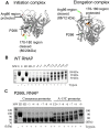
Figure 4. Subdomain H refolding is delayed in the P266L mutant.
(A) T7 RNAP in initiation and elongation conformations have different exposure of Arg 96 (red color) and residues 170–180 (brown color) to trypsin digestion. Formation of an elongation complex is characterized by the disappearance of the 80 kDa fragment and the appearance of the 88 kDa fragment. P266 is colored in green. Limited trypsin digestion (15 s) of WT T7 RNAP after walking to +4 to +19 positions. E- RNAP, ED- RNAP:promoter complex (C) Same experiment as in panel B was carried out with P266L and consensus or A-15C promoter. The digestion pattern with the A-15C promoter shows that transition to EC occurs at +9 in the A-15C promoter, but not with the consensus promoter.
Figure 5
Figure 5. Upstream bubble collapse and transition into elongation are delayed in the P266L mutant.
(A) Real time 2AP fluorescence monitors upstream bubble collapse. Representative time courses of 2-AP fluorescence changes from position −4NT in individual walking experiments (+8, +9, +12, and +15) were observed in the stopped-flow setup. The initial increase indicates rapid opening of the promoter and the decrease indicates bubble collapse. (B) Single molecule FRET histograms measure the rate of promoter unbending by WT and P266L T7 RNAP at +9. The number of DNA molecules that were analyzed to draw the smFRET histograms are as follows: P266L: 3110 at 1 min; 4164 at 5 min; 4883 at 33 min. WT: 3444 at 1 min; 3432 at 5 min; 5855 at 34 min. The x-axis shows corrected FRET (equation 2) and the y-axis represents the frequency of transcription complexes with the respective FRET values. Low FRET is observed in the elongation complex (EC) and high FRET is observed in the initiation complexes (IC). An increase in the low FRET population is observed over time after stalling at +9 using GTP+ATP+CTP+3′dUTP. Concentration of T7 RNAP-DNA, GTP, and NTPs was 10 nM, 1 mM, and 500 µM, respectively. (C) The fraction of EC versus time was fit to a single exponential function. The error bars in Fig 5C is the standard error from fitting smFRET histogram to a single exponential function. The WT (red circles) transitions to EC faster than P266L (black circles) at position +9 (τ = 4.5±1 min for P266L and 1.7±0.3 min for WT). The cartoon shows the layout of the smFRET experiments.
Figure 6
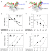
Figure 6. The P266L mutation modifies both rotation and DNA scrunching changes during initiation.
(A) Cartoon illustration of FRET experiments to measure promoter rotation. The polymerase is in gray, the non-template strand in red and the template in green. Fluorescent donor TAMRA (red sphere) was introduced at position −22 in the non-template strand and acceptor Alexa 647 (blue square) at designated downstream positions on the template strand. Transcription complexes were walked to position N (+4 to +13) and FRET efficiency between donor (D) at −22 and acceptor (A) at N+5 was measured to obtain the D-A distances (RDA, discontinuous line). (B and C) Average FRET efficiency and changes in D-A spatial distances are shown for P266L and WT T7 RNAP (D) Cartoon illustration of FRET experiments to measure DNA scrunching. The donor Cy3 (red sphere) was labeled on position −4 and acceptor Cy5 (blue square) labeled on downstream N+5 positions as above. (E and F) Average FRET efficiency and changes in averaged D-A spatial distances between Cy3 at the upstream edge (−4) and Cy5 at the downstream N+5 positions with the P266L and WT T7 RNAP complexes. The error bars of FRET efficiency represent the standard deviations from multiple independent measurements (N≥3).
Figure 7
Figure 7. Distinct transcription initiation pathways of WT and P266L T7 RNAP.
(A) The transcription initiation pathways of WT T7 RNAP (top) and P266L T7 RNAP (bottom) are shown in cartoon format to illustrate the distinct intermediate conformations. The N-terminal domain is shown in blue, C terminal domain in red, DNA in black and RNA in green. Movement of the N terminal domain is marked by the arrow. Both WT and P266L T7 RNAP bind, bend, and open the promoter DNA from −4 to +2 to the same extent. The rigid C-linker of WT favors progressive rotation of the upstream end of the promoter to accommodate the growing hybrid from +4 to +6 positions, which pushes against the N-terminal domain driving the rotation of the promoter. The pushback from the N-terminal domain destabilizes the RNA:DNA hybrid and leads to abortive synthesis in WT. The flexible C-linker of P266L mutant (bottom panel) accommodates RNA extension up to 6 nt without significant promoter/N-terminal domain rotation. The reduced DNA scrunching together with the absence of promoter rotation in the 4–6 nt RNA range in P266L suggests that the growing hybrid is accommodated by an alternative pathway. After 6 nt RNA synthesis, promoter rotation and scrunching resumes in P266L. The weakened promoter interactions in WT after 9 nt RNA synthesis allow release of the N-terminal domain and transition into elongation. Persistent promoter interactions delay the transition in P266L. (B) Template strand scrunching in P266L RNAP with 7 bp RNA:DNA. Template strand from the IC3 (PDB: 1QLN) and IC7 (PDB: 3E2E) crystal structures showing decrease in the distance between C+1 and T-3 in the IC7 structure (brown) compared to the IC3 structure (blue).

Similar articles

Cited by

References

    1. Goldman SR, Ebright RH, Nickels BE (2009) Direct detection of abortive RNA transcripts in vivo. Science 324: 927–928. - PMC - PubMed
    1. Guillerez J, Lopez PJ, Proux F, Launay H, Dreyfus M (2005) A mutation in T7 RNA polymerase that facilitates promoter clearance. Proceedings of the National Academy of Sciences of the United States of America 102: 5958–5963. - PMC - PubMed
    1. Tang GQ, Roy R, Ha T, Patel SS (2008) Transcription initiation in a single-subunit RNA polymerase proceeds through DNA scrunching and rotation of the N-terminal subdomains. Mol Cell 30: 567–577. - PMC - PubMed
    1. Durniak KJ, Bailey S, Steitz TA (2008) The structure of a transcribing T7 RNA polymerase in transition from initiation to elongation. Science 322: 553–557. - PMC - PubMed
    1. Tang GQ, Roy R, Bandwar RP, Ha T, Patel SS (2009) Real-time observation of the transition from transcription initiation to elongation of the RNA polymerase. Proc Natl Acad Sci U S A 106: 22175–22180. - PMC - PubMed

Publication types

MeSH terms