

Therapeutic interaction of systemically-administered mesenchymal stem cells with peri-implant mucosa
- PMID: 24651408
- PMCID: PMC3961234
- DOI: 10.1371/journal.pone.0090681
Therapeutic interaction of systemically-administered mesenchymal stem cells with peri-implant mucosa
Abstract
Objectives: The objective of this study was to investigate the effect of systemically transplanted mesenchymal stem cells (MSCs) on the peri-implant epithelial sealing around dental implants.
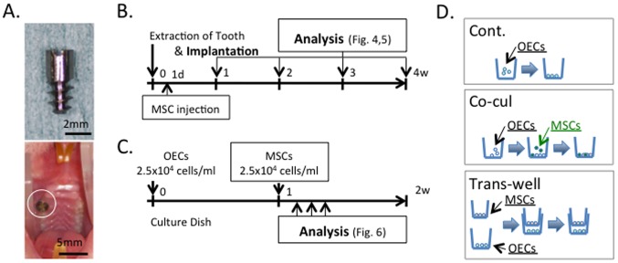
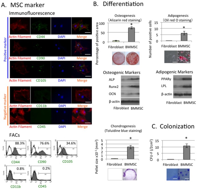
Materials and methods: MSCs were isolated from bone marrow of donor rats and expanded in culture. After recipient rats received experimental titanium dental implants in the bone sockets after extraction of maxillary right first molars, donor rat MSCs were intravenously transplanted into the recipient rats.
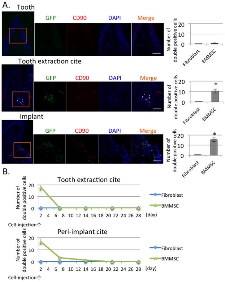
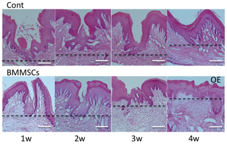
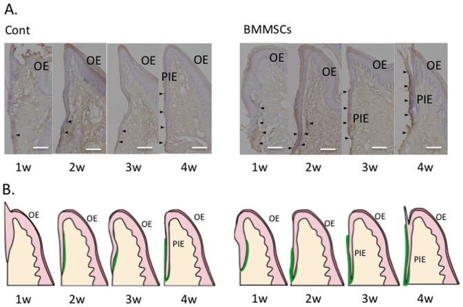
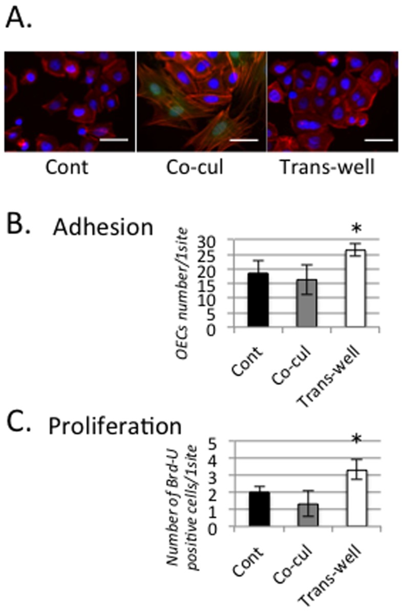
Results: The injected MSCs were found in the oral mucosa surrounding the dental implants at 24 hours post-transplantation. MSC transplantation accelerated the formation of the peri-implant epithelium (PIE)-mediated mucosa sealing around the implants at an early stage after implantation. Subsequently, enhanced deposition of laminin-332 was found along the PIE-implant interface at 4 weeks after the replacement. We also observed enhanced attachment and proliferation of oral mucous epithelial cells.
Conclusion: Systemically transplanted MSCs might play a critical role in reinforcing the epithelial sealing around dental implants.
Conflict of interest statement
Figures
Similar articles
-
The influence of systemically or locally administered mesenchymal stem cells on tissue repair in a rat oral implantation model.Int J Implant Dent. 2018 Jan 13;4(1):2. doi: 10.1186/s40729-017-0112-4. Int J Implant Dent. 2018. PMID: 29332154 Free PMC article.
-
Evaluations of epithelial sealing and peri-implant epithelial down-growth around "step-type" implants.Clin Oral Implants Res. 2012 Apr;23(4):459-66. doi: 10.1111/j.1600-0501.2011.02163.x. Epub 2011 Apr 4. Clin Oral Implants Res. 2012. PMID: 21457351
-
Changes in the distribution of laminin-5 during peri-implant epithelium formation after immediate titanium implantation in rats.Biomaterials. 2005 May;26(14):1751-60. doi: 10.1016/j.biomaterials.2004.05.033. Biomaterials. 2005. PMID: 15576149
-
Dynamics of Mucosal Integration of Machined versus Anodized Titanium Implants.J Dent Res. 2025 Mar;104(3):270-279. doi: 10.1177/00220345241296506. Epub 2024 Dec 20. J Dent Res. 2025. PMID: 39704472
-
Effects of CaCl2 hydrothermal treatment of titanium implant surfaces on early epithelial sealing.Colloids Surf B Biointerfaces. 2015 Jul 1;131:141-7. doi: 10.1016/j.colsurfb.2015.04.025. Epub 2015 Apr 18. Colloids Surf B Biointerfaces. 2015. PMID: 25982317
Cited by
-
Therapeutic interactions between mesenchymal stem cells for healing medication-related osteonecrosis of the jaw.Stem Cell Res Ther. 2016 Aug 17;7(1):119. doi: 10.1186/s13287-016-0367-3. Stem Cell Res Ther. 2016. PMID: 27530108 Free PMC article.
-
Assessment of the Soft-Tissue Seal at the Interface between the Base of the Fixed Denture Pontic and the Oral Mucosa.Materials (Basel). 2021 Jul 16;14(14):3997. doi: 10.3390/ma14143997. Materials (Basel). 2021. PMID: 34300915 Free PMC article.
-
Laminin 332-functionalized coating to regulate the behavior of keratinocytes and gingival mesenchymal stem cells to enhance implant soft tissue sealing.Regen Biomater. 2022 Aug 2;9:rbac054. doi: 10.1093/rb/rbac054. eCollection 2022. Regen Biomater. 2022. PMID: 36072266 Free PMC article.
-
The influence of systemically or locally administered mesenchymal stem cells on tissue repair in a rat oral implantation model.Int J Implant Dent. 2018 Jan 13;4(1):2. doi: 10.1186/s40729-017-0112-4. Int J Implant Dent. 2018. PMID: 29332154 Free PMC article.
-
Enamel matrix derivative enhances the proliferation and osteogenic differentiation of human periodontal ligament stem cells on the titanium implant surface.Organogenesis. 2017 Jul 3;13(3):103-113. doi: 10.1080/15476278.2017.1331196. Epub 2017 Jun 9. Organogenesis. 2017. PMID: 28598248 Free PMC article.
References
-
- Brånemark PI, Adell R, Albrektsson T, Lekholm U, Lundkvist S, et al. (1983) Osseointegrated titanium fixtures in the treatment of edentulousness. Biomaterials 4: 25–28. - PubMed
-
- Buser D, Weber HP, Donath K, Fiorellini JP, Paquette DW, et al. (1992) Soft tissue reactions to non-submerged unloaded titanium implants in beagle dogs. J Periodontol 63: 225–235. - PubMed
-
- Gould TR, Westbury L, Brunette DM (1984) Ultrastructural study of the attachment of human gingiva to titanium in vivo. J Prosthet Dent 52: 418–420. - PubMed
-
- Schroeder A, van der Zypen E, Stich H, Sutter F (1981) The reactions of bone, connective tissue, and epithelium to endosteal implants with titanium-sprayed surfaces. J Maxillofac Surg 9: 15–25. - PubMed
-
- Furuhashi A, Ayukawa Y, Atsuta I, Okawachi H, Koyano K (2012) The difference of fibroblast behavior on titanium substrata with different surface characteristics. Odontology 100: 199–205. - PubMed
Publication types
MeSH terms
Substances
LinkOut - more resources
Full Text Sources
Other Literature Sources
