. 2014 Mar 20;5(3):e1139.
doi: 10.1038/cddis.2014.97.

Heme oxygenase-1 derived carbon monoxide permits maturation of myeloid cells

Affiliations

Heme oxygenase-1 derived carbon monoxide permits maturation of myeloid cells

B Wegiel et al. Cell Death Dis. .

Abstract

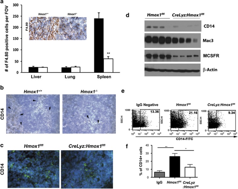
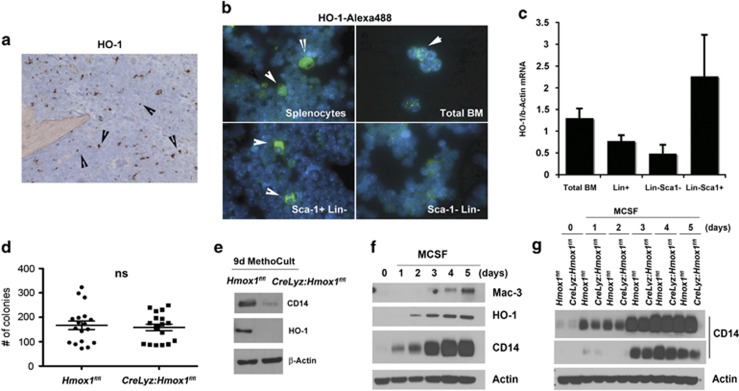
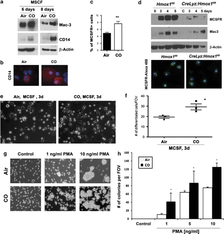
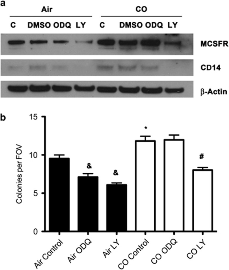
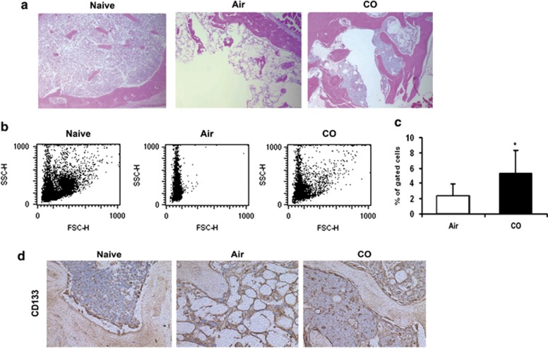
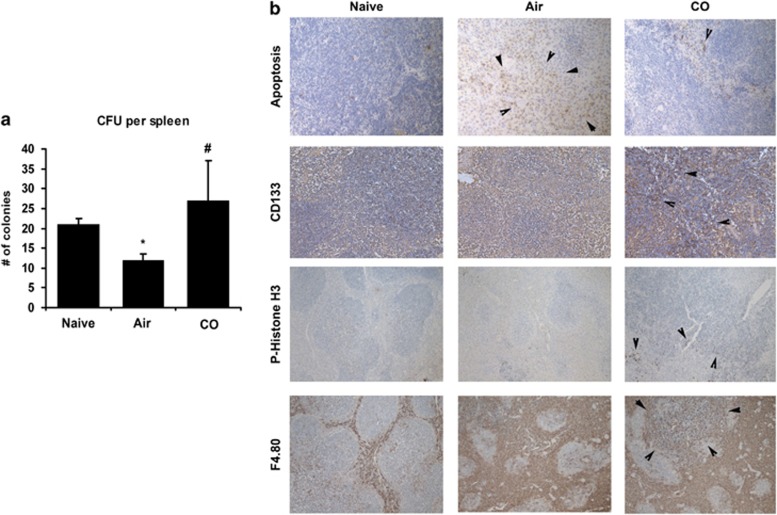
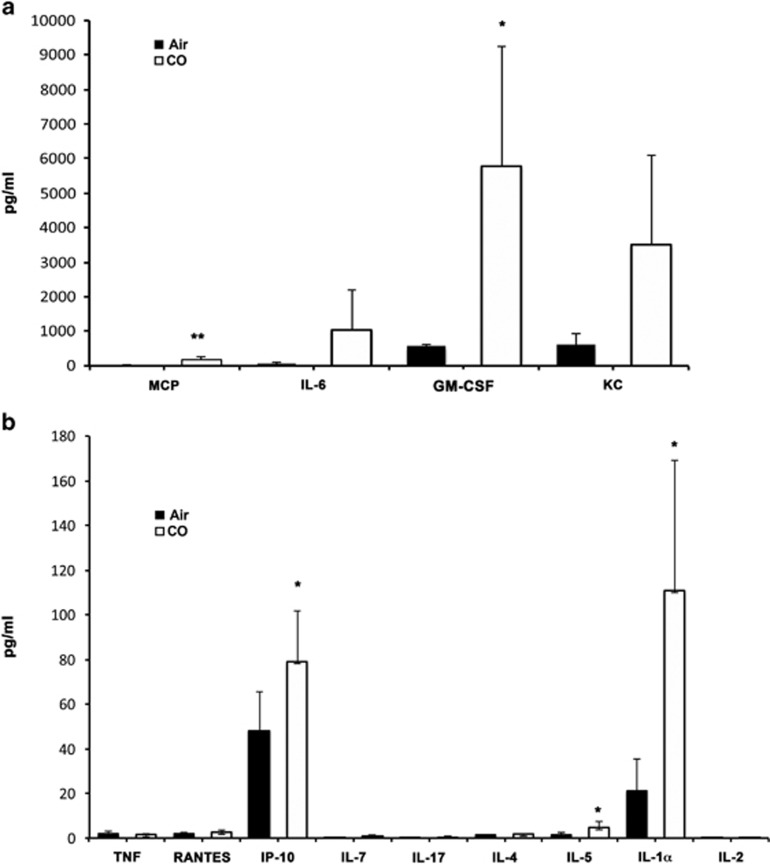
Critical functions of the immune system are maintained by the ability of myeloid progenitors to differentiate and mature into macrophages. We hypothesized that the cytoprotective gas molecule carbon monoxide (CO), generated endogenously by heme oxygenases (HO), promotes differentiation of progenitors into functional macrophages. Deletion of HO-1, specifically in the myeloid lineage (Lyz-Cre:Hmox1(flfl)), attenuated the ability of myeloid progenitors to differentiate toward macrophages and decreased the expression of macrophage markers, CD14 and macrophage colony-stimulating factor receptor (MCSFR). We showed that HO-1 and CO induced CD14 expression and efficiently increased expansion and differentiation of myeloid cells into macrophages. Further, CO sensitized myeloid cells to treatment with MCSF at low doses by increasing MCSFR expression, mediated partially through a PI3K-Akt-dependent mechanism. Exposure of mice to CO in a model of marginal bone marrow transplantation significantly improved donor myeloid cell engraftment efficiency, expansion and differentiation, which corresponded to increased serum levels of GM-CSF, IL-1α and MCP-1. Collectively, we conclude that HO-1 and CO in part are critical for myeloid cell differentiation. CO may prove to be a novel therapeutic agent to improve functional recovery of bone marrow cells in patients undergoing irradiation, chemotherapy and/or bone marrow transplantation.


Figures

Figure 1
Figure 1
Lack of HO-1 in myeloid cells results in lower number of mature macrophages. (a) Immunohistochemistry with antibody against F4.80 in the spleens, lungs, and livers from Hmox1−/− and Hmox1+/+ mice. Number of F4.80-positive cells per field of view (n=4–6) was evaluated. Inset: Representative staining is shown. n=3 per group. **P<0.001 Hmox1−/− versus Hmox1+/+. (b) Immunohistochemistry of the spleens from Hmox1−/− and Hmox1+/+ mice with staining against CD14. Representative sections are shown. (c) Immunostaining with antibodies against CD14 (Alexa-488) was performed on the spleen sections from Hmox1flfl and CreLyz:Hmox1flfl mice. n=3–4 per group. (d) Immunoblotting in the lysates of the spleens from Hmox1flfl and CreLyz:Hmox1flfl mice. Antibodies against CD14, Mac3, and MCFR were applied to test for macrophages maturation. β-Actin was used as loading control. n=4 mice per group. (e, f) Blood cells were isolated by Ficol extraction from Hmox1flfl and CreLyz:Hmox1flfl mice (n=2–3 per group) and stained against CD14 conjugated with FITC. Appropriate IgG-FITC control antibodies were used. Representative flow cytometry plots are shown in e, and quantitation of fluorescent mean is shown in f. ANOVA, P=0.0026; Tukey's test: **P<0.01 Hmox1flfl versus IgG and *P<0.05 Hmox1flfl versus CreLyz:Hmox1flfl
Figure 2
Figure 2
HO-1 dictates bone marrow differentiation toward macrophage linage. (a) Immunohistochemistry with antibody against HO-1 in bone marrow of wild-type mice. Note the single positive cells in the bone marrow progenitor fraction. (b) Immunofluorescent staining of HO-1 on enriched progenitor cells from the bone marrow in a C57/BL6 mouse. Isolated splenocytes (positive control for HO-1 staining), total bone marrow cells, sca1+/lin- and sca1-/lin- populations were subjected to cytospin and stained with antibody against HO-1. Representative pictures from 2 to 3 experiments are shown. (c) Real time PCR was performed in the cell fractions as in b. HO-1 expression per β-Actin is presented as fold change over total BM levels of HO-1. (d) Total BM cells from Hmox1flfl and CreLyz:Hmox1flfl mice were isolated and cultured in methylcellulose medium for 9 days following manufacturer's protocol. Colony counts in methylcellulose medium were performed on day 9. P>0.05, NS (e). Total BM cells from Hmox1flfl and CreLyz:Hmox1flfl mice were isolated and cultured in methylcellulose medium for 9 days following manufacturer's protocol. Immunoblotting with antibody against CD14 and HO-1 was performed on cell lysates. Data are representative for 2–3 experiments. (f) BM cells were induced toward macrophage differentiation using MCSF (20 ng/ml). Detection of HO-1, Mac3, and CD14 was performed by immunoblotting. Cells were harvested on days 0–5 during differentiation. Data are representative for at least three experiments. (g) BM cells isolated from Hmox1flfl and CreLyz:Hmox1flfl mice were subjected to MCSF-induced differentiation as in f, and the levels of CD14 were measured by immunoblotting. Note that the levels of CD14 are lower in BM from CreLyz:Hmox1flfl mice as early as day 1. Two different exposures of the same CD14 blot are presented to better appreciate the changes in early and later time points
Figure 3
Figure 3
CO accelerates differentiation and maturation of macrophages. (a) BM was isolated from wild-type mice and stimulated with MCSF in the presence or absence of CO (250 p.p.m.) for 6 and 8 days. Immunoblotting with antibody against Mac3 and CD14 was performed in the lysates of differentiated macrophages. (b) Immunostaining with antibody against CD14 in CO differentiated cells cultured in methylcellulose for 9 days. Data are representative for two independent experiments performed in triplicates. (c) Flow cytometry of BMDM treated with CO for 24 h and cell surface MCSFR was detected. n=3 in duplicates, **P<0.01. (d) Immunoblotting and immunostaining with antibodies against MCSFR and Mac3 in total BM cells from Hmox1flfl and CreLyz:Hmox1flfl mice differentiated with MCSF (20 ng/ml) for 2, 4, and 5 days. Data are representative for three independent experiments. Densitometric quantitation is shown in Supplementary Figure 6. (e, f). Macrophage colony formation was tested at suboptimal levels of MCSF (1 ng/ml) in the presence or absence of CO (250 p.p.m.). Significant increase in the number of CO-induced BMDM was observed at day 3. Data are representative for three independent experiments. *P<0.05. (g, h) U937 cells were stimulated with PMA (1, 5, or 10 ng/ml) to induce differentiation toward macrophages. Representative data are shown in g and quantitation is presented in h. n=3 experiments in triplicates. *P<0.05. CO versus Air at each time point
Figure 4
Figure 4
CO-induced differentiation and maturation of macrophages is partially mediated through PI3K-Akt pathway. (a) Immunoblotting with antibodies against MCSFR and CD14 at stimulation with suboptimal levels of MCSF (1 ng/ml) in the presence or absence of CO (250 p.p.m.). Cells were pretreated with LY24009 or ODQ for 1 h prior treatment with CO. Data are representative for two independent experiments. (b) Macrophage colony formation was tested in cells treated as above at day 3. ANOVA, P<0.0001; Tukey's test: &P<0.01 Air+ODQ or Air+LY versus Air, *P<0.05 CO versus Air, #P<0.001 CO+LY versus CO, NS CO versus CO+ODQ
Figure 5
Figure 5
CO treatment enhances survival and bone marrow engraftment in marginal bone marrow transplant. (ac) Representative section of bones and flow cytometry differential counts of bone marrow cells at day 7 are shown; n=3–4 per group. *P<0.05. (d) Immunohistochemical staining with antibodies against CD133 in the group of CO and Air-treated mice after marginal bone marrow transplantation at day 7
Figure 6
Figure 6
CO induces early engraftment and expansion of myeloid progenitors in the spleen during marginal bone marrow transplant. (a) Number of colony-forming units in the spleen in the mice after marginal BM transplant as in Figure 5. *P<0.05 Air versus Naive and #P<0.05 CO versus Air. (b) Immunohistochemistry with antibody against CD133 (progenitor marker), P-Histone-H3 (proliferation marker), F4.80 (macrophage marker) as well as TUNEL staining (apoptosis) was performed in the spleens as in a
Figure 7
Figure 7
Luminex cytokine assay was performed in serum from mice treated with CO (250 p.p.m.) or Air for 1 h. n=5 mice per group. Highly elevated and mildly elevated cytokines and chemokines are presented in a and b, respectively. *P<0.05 CO versus Air, **P<0.01 CO versus Air

References

    1. Kumagai Y, Takeuchi O, Akira S. Pathogen recognition by innate receptors. J Infect Chemother. 2008;14:86–92. - PubMed
    1. Galli SJ, Borregaard N, Wynn TA. Phenotypic and functional plasticity of cells of innate immunity: macrophages, mast cells and neutrophils. Nat Immunol. 2011;12:1035–1044. - PMC - PubMed
    1. Toobiak S, Shaklai M, Shaklai N. Carbon monoxide induced erythroid differentiation of K562 cells mimics the central macrophage milieu in erythroblastic islands. PloS One. 2012;7:e33940. - PMC - PubMed
    1. Kozakowska M, Ciesla M, Stefanska A, Skrzypek K, Was H, Jazwa A, et al. Heme oxygenase-1 inhibits myoblast differentiation by targeting myomirs. Antioxid Redox Signal. 2012;16:113–127. - PMC - PubMed
    1. Vanella L, Kim DH, Asprinio D, Peterson SJ, Barbagallo I, Vanella A, et al. HO-1 expression increases mesenchymal stem cell-derived osteoblasts but decreases adipocyte lineage. Bone. 2010;46:236–243. - PMC - PubMed

Publication types

MeSH terms