. 2014 Mar 20;9(3):e90714.
doi: 10.1371/journal.pone.0090714. eCollection 2014.

High genetic diversity and adaptive potential of two simian hemorrhagic fever viruses in a wild primate population

Affiliations

High genetic diversity and adaptive potential of two simian hemorrhagic fever viruses in a wild primate population

Adam L Bailey et al. PLoS One. .

Erratum in

  • PLoS One. 2014;9(7):e102939

Abstract

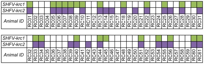
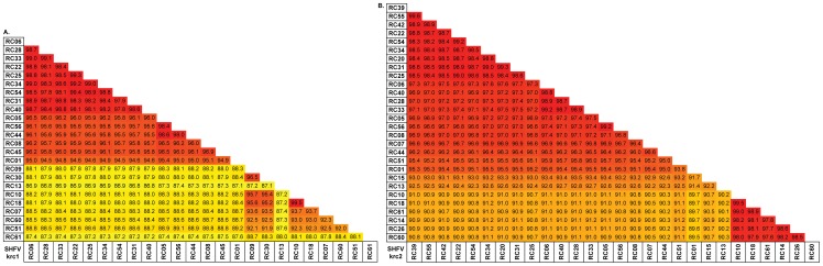
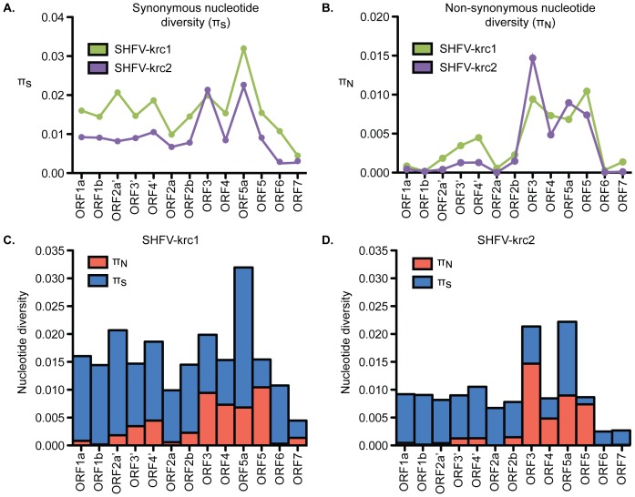
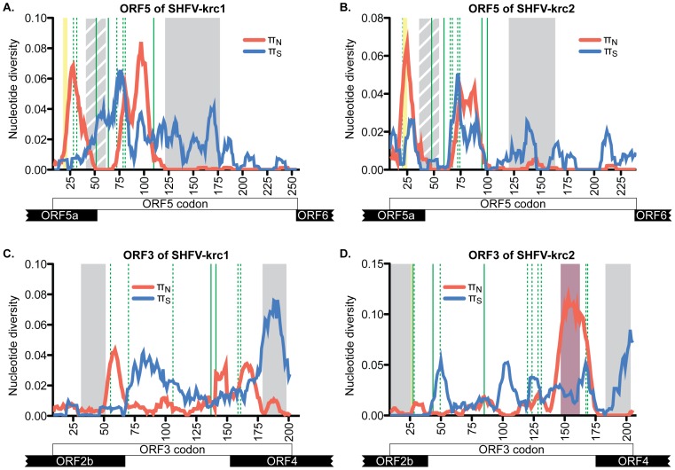
Key biological properties such as high genetic diversity and high evolutionary rate enhance the potential of certain RNA viruses to adapt and emerge. Identifying viruses with these properties in their natural hosts could dramatically improve disease forecasting and surveillance. Recently, we discovered two novel members of the viral family Arteriviridae: simian hemorrhagic fever virus (SHFV)-krc1 and SHFV-krc2, infecting a single wild red colobus (Procolobus rufomitratus tephrosceles) in Kibale National Park, Uganda. Nearly nothing is known about the biological properties of SHFVs in nature, although the SHFV type strain, SHFV-LVR, has caused devastating outbreaks of viral hemorrhagic fever in captive macaques. Here we detected SHFV-krc1 and SHFV-krc2 in 40% and 47% of 60 wild red colobus tested, respectively. We found viral loads in excess of 10(6)-10(7) RNA copies per milliliter of blood plasma for each of these viruses. SHFV-krc1 and SHFV-krc2 also showed high genetic diversity at both the inter- and intra-host levels. Analyses of synonymous and non-synonymous nucleotide diversity across viral genomes revealed patterns suggestive of positive selection in SHFV open reading frames (ORF) 5 (SHFV-krc2 only) and 7 (SHFV-krc1 and SHFV-krc2). Thus, these viruses share several important properties with some of the most rapidly evolving, emergent RNA viruses.


Conflict of interest statement

Competing Interests: Dr. Jens H. Kuhn is employed by a commercial company, Tunnell Government Services, Inc. Tunnell is a subcontractor to Battelle Memorial Institute, which is the primary contractor to NIH to run the NIH facility “Integrated Research Facility at Fort Detrick”. As the authors pointed out in the disclaimer, Tunnell's involvement with Battelle, and Battelle's involvement with NIH is stipulated in US Department of Health and Human Services contract HHSN272200700016I. All work performed by Jens H. Kuhn (and all other Tunnell IRF employees) is property of the US government under the contract. There are no patents, products in development or marketed products to declare. This does not alter the authors' adherence to all the PLOS ONE policies on sharing data and materials.

Figures

Figure 1
Figure 1. Schematic of the SHFV genome.
(A) ORFs as they are referred to in Lauck et al., 2011 , labeled sequentially 5′-3′: ORF1a-ORF9. Asterisks denote ORFs identified in SHFV-krc1 and SHFV-krc2 not reported in Lauck et al., 2011 . (B) ORFs as they are named in Snijder et al., 2013 , labeled 5′-3′: ORF1a-ORF7, with duplicated ORFs designated by a “prime” (e.g. ORF2a’). Expression products are given in bold.
Figure 2
Figure 2. Infection frequency of SHFV-krc1 and SHFV-krc2 in the Kibale red colobus.
SHFV-krc1 (green) and SHFV-krc2 (purple) infections were identified by “unbiased” deep sequencing and confirmed by strain-specific qRT-PCR.
Figure 3
Figure 3. Viral loads of SHFV-krc1 and SHFV-krc2 in the Kibale red colobus.
Comparison of SHFV-krc1 (green) and SHFV-krc2 (purple) viral loads from all animals positive for either virus (A) and viral loads from mono-infections vs. co-infections of SHFV-krc1 (B) and SHFV-krc2 (C). RNA was isolated from blood plasma and quantitative RT-PCR was performed using strain-specific primers and probes designed from deep sequencing data. Statistical significance was assessed using a two-tailed t-test performed on log-transformed values (CI = 95%).
Figure 4
Figure 4. Pairwise comparison of nucleotide identity among variants of SHFV-krc1 and SHFV-krc2 from Kibale red colobus (RC).
Full coding sequences for each isolate were aligned using CLC Genomics Workbench. Numbers show percent nucleotide identity between two variants within (A) SHFV-krc1 or (B) SHFV-krc2. Colors highlight similarity, with red representing the most similar sequences and yellow representing sequences with the lowest degree of nucleotide identity. The same color scale was used for (A) and (B).
Figure 5
Figure 5. Overall nucleotide diversity of SHFV-krc1 and SHFV-krc2.
Mean (± S.E.) πS (A), πN (B), and πNS (C) in monkeys infected with SHFV-krc1 (green) and SHFV-krc2 (purple). Paired t-tests were performed to compare mean values between SHFV-krc1 and SHFV-krc2.
Figure 6
Figure 6. Nucleotide diversity of SHFV-krc1 and SHFV-krc2 by ORF.
Interaction graphs comparing mean πS (A) and πN (B) in ORFs from SHFV-krc1 (green) and SHFV-krc2 (purple). In the case of πN there was a significant ORF-by-virus interaction (F13, 459 = 4.39; p<0.001). Comparison of mean πs (blue) to πN (red) within ORFs of SHFV-krc1 (C) and SHFV-krc2 (D) revealed substantial differences among ORFs within each virus.
Figure 7
Figure 7. Nucleotide diversity across ORF3 and ORF5 of SHFV-krc1 and SHFV-krc2.
Mean πS (blue) and πN (red) in sliding windows of 9 codons across the coding region of ORF5 (A,B) and ORF3 (C,D). Overlapping ORFs are shown at the bottom. Grey boxes represent predicted transmembrane domains, with striped grey boxes representing a hydrophobic region unique to the SHFVs. Green lines depict putative sites of N-glycosylation, with dashed green lines showing sites that are variably glycosylated. Yellow boxes show predicted signal peptide cleavage sites that vary in location in GP5 of SHFV-krc1 and SHFV-krc2 and were not found in GP3 of SHFV-krc1. The purple box corresponds to the unique region of highly variable acidic residues found only in ORF3 of SHFV-krc2.
Figure 8
Figure 8. Relationship between viral load and nucleotide diversity.
(A) Synonymous nucleotide diversity (πS) and (B) nonsynonymous nucleotide diversity (πN) were plotted against log-transformed viral loads for both SHFV-krc1 (green) and SHFV-krc2 (purple) infections. A significant correlation between nucleotide diversity and viral load was found for both πS (r2 = 0.2465, p = 0.0015) and πN (r2 = 0.1749, p = 0.0090).

Similar articles

Cited by

References

    1. Holmes EC, Grenfell BT (2009) Discovering the phylodynamics of RNA viruses. PLoS computational biology 5: e1000505. - PMC - PubMed
    1. Parrish CR, Holmes EC, Morens DM, Park E-C, Burke DS, et al. (2008) Cross-species virus transmission and the emergence of new epidemic diseases. Microbiology and molecular biology reviews: MMBR 72: 457–470. - PMC - PubMed
    1. Grenfell BT, Pybus OG, Gog JR, Wood JLN, Daly JM, et al. (2004) Unifying the epidemiological and evolutionary dynamics of pathogens. Science (New York, NY) 303: 327–332. - PubMed
    1. King DA, Peckham C, Waage JK, Brownlie J, Woolhouse MEJ (2006) Epidemiology. Infectious diseases: preparing for the future. Science (New York, NY) 313: 1392–1393. - PubMed
    1. Woolhouse M, Scott F, Hudson Z, Howey R, Chase-Topping M (2012) Human viruses: discovery and emergence. Philosophical transactions of the Royal Society of London Series B, Biological sciences 367: 2864–2871. - PMC - PubMed

Publication types

LinkOut - more resources