. 2014 Mar 20;9(3):e92640.
doi: 10.1371/journal.pone.0092640. eCollection 2014.

ATM alters the otherwise robust chromatin mobility at sites of DNA double-strand breaks (DSBs) in human cells

Affiliations

ATM alters the otherwise robust chromatin mobility at sites of DNA double-strand breaks (DSBs) in human cells

Annabelle Becker et al. PLoS One. .

Abstract

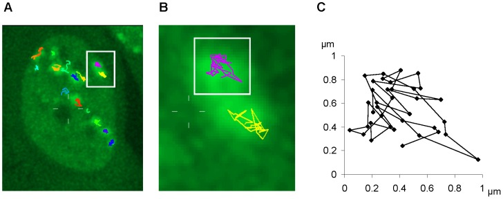
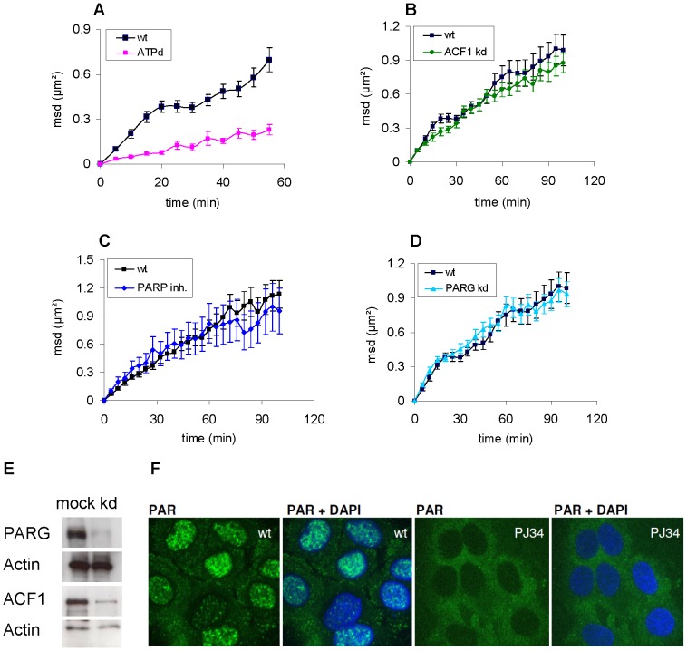
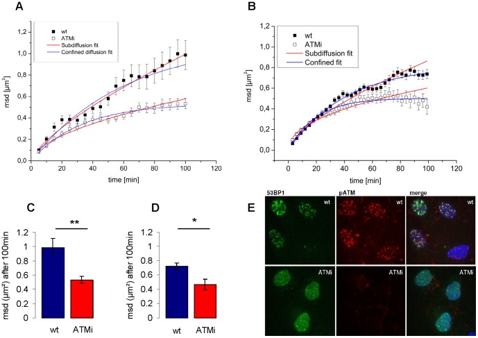
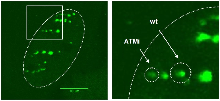
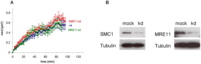
Ionizing radiation induces DNA double strand breaks (DSBs) which can lead to the formation of chromosome rearrangements through error prone repair. In mammalian cells the positional stability of chromatin contributes to the maintenance of genome integrity. DSBs exhibit only a small, submicron scale diffusive mobility, but a slight increase in the mobility of chromatin domains by the induction of DSBs might influence repair fidelity and the formation of translocations. The radiation-induced local DNA decondensation in the vicinity of DSBs is one factor potentially enhancing the mobility of DSB-containing chromatin domains. Therefore in this study we focus on the influence of different chromatin modifying proteins, known to be activated by the DNA damage response, on the mobility of DSBs. IRIF (ionizing radiation induced foci) in U2OS cells stably expressing 53BP1-GFP were used as a surrogate marker of DSBs. Low angle charged particle irradiation, known to trigger a pronounced DNA decondensation, was used for the defined induction of linear tracks of IRIF. Our results show that movement of IRIF is independent of the investigated chromatin modifying proteins like ACF1 or PARP1 and PARG. Also depletion of proteins that tether DNA strands like MRE11 and cohesin did not alter IRIF dynamics significantly. Inhibition of ATM, a key component of DNA damage response signaling, resulted in a pronounced confinement of DSB mobility, which might be attributed to a diminished radiation induced decondensation. This confinement following ATM inhibition was confirmed using X-rays, proving that this effect is not restricted to densely ionizing radiation. In conclusion, repair sites of DSBs exhibit a limited mobility on a small spatial scale that is mainly unaffected by depletion of single remodeling or DNA tethering proteins. However, it relies on functional ATM kinase which is considered to influence the chromatin structure after irradiation.


Conflict of interest statement

Competing Interests: The authors have declared that no competing interests exist.

Figures

Figure 1
Figure 1. Mobility measurement of 53BP1 IRIF in U2OS-cells.
A) 2D maximum projection of a U2OS cell nucleus after irradiation with Cr ions (LET: 2360 keV/μm) with 53BP1-GFP accumulation (green foci) along the ion trajectory at sites of DSBs. Colored tracks represent movement of 53BP1 foci measured by 2D tracking within three hours of observation. B) Magnification of two exemplary tracks of 53BP1 mobility indicated by the white square in A. C) Spatial mobility of the in B) selected 53BP1 track over three hours.
Figure 2
Figure 2. Influence of repair-related chromatin modifying proteins on mobility of 53BP1-GFP foci in irradiated U2OS cells.
A) Plot of the mean square displacement (msd) of IRIF in control cells (wt) and cells which were depleted for ATP 30 min prior to carbon ion (LET: 170 keV/μm) irradiation (n = 7). Errors represent SEM in all plots. B) Msd of IRIF in cells after knockdown of ACF1 (n = 23) and non treated controls (wt) (n = 11) after irradiation with Cr (LET: 2360 keV/μm). C) Comparison of the msd of IRIF in cells after inhibition of PARP (10 μM PJ34) and controls (wt) (n = 15). Cells were irradiated with C (LET: 170 keV/μm). D) Msd of IRIF in cells after knockdown of PARG (n = 15) and in non treated controls (wt) (n = 11). Cells were irradiated with Cr (LET: 2360 keV/μm). E) Western Blot showing the ACF1 and PARG knockdown efficiency with actin as loading control. F) U2OS cells treated with 20 mM H2O2 for 10 min, fixed and stained for PAR (green) and DNA (blue) show efficiency of PARP1 inhibition with 10 μM PJ34.
Figure 3
Figure 3. Inhibition of ATM constricts mobility of 53BP1 foci induced by heavy ion or photon irradiation.
Irradiation of U2OS cells was performed by Cr (LET: 2630 keV/μm) for plots A and C and by 1 Gy X-rays for plots B and D. The mean square displacement (msd) of IRIF is plotted over time. Errors represent SEM. A, B) Msd plots of control (solid squares) (Cr n = 11, X-ray n = 21) and ATM inhibited cells (KU55933 open squares) (Cr n = 31, X-ray n = 11) fitted for subdiffusion (red line) and confined diffusion (blue line). C,D) Bar graphs of the average msd after 100 min observation time by live cell microscopy for control and ATM inhibited cells (KU55933) after irradiation with Cr C) and after irradiation with 1 Gy X-rays D). E) U2OS-53BP1-GFP cells irradiated with 1 Gy X-rays, fixed after 30 min and stained for pATM (red) and DNA (blue). Wt compared to cells treated with 15 μM KU55933 for 2 hours (ATMi) show efficiency of ATM kinase inhibition.
Figure 4
Figure 4. Reduced confinement radius of IRIF in U2OS cells after inhibition of ATM.
A) U2OS cell stably expressing 53BP1 after low angle irradiation with C (170 keV/μm). B) Magnification of the cell nucleus. Cells and calculated confinement areas (see eq. 2) are marked by dotted lines. For simplification, both radii were exemplarily shown in the same nucleus. 3D Confinement volumes in ATM inhibited cells are decreased by a factor of 3 compared to the reference volumes of non treated cells.
Figure 5
Figure 5. Knockdown of SMC1 or MRE11 does not influence mobility of IRIF.
A) Mean square displacement (msd) of 53BP1 foci after irradiation with Pb (LET: 13500 keV/μm) is plotted against time for wt (blue line), SMC1 knockdown (red line) and MRE11 knockdown cells (green line). B) Western blots of U2OS cells 48 h after knockdown of SMC1 and MRE11 with tubulin as loading control.

References

    1. Sulli G, Di Micco R, d'Adda di Fagagna F (2012) Crosstalk between chromatin state and DNA damage response in cellular senescence and cancer. Nat Rev Cancer 12: 709–720. - PubMed
    1. Lukas J, Lukas C, Bartek J (2011) More than just a focus: The chromatin response to DNA damage and its role in genome integrity maintenance. Nat Cell Biol 13: 1161–1169. - PubMed
    1. Shi L, Oberdoerffer P (2012) Chromatin dynamics in DNA double-strand break repair. Biochim Biophys Acta 1819: 811–819. - PMC - PubMed
    1. Goodarzi A a, Noon AT, Deckbar D, Ziv Y, Shiloh Y, et al. (2008) ATM signaling facilitates repair of DNA double-strand breaks associated with heterochromatin. Mol Cell 31: 167–177. - PubMed
    1. Dion V, Gasser SM (2013) Chromatin movement in the maintenance of genome stability. Cell 152: 1355–1364. - PubMed

Publication types

MeSH terms