

Trehalose maintains vitality of mouse epididymal epithelial cells and mediates gene transfer
- PMID: 24651491
- PMCID: PMC3961358
- DOI: 10.1371/journal.pone.0092483
Trehalose maintains vitality of mouse epididymal epithelial cells and mediates gene transfer
Abstract
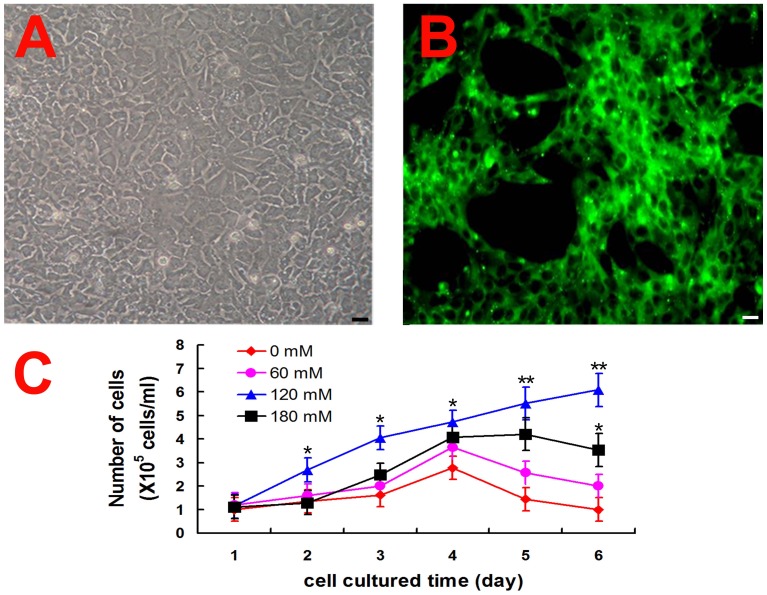
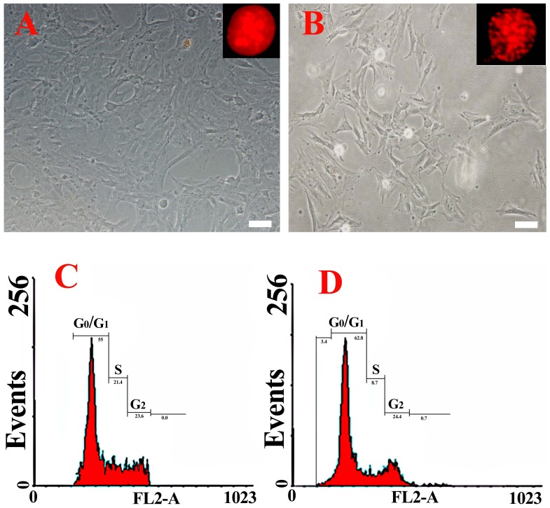
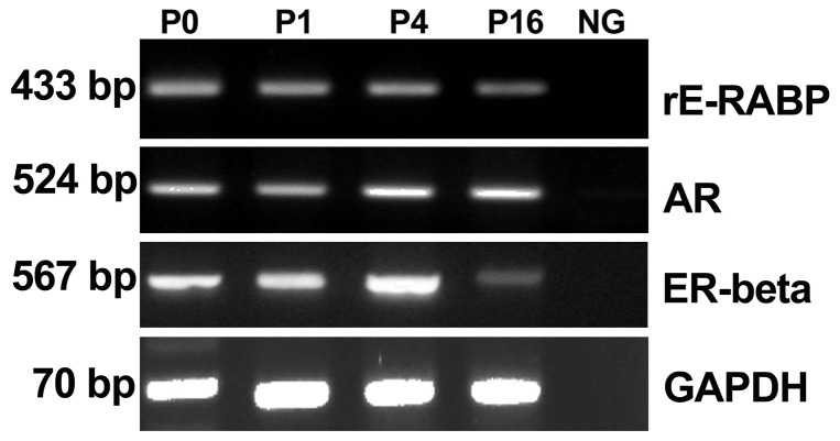
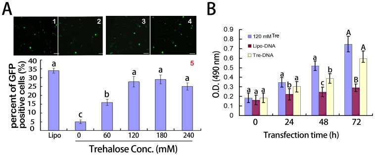
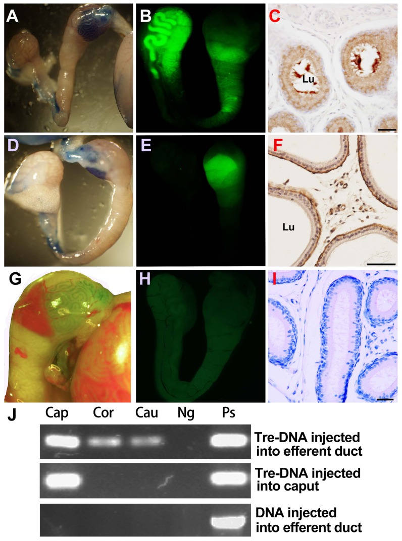
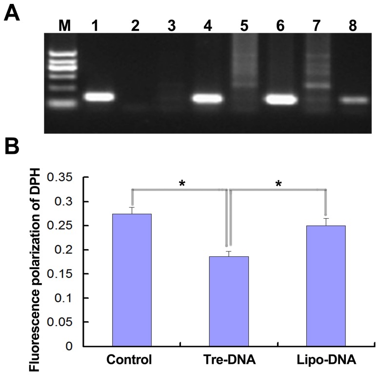
In the present study, trehalose was utilized to improve primary culture of mouse epididymal epithelial cells in vitro, and to enhance naked DNA delivery in epididymis in vivo. During the six-day culture, the proliferation activity of the cells in the medium with addition of trehalose was higher than that of those cells cultured in absence of trehalose (p<0.01). To determine the optimal concentration for cell proliferation, a series of trehalose concentrations (0, 60, 120, 180 mM) were tested, and the result indicated that the cell in the medium with 120 mM trehalose showed the highest proliferation potential. The epididymis epithelial cells were cultured in the medium containing 120 mM trehalose upon 16th passage, and they continued expressing markers of epididymal epithelial cell, such as rE-RABP, AR and ER-beta. Our study also indicated that trehalose concentrations of 120-240 mM, especially 180 mM, could effectively enhance DNA delivery into the mouse epididymis epithelial cell in vitro. Moreover, trehalose could induce in vivo expression of exogenous DNA in epididymal epithelial cells and help to internalize plasmid into sperm,which did not influence motility of sperm when the mixture of trehalose (180 mM) and DNA was injected into epididymal lumen through efferent tubule. This study suggested that trehalose, as an effective and safer reagent, could be employed potentially to maintain vitality of mouse epididymal epithelial cells during long-term culture in vitro and to mediate in vitro and in vivo gene transfer.
Conflict of interest statement
Figures
Similar articles
-
Direct injection of foreign DNA into mouse testis as a possible in vivo gene transfer system via epididymal spermatozoa.Mol Reprod Dev. 2002 Jan;61(1):49-56. doi: 10.1002/mrd.1130. Mol Reprod Dev. 2002. PMID: 11774375
-
Epididymal epithelial cells cultured in vitro prolong the motility of bovine sperm.J Androl. 2000 Nov-Dec;21(6):842-7. J Androl. 2000. PMID: 11105910
-
Junctional adhesion molecule A: expression in the murine epididymal tract and accessory organs and acquisition by maturing sperm.Mol Hum Reprod. 2017 Feb 10;23(2):132-140. doi: 10.1093/molehr/gaw082. Mol Hum Reprod. 2017. PMID: 28062807 Free PMC article.
-
Profiling of epididymal small non-protein-coding RNAs.Andrology. 2019 Sep;7(5):669-680. doi: 10.1111/andr.12640. Epub 2019 Apr 25. Andrology. 2019. PMID: 31020794 Review.
-
Contribution of epididymal factors to sperm maturation and storage.Andrologia. 1998 Aug-Sep;30(4-5):233-9. doi: 10.1111/j.1439-0272.1998.tb01165.x. Andrologia. 1998. PMID: 9739420 Review.
Cited by
-
Advancement and Potential Applications of Epididymal Organoids.Biomolecules. 2024 Aug 17;14(8):1026. doi: 10.3390/biom14081026. Biomolecules. 2024. PMID: 39199413 Free PMC article. Review.
-
Testosterone promotes GPX5 expression of goat epididymal epithelial cells cultured in vitro.In Vitro Cell Dev Biol Anim. 2019 Oct;55(9):677-685. doi: 10.1007/s11626-019-00391-y. Epub 2019 Aug 19. In Vitro Cell Dev Biol Anim. 2019. PMID: 31429037
-
Trehalose can effectively protect sheep epididymis epithelial cells from oxidative stress.Arch Anim Breed. 2021 Aug 18;64(2):335-343. doi: 10.5194/aab-64-335-2021. eCollection 2021. Arch Anim Breed. 2021. PMID: 34458560 Free PMC article.
-
Exploring Trehalose on the Release of Levonorgestrel from Implantable PLGA Microneedles.Polymers (Basel). 2020 Jan 1;12(1):59. doi: 10.3390/polym12010059. Polymers (Basel). 2020. PMID: 31906331 Free PMC article.
-
Conditioned medium and secretome from epididymal epithelial cell cultures improve sperm kinetics and capacitation.Vet World. 2023 Jun;16(6):1325-1332. doi: 10.14202/vetworld.2023.1325-1332. Epub 2023 Jun 13. Vet World. 2023. PMID: 37577187 Free PMC article.
References
-
- Esponda P (2011) Gene transfer to the mammalian reproductive tract. Zygote 19: 287–295. - PubMed
-
- Bongso A, Trounson A (1996) Evaluation of motility, freezing ability and embryonic development of murine epididymal sperm after coculture with epididymal epithelium. Hum Reprod 11: 1451–1456. - PubMed
-
- Bassols J, Kadar E, Briz MD, Pinart E, Sancho S, et al. (2004) In vitro culture of epithelial cells from the caput, corpus, and cauda epididymis of Sus domesticus. Theriogenology 62: 929–942. - PubMed
-
- Moore HD, Hartman TD, Smith CA (1986) In-vitro culture of hamster epididymal epithelium and induction of sperm motility. J Reprod Fertil 78: 327–336. - PubMed
-
- Moore HD, Samayawardhena LA, Brewis IA (1998) Sperm maturation in vitro: co-culture of spermatozoa and epididymal epithelium. J Reprod Fertil Suppl 53: 23–31. - PubMed
Publication types
MeSH terms
Substances
LinkOut - more resources
Full Text Sources
Other Literature Sources
Research Materials
