Review
. 2014 Apr;15(4):225-42.
doi: 10.1038/nrm3775.

Organization and execution of the epithelial polarity programme

Affiliations
Review

Organization and execution of the epithelial polarity programme

Enrique Rodriguez-Boulan et al. Nat Rev Mol Cell Biol. 2014 Apr.

Abstract

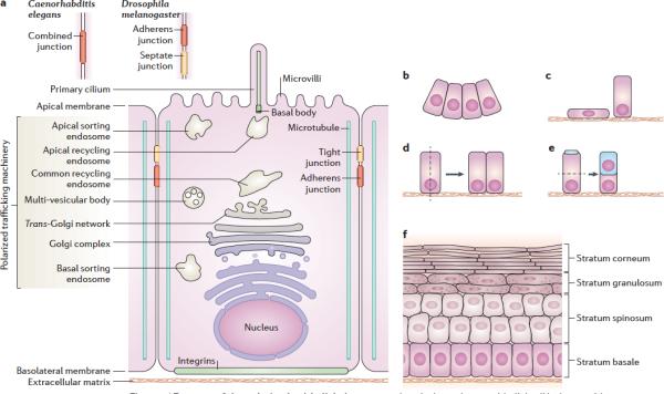
Epithelial cells require apical-basal plasma membrane polarity to carry out crucial vectorial transport functions and cytoplasmic polarity to generate different cell progenies for tissue morphogenesis. The establishment and maintenance of a polarized epithelial cell with apical, basolateral and ciliary surface domains is guided by an epithelial polarity programme (EPP) that is controlled by a network of protein and lipid regulators. The EPP is organized in response to extracellular cues and is executed through the establishment of an apical-basal axis, intercellular junctions, epithelial-specific cytoskeletal rearrangements and a polarized trafficking machinery. Recent studies have provided insight into the interactions of the EPP with the polarized trafficking machinery and how these regulate epithelial polarization and depolarization.


Figures

Figure 1
Figure 1. Features of the polarized epithelial phenotype
(a) A typical vertebrate epithelial cell is shown with components of the polarized vesicle sorting machinery and the apical junctional complex depicted. Note that invertebrate (for example in Drosophila) epithelial cells lack primary cilia and the junctional complex is organized differently with adherens junctions located more apically than the sealing junction (named septate junction instead of tight junction) (top right insert). In C. elegans, adherens junctions and sealing junctions are combined into a single structure (top left insert). (b - f) Epithelial cells organize into different structures through their cytoskeleton and through oriented cell division. (b) actin-mediated constriction of the apical domain causes furrowing; (c) reducing the size of the lateral membrane produces squamous epithelia; (d) cell division (indicated by the dashed line) in the plane of the epithelium expands the sheet; whereas divisions perpendicular to the plane may generate different cell lineages in the case of stem cells (e), or may give rise to stratified epithelia (f)
Figure 2
Figure 2. The EPP players. (a) Feed-back loops between polarity proteins
The differential localization of polarity proteins at the cell cortex is regulated through binding interactions (dashed lines) and phosphorylations (solid arrows); dashed arrows indicate change to or from a phosphorylated state. The apical polarity protein Crumbs (Crb) recruits Pals1 through its C-terminal PDZ-binding domain (ERLI) which recruits Par6 to phosphorylate Par3, the kinase Par1 and LGL and exclude them (together with other members of the Scribble complex (Dlg Scribble) from the apical domain. Members of the Scribble complex interact genetically with each other but there is no evidence of physical interaction. Conversely, Par1-mediated phosphorylation events prevent basal invasion by the apical polarity determinants, such as Par3. Phosphorylated proteins, including Par1, LGL and Par3, bind Par5 during relocation to their resident domain. Polarity lipids also help generate membrane asymmetries. Specifically, PTEN, recruited to the junctional area through interaction with Par3, generates ptdIns(4,5)P2 (PIP2), which helps recruit Cdc42 via annexin 2. Cdc42 participates in the activation of aPKC via Par6. Basolateral PI3K, recruited to the junctional area through E-cadherin, recruits Dlg and generates PIP3, which additionally contributes to basal membrane identity through the recruitment of Scribble. Lgl contributes to basal identity through interaction with Syntaxin 4, which promotes basolateral secretion. (b) Modular organization of the EPP players. EPP proteins are made up of several modular domains, which enable key interactions to occur between among EPP players. They also allow interactions to occur between EPP players and other proteins that are necessary for polarity. Solid arrows indicate phosphorylation events, dashed lines represent binding interactions.
Figure 3
Figure 3. Execution of the EPP
The epithelium is the first tissue to appear during development. (A) In mammalian embryos, an epithelium arises when morula cells compact and form a lumen upon expression of E-cadherin, to form a blastocyst. (Ba) In the bilaminar embryo, epiblast epithelial cells at the primitive streak differentiate into mesenchymal cells (epithelial-mesenchymal transition (EMT)) that migrate to form the intermediate mesoderm. In turn, mesoderm cells convert into epithelial cells (mesenchymal-epithelial transition (MET)), for example during formation of the kidney. (Bb) During MET, epithelial cells express epithelial signature markers such as E-cadherin (left), laminin receptors (integrins), Crumb complex proteins and undergo a dramatic cytoskeletal re-organization and organelle repositioning , , , -.The Crumbs complex, the Cdc42-Par3-Par6-aPKC complex and the Scribble complex cooperate to form an immature apical junctional complex (left plasma membrane), which matures into segregated tight and adherens junctions (right plasma membrane). Junction formation involves the delivery of E-cadherin from apical recycling endosomes to form spot adherens junctions and the exocytosis of the tight junction components occludin and claudins by the basolateral sorting machinery to form tight junctions. Rac1 interacts with Par 3 through Tiam1 and thus contributes to the organization of the peri-junctional actin cytoskeleton required for the coalescence of spot into belt adherens junctions. RhoA and myosin2 contribute as well to the formation of an actomyosin belt that enhances cell adhesion , . At the basal pole, Rac1 stimulates the secretion of laminin that interacts with basal integrin receptors; this contributes to the orientation of the cell along an apical–basal axis.. The relocalization of the centrosome to the apical pole, the developing junctions, the polarity protein Par1, together with APC, the kinesin KIF17 and EB1 contribute to the reorganization of microtubules, In turn this contributes to the polarized organization of endosomal compartments and the Golgi complex. EMT (right) is promoted by transcriptional events that often involve TGFβ receptors and culminate with the loss of E-cadherin resulting in the disassembly of adherens junctions . The polarized movement of mesenchymal cells along cytokine gradients is regulated by many polarity proteins and lipids that are also part of the EPP. At the front of the cell, activated CDC42 recruits Par6-aPKC, and ultimately GSK3b and APC to control the position of the nucleus and centrosome. In turn, the centrosome nucleates a centrifugal array of microtubules with peripheral plus ends that mediates: the juxta-nuclear localization of the Golgi apparatus, TGN and recycling endosomes , and the peripheral localization of sorting endosomes via plus-end kinesins,; and the formation of an actin-based leading edge. Microtubules facing the direction of forward motion are stabilized by formins and activate Rac1 (activated also by aPKC), which, in turn promotes actin polymerization and the formation of a actin-driven frontal lamellipodium through Arp 2/3 , RhoA is activated at the back end to control the generation of contractile forces through regulation of actomyosin filament assembly and contraction (C) Blastoderm cellularization in Drosophila melanogaster. This process illustrates an alternative strategy to generate an epithelial cell. Embryonic development in flies begins with a rapid series of nuclear divisions without cytokinesis that originate a syncytial embryo with the nuclei present at the periphery. Nuclei are segregated from other nuclei by a compartmentalization process that involves the formation of membrane cleavage furrows between cells. E-cadherin, found initially in puncta at apical surface is displaced to the tip of the growing cleavage furrows and is progressively recruited more apically to form belt-like adherens junctions in a process promoted by Par3 and the actin cytoskeleton. Septate junctions form more basally stimulated by the Scribble complex.
Figure 4
Figure 4. Trafficking of EPP players during polarization of epithelial cells
a. Polarization of MDCK cells in 3D cultures. The steps that lead to the establishment of apical–basal polarity have been best characterized in MDCK cell cysts, generated by cell division from individual cells in collagen-rich matrigel gels. (a) Polarization starts at the two-cell stage with the accumulation of E-cadherin, occludin and exocyst components (such as Sec10)at the site of cell–cell contact called apical membrane initiation site (AMIS). (b) The AMIS progresses into a pre-apical patch through a series of trafficking events that include transcytosis of Crumbs and podocalyxin from the periphery to apical recycling endosomes and their vesicular delivery to AMIS mediated by Rab11a, exocyst (Sec10) and the t-SNARE syntaxin 3. Exocytosis also functions in the initial recruitment of Par3 and aPKC (to the pre-apical patch, where they contribute to form an immature junctional complex. (c) The mature cyst exhibits segregated tight junctions and adherens junctions, a fully developed lumen expanded through polarized fluid transport and polarized cytoskeleton and organelles. (d) Control of Crumbs recycling. The localization of Crumbs is maintained through endocytosis and recycling. Crumbs interacts with a FERM domain protein, Expanded (Exp), and the Pals1–Par6–aPKC complex. The FERM domain protein links Crb to the cortical actin cytoskeleton. A Hippo pathway protein, Kibra, can bind to Exp and inhibits aPKC activity. Kibra is in turn inhibited by a lateral polarity protein, Lgl. Lgl is phosphorylated by aPKC, which causes to dissociate from the membrane. Endocytosis of Crb is followed by retromer-mediated retrograde transport to the trans-Golgi network (TGN), from where it can be recycled back to the apical cortex.
Figure 5
Figure 5. The primary cilium and Hedgehog signaling
a. Primary cilium structure and signaling components. Multiple types of diffusion barrier separate the ciliary membrane and apical membrane to control the access to the primary cilium, creating a distinct polarized membrane domain Transition fibers that connect the basal body to the ciliary membrane blocks vesicle access to the cilium, and nuclear pore components are also found in this region and have been implicated to function as a diffusion barrier, perhaps functioning as a diffusion barrier to the entry and exit of soluble proteins. Patched (Ptc) is inactive in the primary cilium, but moves out of this organelle upon binding of Hedgehog (HH) (not shown). Other components of the HH signalling cascade, Smoothened (Smo), which is located on intracellular vesicles, and Gli, also move in and out of the cilium in a dynamic manner, and is recruited to the tip of the cilium by a kinesin. Activation of Ptc triggers the translocation of Smo to the ciliary membrane, where it activates Gli. After a series of post-translational modifications, GLi moves to the nucleus to activate transcription of target genes. b. Transcytosis of HH. HH is produced by endothelial or mesenchymal cells underlying epithelial cells. Upon binding of HH to its receptor Patched on the surface of the primary cilium, the transcription factor Gli is activated and transported to the nucleus to activate epithelial differentiation genes. The transcytotic pathway of HH has not yet been characterized.

References

    1. Dickinson DJ, Nelson WJ, Weis WI. A polarized epithelium organized by beta- and alpha-catenin predates Cadherin and metazoan origins. Science. 2011;331:1336–9. - PMC - PubMed
    1. Cereijido M, Contreras RG, Shoshani L. Cell adhesion, polarity, and epithelia in the dawn of metazoans. Physiol Rev. 2004;84:1229–62. - PubMed
    1. Magie CR, Martindale MQ. Cell-cell adhesion in the cnidaria: insights into the evolution of tissue morphogenesis. Biol Bull. 2008;214:218–32. - PubMed
    1. Alberts B. Cell biology: the endless frontier. Mol Biol Cell. 2010;21:3785. - PMC - PubMed
    1. Bryant DM, Mostov KE. From cells to organs: building polarized tissue. Nat Rev Mol Cell Biol. 2008;9:887–901. - PMC - PubMed

Publication types