Review
. 2014 Apr;15(4):243-56.
doi: 10.1038/nrm3772.

Metabolic requirements for the maintenance of self-renewing stem cells

Affiliations
Review

Metabolic requirements for the maintenance of self-renewing stem cells

Keisuke Ito et al. Nat Rev Mol Cell Biol. 2014 Apr.

Abstract

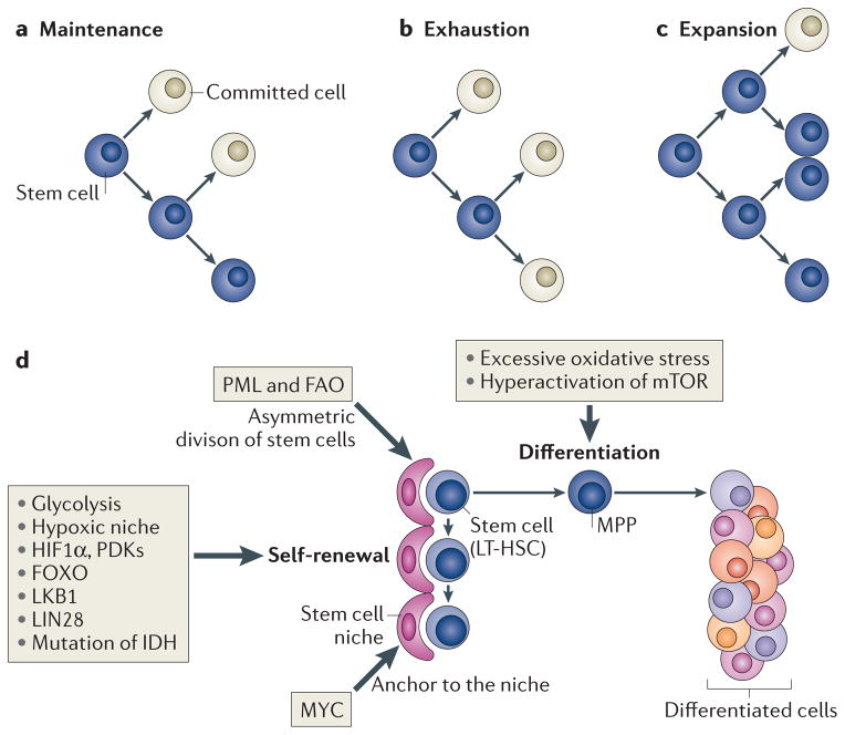
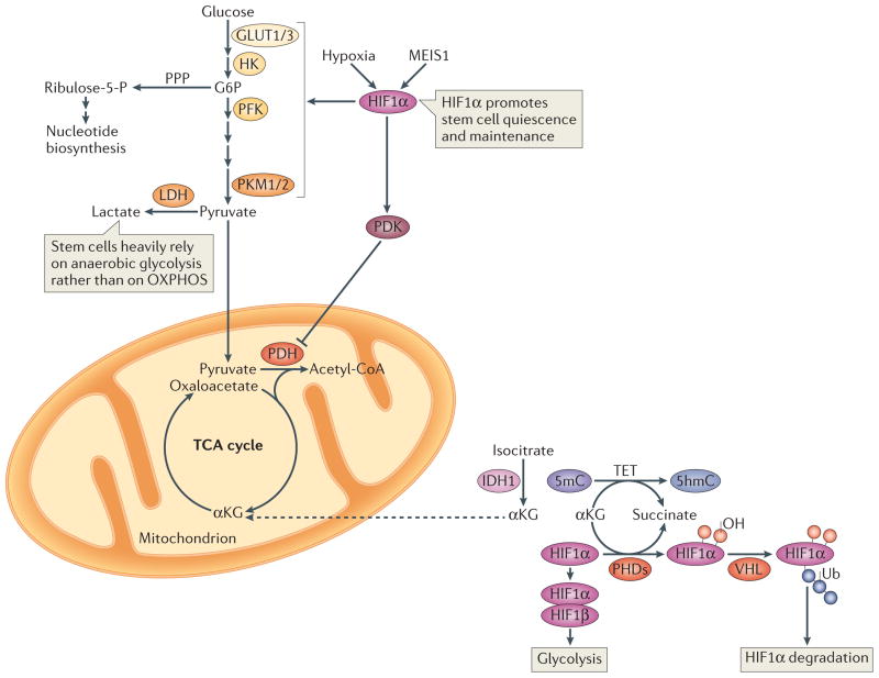
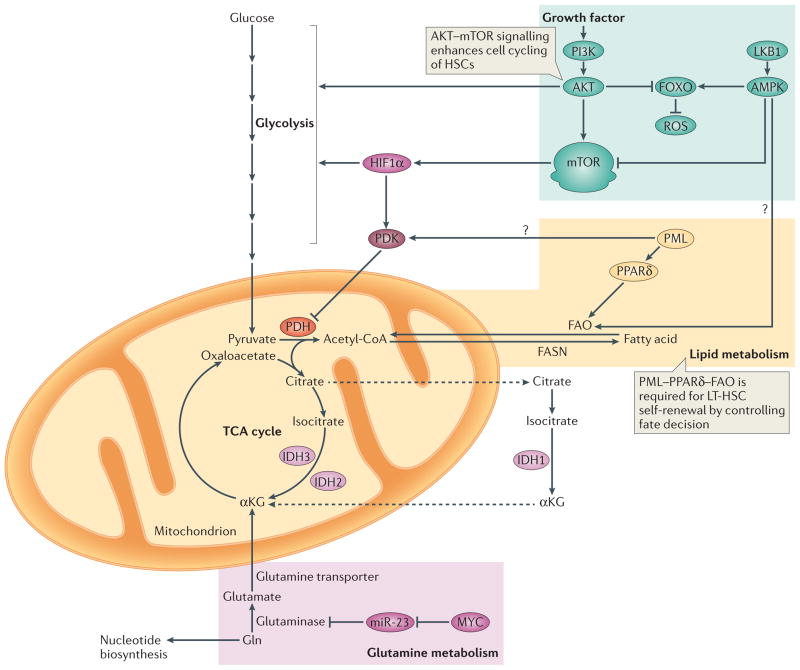
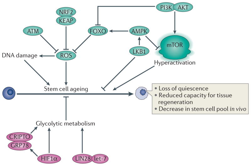
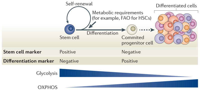
A distinctive feature of stem cells is their capacity to self-renew to maintain pluripotency. Studies of genetically-engineered mouse models and recent advances in metabolomic analysis, particularly in haematopoietic stem cells, have deepened our understanding of the contribution made by metabolic cues to the regulation of stem cell self-renewal. Many types of stem cells heavily rely on anaerobic glycolysis, and stem cell function is also regulated by bioenergetic signalling, the AKT-mTOR pathway, Gln metabolism and fatty acid metabolism. As maintenance of a stem cell pool requires a finely-tuned balance between self-renewal and differentiation, investigations into the molecular mechanisms and metabolic pathways underlying these decisions hold great therapeutic promise.


Conflict of interest statement

Competing interests statement

The authors declare no competing interests.

Figures

Figure 1
Figure 1. Two specific potentials and cell fates of stem cells
Stem cells exhibit both self-renewal capacity and pluripotency (parts a,b,c). Asymmetric cell division has been suggested as a regulator of stem cell-fate decisions and is essential for the maintenance of the stem cell compartment (part a). Alterations in the equilibrium of self-renewal and commitment of adult stem cells can affect tissue homeostasis and can lead to stem cell exhaustion (part b) or expansion (part c). Several tissue stem cells (part d) (for example, long-term haematopoietic stem cells (LT-HSCs) in the bone marrow niche) maintain a quiescent state, as this is essential for preserving their self-renewal capacity. Many types of stem cells heavily rely on anaerobic glycolysis to maintain such a quiescent state and are more sensitive to oxidative stress. In hypoxic conditions (such as those found in the stem cell niche), the transcription factor hypoxia-inducible factor 1α (HIF 1α) promotes glycolysis as it induces the expression of pyruvate dehydrogenase kinases (PDKs), which prevent pyruvate from entering the tricarboxylic acid cycle, thus blocking mitochondrial respiration. Forkhead box O (FOXO), liver kinase B1 (LKB1) and LIN28 are crucial to maintain stem cells, and mutation of the gene encoding isocitrate dehydrogenase (IDH) leads to enhanced self-renewal capacity of HSCs. Nutrient-sensitive PI3K–AKT–mTOR pathways, Gln metabolism and fatty acid metabolism also have a crucial role in regulating the balance between quiescence and proliferation of stem cells. The boxes indicate how or which potentials of stem cells are regulated by these factors. FAO, fatty acid oxidation; MPP, multipotent progenitor cell; PML, promyelocytic leukaemia.
Figure 2
Figure 2. Glycolysis and hypoxia-inducible factor 1α
Hypoxic conditions and myeloid ecotropic viral integration site 1 (MEIS1) activate hypoxia-inducible factor 1α (HIF1α), which promotes glycolysis and HIF1α-dependent pyruvate dehydrogenase kinase (PDK) activation. PDK in turn prevents pyruvate oxidation by suppressing the pyruvate dehydrogenase (PDH) complex. Such hypoxic conditions are crucial for stem cell maintenance. In normoxic conditions, prolyl hydroxylases (PHDs) catalyse HIF1α hydroxylation in the presence of iron and α-ketoglutarate (αKG), and generate succinate in the process of hydroxylating Pro residues in HIF1α. Hydroxylated HIF1α is targeted for degradation by the von Hippel–Lindau (VHL) ubiquitin ligase complex. A group of αKG-dependent dioxygenases, the ten-eleven translocation (TET) proteins, catalyze the oxidation of 5-methylcytosine (5mC) to 5-hydroxymethylcytosine (5hmC) in the genome. TET proteins require iron for catalysis and oxidize prime substrates (such as 5mC into 5hmC) with coupled oxidation of αKG (the co-substrate) into succinate and CO2. TET deficiency in mice leads to skewed differentiation and enhanced repopulating capacity of haematopoietic stem cells (HSCs). Isocitrate dehydrogenase 1 (IDH1) catalyzes the oxidative decarboxylation of isocitrate to αKG and CO2. Dashed arrows indicate transport between mitochondria and the cytosol. G6P, glucose-6-phosphate; GLUT, glucose transporter; HK, hexokinase; LDH, lactate dehydrogenase; PFK, phosphofructokinase; PKM, pyruvate kinase muscle isozyme; PPP, pentose phospate pathway; OXPHOS, oxidative phosphorylation; TCA cycle, tricarboxylic acid cycle.
Figure 3
Figure 3. Overview of metabolic requirements in stem cells
PI3K–AKT signalling promotes the production of reactive oxygen species (ROS) by repressing the forkhead box O (FOXO)-mediated stress response. This is accompanied by increased glycolysis and activation of mTOR, which promotes cell cycling of haematopoietic stem cells (HSCs) (indicated by green background). MYC controls the balance between HSC self-renewal and differentiation, (indicated by pink background). Gln is converted to glutamate by glutaminase, which is under the control of MYC, in part through microRNA-23 (miR-23), and its role in stem cell homeostasis is currently being elucidated. The promyelocytic leukaemia protein (PML)–peroxisome proliferator activator receptor-δ (PPARδ) pathway for fatty acid oxidation (FAO) is required for long-term haematopoietic stem cell (LT-HSC) self-renewal and quiescence (indicated by orange background). Isocitrate dehydrogenases (IDHs) are a family of enzymes that are responsible for catalyzing the oxidative decarboxylation of isocitrate into α-chetoglutarate (αKG). IDH1 functions in the cytosol and peroxisomes, whereas IDH2 and IDH3 are both localized in the mitochondria. Dashed arrows indicate transport between mitochondria and the cytosol. Question marks indicate interesting links between metabolic programmes, although these connections have not been formally tested in stem cells. Acetyl-CoA, acetyl-coenzyme A; FASN, fatty acid synthase; LKB1, liver kinase B1; PDH, pyruvate dehydrogenase; PDK, pyruvate dehydrogenase kinase; TCA cycle, tricarboxylic acid cycle.
Figure 4
Figure 4. A ROS rheostat tightly regulates cellular ROS to maintain stemness and inhibit stem cell ageing
Increased levels of reactive oxygen species (ROS) activate the p38 MAPK–p16 pathway, which results in defective self-renewal activity and increased stem cell senescence. Therefore, the cellular reduction–oxidation (redox) status is crucial to stem cell function and low oxidation levels are required to maintain the quiescent state of long-term haematopoietic stem cells (LT-HSCs) and to prevent their exhaustion. The ‘ROS rheostat’ has been proposed to function by inhibiting excess ROS, maintaining important stem cell characteristics (that is, quiescence and self-renewal potential) and preventing stem cell ageing. This rheostat would include an immense cellular antioxidant defence system (for example, forkhead box O (FOXO), ataxia telangiectasia mutated (ATM)–BH3-interacting domain death agonist (BID) pathway) and a relatively inactive PI3K–AKT–mTOR pathway, all operating under hypoxic niche conditions. GRP78, 78 kDa glucose-regulated protein; HIF1α, hypoxia-inducible factor 1α; KEAP, Kelch-like ECH-associated protein 1; LKB1, liver kinase B1; NRF2, nuclear factor erythroid 2-related factor 2.
Figure 5
Figure 5. Coordinated regulation of stem cell function by metabolism
Stem cell equilibrium is bioenergetically and biosynthetically balanced through proper regulation of the flux of pathways that metabolize glucose, Gln and/or fatty acids. Mitochondria in stem cells are relatively inactive and stem cells heavily rely on anaerobic glycolysis, whereas oxidative phosphorylation (OXPHOS) is associated with differentiation, as well as impaired stem cell function. Fatty acid metabolism has recently been identified as a critical factor in the self-renewal of haematopoietic stem cells (HSCs) through the control it exerts over stem cell fate decisions. FAO, fatty acid oxidation.

References

    1. Weissman IL, Anderson DJ, Gage F. Stem and progenitor cells: origins, phenotypes, lineage commitments, and transdifferentiations. Annu Rev Cell Dev Biol. 2001;17:387–403. - PubMed
    1. Seita J, Weissman IL. Hematopoietic stem cell: self-renewal versus differentiation. Wiley Interdiscip Rev Syst Biol Med. 2010;2:640–653. - PMC - PubMed
    1. Zon LI. Intrinsic and extrinsic control of haematopoietic stem-cell self-renewal. Nature. 2008;453:306–313. - PubMed
    1. He S, Nakada D, Morrison SJ. Mechanisms of stem cell self-renewal. Annu Rev Cell Dev Biol. 2009;25:377–406. - PubMed
    1. van der Lugt NM, et al. Posterior transformation, neurological abnormalities, and severe hematopoietic defects in mice with a targeted deletion of the bmi-1 proto-oncogene. Genes Dev. 1994;8:757–769. - PubMed

Publication types