. 2014 Mar 20;9(3):e92545.
doi: 10.1371/journal.pone.0092545. eCollection 2014.

Activation of 2' 5'-oligoadenylate synthetase by stem loops at the 5'-end of the West Nile virus genome

Affiliations

Activation of 2' 5'-oligoadenylate synthetase by stem loops at the 5'-end of the West Nile virus genome

Soumya Deo et al. PLoS One. .

Abstract

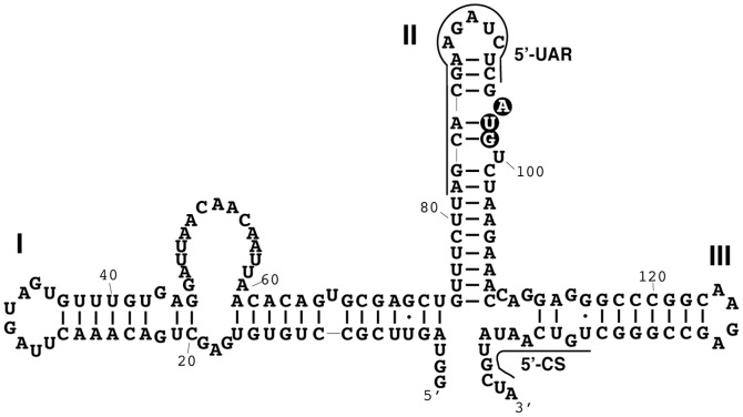
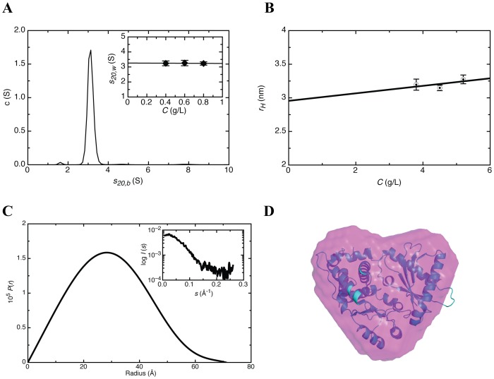
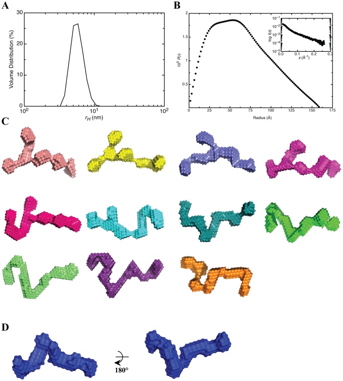
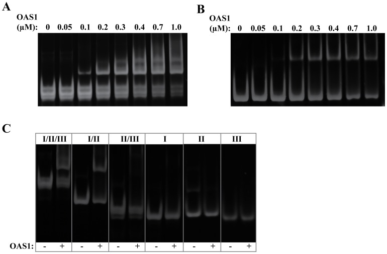
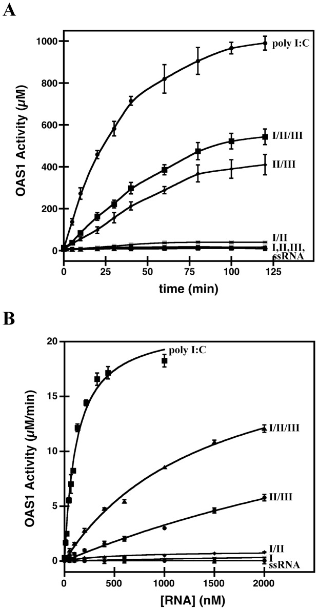
West Nile virus (WNV) has a positive sense RNA genome with conserved structural elements in the 5' and 3' -untranslated regions required for polyprotein production. Antiviral immunity to WNV is partially mediated through the production of a cluster of proteins known as the interferon stimulated genes (ISGs). The 2' 5'-oligoadenylate synthetases (OAS) are key ISGs that help to amplify the innate immune response. Upon interaction with viral double stranded RNA, OAS enzymes become activated and enable the host cell to restrict viral propagation. Studies have linked mutations in the OAS1 gene to increased susceptibility to WNV infection, highlighting the importance of OAS1 enzyme. Here we report that the region at the 5'-end of the WNV genome comprising both the 5'-UTR and initial coding region is capable of OAS1 activation in vitro. This region contains three RNA stem loops (SLI, SLII, and SLIII), whose relative contribution to OAS1 binding affinity and activation were investigated using electrophoretic mobility shift assays and enzyme kinetics experiments. Stem loop I, comprising nucleotides 1-73, is dispensable for maximum OAS1 activation, as a construct containing only SLII and SLIII was capable of enzymatic activation. Mutations to the RNA binding site of OAS1 confirmed the specificity of the interaction. The purity, monodispersity and homogeneity of the 5'-end (SLI/II/III) and OAS1 were evaluated using dynamic light scattering and analytical ultra-centrifugation. Solution conformations of both the 5'-end RNA of WNV and OAS1 were then elucidated using small-angle x-ray scattering. In the context of purified components in vitro, these data demonstrate the recognition of conserved secondary structural elements of the WNV genome by a member of the interferon-mediated innate immune response.


Conflict of interest statement

Competing Interests: The authors have declared that no competing interests exist.

Figures

Figure 1
Figure 1. Secondary structure of the WNV 5′-end.
Highlighted are SLI, SLII, SLIII, the AUG start codon (black circles), the upstream AUG region (SLI/II/III, solid line) and the conserved sequence element (5′-CS, solid line) .
Figure 2
Figure 2. Recombinant human OAS1 adopts a globular fold.
(A) Sedimentation velocity (SV) distribution analysis in terms of c(S) at 0.4 mg/mL. In-set is the resultant concentration dependence of the SV distribution. (B) Concentration dependence of hydrodynamic radius obtained from DLS measurements. (C) The pair distribution function versus particle radius obtained from the GNOM analysis. In-set is the merged scattering data obtained from multiple concentrations. (D) Superimposition of the human OAS1 (PDB 4IG8) high-resolution structure on the ab initio model generated using DAMMIF on the data obtained from SAXS experiments on human OAS1.
Figure 3
Figure 3. Solution conformations of the WNV SLI/II/III from SAXS.
(A) Dynamic light scattering profile of SLI/II/III at 2 mg/mL. (B) Pair distribution function of SLI/II/III obtained from merged data of multiple concentrations. In-set is the merged SAXS data obtained from multiple concentrations. (C) Individual ab-initio models calculated from the SAXS data using DAMMIF program demonstrating two distinct subpopulations of the RNA molecule. (D) Averaged model of SLI/II/III obtained from individual models presented in Fig. 3C.
Figure 4
Figure 4. The WNV SLI/II/III forms a direct interaction with human OAS1.
(A) EMSA for OAS1 (100 nM) binding to the SLI/II/III under non-denaturing conditions. (B) EMSA for OAS1 (100 nM) binding to SLI+II under non-denaturing conditions. (C) Non-denaturing gel electrophoresis of SLI/II/III truncations (100 nM) in the presence or absence of OAS1 (400 nM). In all cases, 8% native TBE gels were used and stained with Sybr Gold (Invitrogen, USA) to visualize RNA-containing species.
Figure 5
Figure 5. Catalytic activation of OAS1 by the SLI/II/III and its truncations.
(A) Purified OAS1 (300 nM) and RNA (300 nM) were incubated in the presence of ATP (2 mM) and MgCl2 (5 mM) at 37°C, quenched at time points from 0–180 minutes, and 2′-5′(A) chain formation quantitated by PPi detection. In all cases, errors represent the standard deviation from at least 3 replicates, and ssRNA represents a single-stranded negative control. (B) Enzymatic activity of OAS1 (400 nM) shown as a function of RNA concentration. Linear regression analysis of the initial velocity was used to determine OAS1 activity and the error in the analysis represented as error bars.
Figure 6
Figure 6. Analysis of OAS1 mutants.
(A) Pair distribution function versus particle radius obtained from GNOM analysis for wild-type OAS1 (red), R195E (blue) and K199E (green). Inset, is a SDS-polyacrylamide gel presenting wild type and mutant OAS1s that suggests that all constructs have similar molecular weight. (B) EMSA for OAS1 and mutants (100 nM) binding to WNV SLI/II/III under non-denaturing conditions. (C) Reactions containing purified OAS1 or OAS1 mutants (300 nM) and RNA (300 nM) quenched at time points from 0–180 minutes followed by quantification of PPi production. In all cases, errors represent the standard deviation from at least 3 replicates, and ssRNA represents a single-stranded negative control. (D) Enzymatic activity of OAS1 or OAS1 mutants (400 nM) shown as a function of RNA concentration. Linear regression analysis of the initial velocity was used to determine OAS1 activity and the error in the analysis represented as error bars.

References

    1. Gubler DJ (2007) The continuing spread of West Nile virus in the western hemisphere. Clin Infect Dis 45: 1039–1046. - PubMed
    1. Asnis DS, Conetta R, Teixeira AA, Waldman G, Sampson BA (2000) The West Nile virus outbreak of 1999 in New York: the Flushing Hospital experience. (vol 30, pg 413, 2000). Clinical Infectious Diseases 30: 841–841. - PubMed
    1. Savage HM, Ceianu C, Nicolescu G, Karabatsos N, Lanciotti R, et al. (1999) Entomologic and avian investigations of an epidemic of West Nile fever in Romania in 1996, with serologic and molecular characterization of a virus isolate from mosquitoes. Am J Trop Med Hyg 61: 600–611. - PubMed
    1. Lanciotti RS, Ebel GD, Deubel V, Kerst AJ, Murri S, et al. (2002) Complete genome sequences and phylogenetic analysis of West Nile virus strains isolated from the United States, Europe, and the Middle East. Virology 298: 96–105. - PubMed
    1. Calisher CH (2000) West Nile virus in the New World: appearance, persistence, and adaptation to a new econiche—an opportunity taken. Viral Immunol 13: 411–414. - PubMed

Publication types

LinkOut - more resources