. 2014 Mar 20;10(3):e1004026.
doi: 10.1371/journal.ppat.1004026. eCollection 2014 Mar.

The role of host and microbial factors in the pathogenesis of pneumococcal bacteraemia arising from a single bacterial cell bottleneck

Affiliations

The role of host and microbial factors in the pathogenesis of pneumococcal bacteraemia arising from a single bacterial cell bottleneck

Alice Gerlini et al. PLoS Pathog. .

Abstract

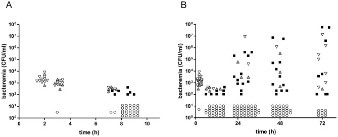
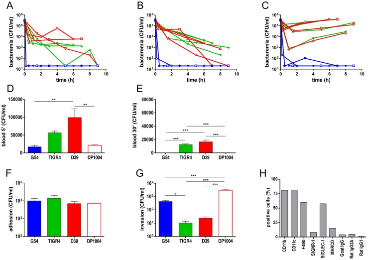
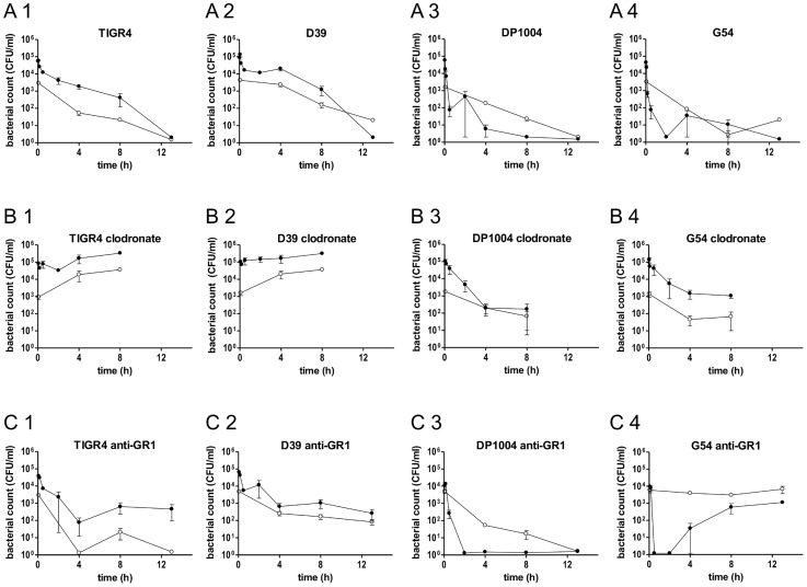
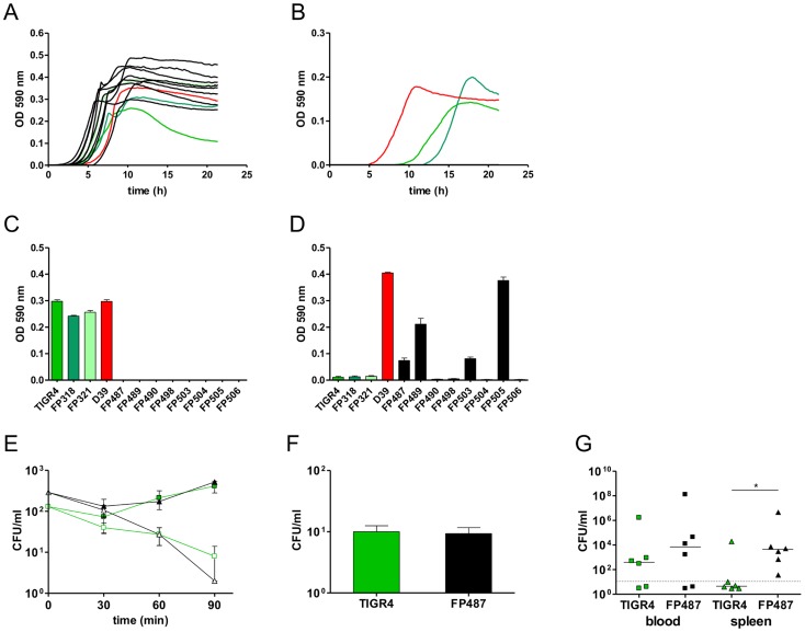
The pathogenesis of bacteraemia after challenge with one million pneumococci of three isogenic variants was investigated. Sequential analyses of blood samples indicated that most episodes of bacteraemia were monoclonal events providing compelling evidence for a single bacterial cell bottleneck at the origin of invasive disease. With respect to host determinants, results identified novel properties of splenic macrophages and a role for neutrophils in early clearance of pneumococci. Concerning microbial factors, whole genome sequencing provided genetic evidence for the clonal origin of the bacteraemia and identified SNPs in distinct sub-units of F0/F1 ATPase in the majority of the ex vivo isolates. When compared to parental organisms of the inoculum, ex-vivo pneumococci with mutant alleles of the F0/F1 ATPase had acquired the capacity to grow at low pH at the cost of the capacity to grow at high pH. Although founded by a single cell, the genotypes of pneumococci in septicaemic mice indicate strong selective pressure for fitness, emphasising the within-host complexity of the pathogenesis of invasive disease.


Conflict of interest statement

The authors have declared that no competing interests exist.

Figures

Figure 1
Figure 1. Co-infection of CD1 mice with three isogenic variants of S. pneumoniae TIGR4.
A mixture of three isogenic S. pneumoniae TIGR4 variants (3×10⁵ CFU/each strain) was given to CD1 mice (n = 68) by the i.v. route. Bacterial counts were performed collecting blood at various time points. (A) Blood counts in the first 10 h after challenge. (B) Blood counts up to 72 h post-challenge, (including data reported in A). Each symbol indicates a single mouse. Blood cultures yielding all three variants are shown in downward white triangles, those yielding two variants as upward grey triangles, and samples yielding a monoclonal blood culture are shown as black squares. Samples from mice with negative blood cultures are shown as open circles. The ratio of infected over un-infected mice was 0.31 at 24 h, 0.37 at 48 h and 0.67 at 72 h. The cut off for detection is 100 CFU/ml. Data of two independent experiments are reported.
Figure 2
Figure 2. Pneumococcal sepsis and phagocytosis by splenic macrophages.
Intravenous challenge of four outbred CD1 mice (A),four inbred BALB/c mice (B) and four inbred CBA/Ca mice (C) was performed by co-infection with D39 (red), G54 (blue) and TIGR4 (green). Bacterial counts in blood collected at different time points from each single mouse are reported (A–C). Cut off is 100 CFU/ml). Blood counts at 5 min (D) and 30 min (E) after i.v. infection of BALB/c mice (n = 6–12) with four pneumococcal strains: D39 (red bar), G54 (blue bar), TIGR4 (green bar) and a rough D39 derivative DP1004 (red open bar). Adhesion (F) and invasion (phagocytosis) (G) of pneumococcal strains in primary spleen macrophages from BALB/c mice. Independent experiments were run in triplicate and reported as mean ± SEM. Statistical analysis were performed with Kruskal-Wallis and Dunn's test. (H) Cytoflurimetric characterisation of surface markers of the primary spleen macrophages. A representative experiment is reported. Comparable data on splenic macrophages isolates from C57BL/6 mice are in Figure S4.
Figure 3
Figure 3. Bacterial counts in blood and spleen of BALB/c mice depleted of neutrophils or macrophages.
Twelve groups of BALB/c mice (n = 3–9) were infected by the i.v. route with four different strains of S. pneumoniae (TIGR4, D39, DP1004 and G54) at the challenge dose of 2.5×10⁵ CFU/each strain. Bacterial counts over time of each pneumococcal strain in the blood (black lines) and in the spleen (grey lines) were reported for untreated mice (A1, A2, A3 and A4), clodronate liposomes treated mice (B1, B2, B3 and B4) and anti-GR1 mAb treated mice (C1, C2, C3 and C4). Samples were collected over 13 h, with the exception of mice treated with clodronate (8 h). The cut off is 20 CFU/ml. Data are reported as the mean ± SD of bacterial counts.
Figure 4
Figure 4. Phenotypes of single-colony blood culture isolates.
Growth profiles of wild type strains (coloured lines) and ATPase mutants (black lines) in standard laboratory media THY (A) and TSB (B). D39 is indicated in red while TIGR4, FP318 and FP321 in different shade of green. (C, D) Maximum OD590 reached by wild type and ATPase mutants during growth with 80 mM K2HPO4 and pH 8.0 (D) and growth at pH 6.6 (E). Kinetics of opsono-phagocytosis assays in rotating blood with anti-type 4 serum (open symbols) and without antibodies (filled symbols) of parental strains (green) compared to a SP1507 atpC mutant (black). (F) Phagocytosis with primary cultures of spleen macrophages of TIGR4 and FP487 atpC mutant. (G) Bacterial counts in blood (squares) and spleen (triangles) at 72 h after i.v. infection of BALB/c mice (n = 6) with TIGR4 (green) and FP487 carrying a frame shifted SP1507 atpC gene (black). Data points below the cut off are negative. All data were analyzed by Student's t-test (P<0.05).

References

    1. Gray BM, Converse GM, Dillon HCJ (1980) Epidemiologic studies of Streptococcus pneumoniae in infants: acquisition, carriage, and infection during the first 24 months of life. J Infect Dis 142: 923–933. - PubMed
    1. Hogberg L, Geli P, Ringberg H, Melander E, Lipsitch M, et al. (2007) Age- and serogroup-related differences in observed durations of nasopharyngeal carriage of penicillin-resistant pneumococci. J Clin Microbiol 45: 948–952. - PMC - PubMed
    1. Melegaro A, Edmunds WJ, Pebody R, Miller E, George R (2006) The current burden of pneumococcal disease in England and Wales. Journal of Infection 52: 37–48. - PubMed
    1. Ispahani P, Slack RCB, Donald FE, Weston WC, Rutter N (2004) Twenty year surveillance of invasive pneumococcal disease in Nottingham: serogroups responsible and implications for immunisation. Arch Dis Child 89: 757–762. - PMC - PubMed
    1. Rudan I, Boschi-Pinto C, Mulholland K, Campbell H (2008) Epidemiology and ethiology of childhood pneumoniae. Bull World Health Organ 86: 408–416. - PMC - PubMed

Publication types

MeSH terms