. 2014 May 9;289(19):13295-307.
doi: 10.1074/jbc.M114.550541. Epub 2014 Mar 20.

Effect of thrombin on human amnion mesenchymal cells, mouse fetal membranes, and preterm birth

Affiliations

Effect of thrombin on human amnion mesenchymal cells, mouse fetal membranes, and preterm birth

Haruta Mogami et al. J Biol Chem. .

Abstract

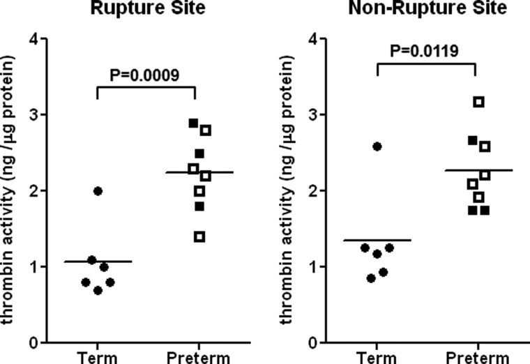
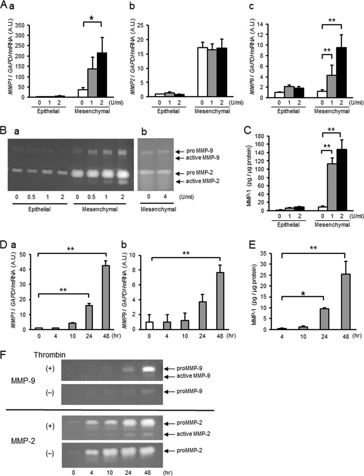
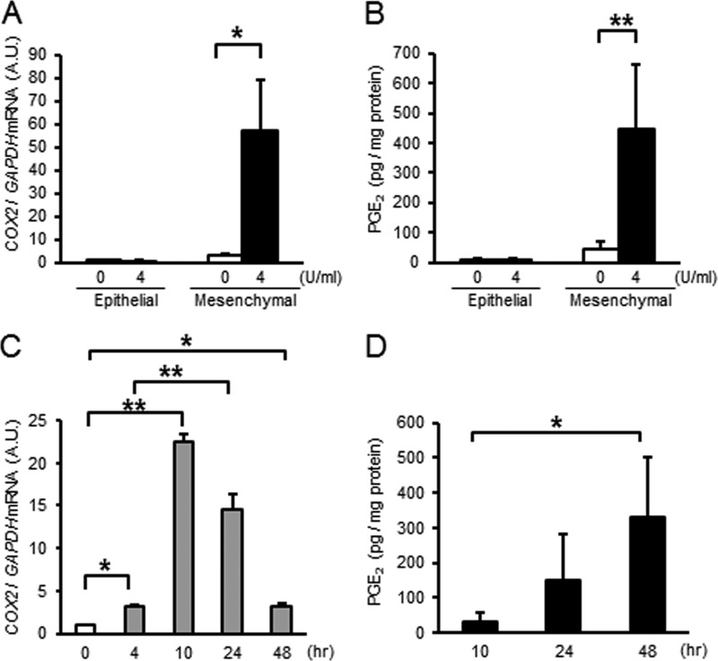
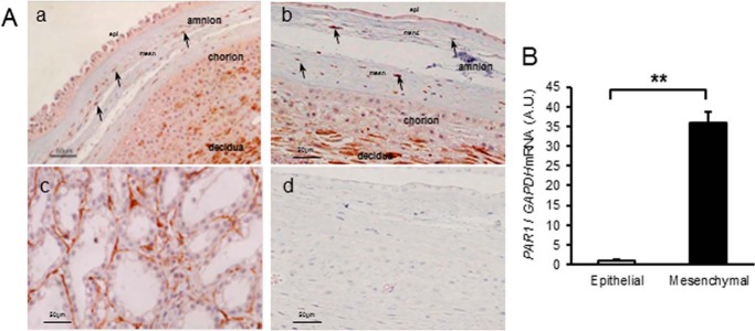
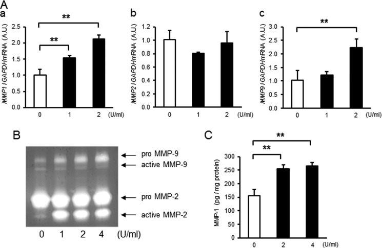
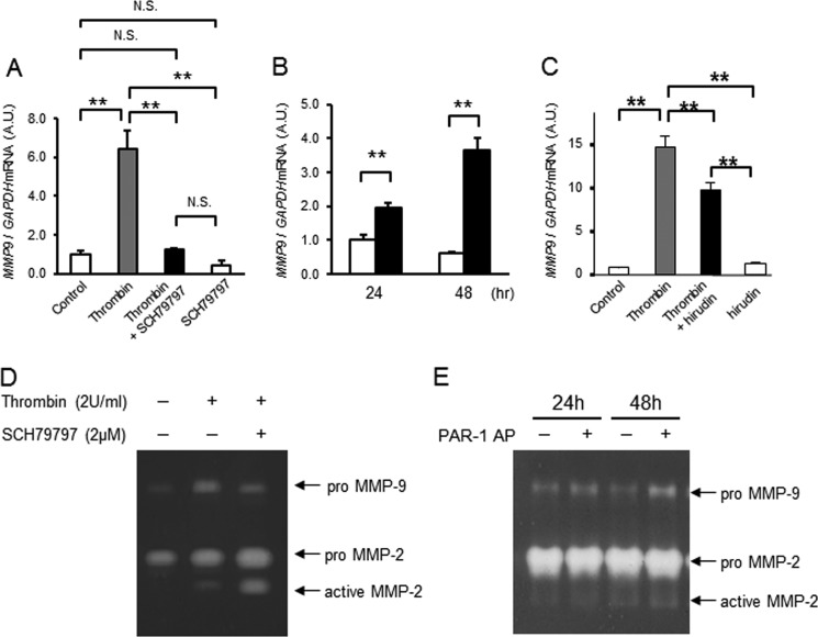
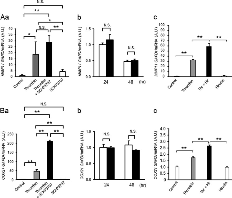
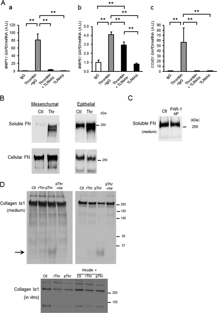
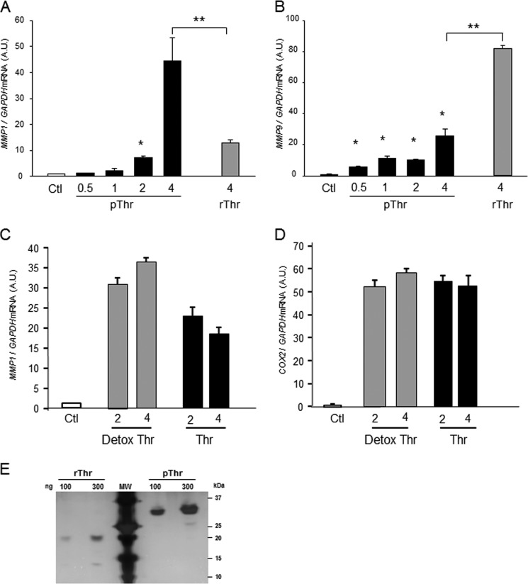
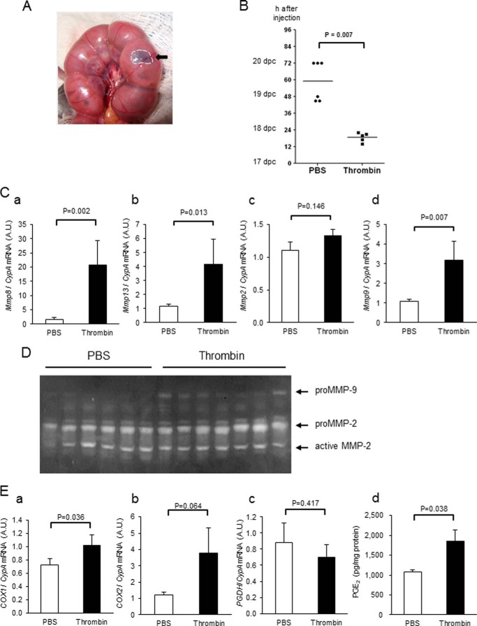
Here, we investigated the effects of thrombin on matrix metalloproteinases (MMPs) and prostaglandin (PG) synthesis in fetal membranes. Thrombin activity was increased in human amnion from preterm deliveries. Treatment of mesenchymal, but not epithelial, cells with thrombin resulted in increased MMP-1 and MMP-9 mRNA and enzymatic activity. Thrombin also increased COX2 mRNA and PGE2 in these cells. Protease-activated receptor-1 (PAR-1) was localized to amnion mesenchymal and decidual cells. PAR-1-specific inhibitors and activating peptides indicated that thrombin-induced up-regulation of MMP-9 was mediated via PAR-1. In contrast, thrombin-induced up-regulation of MMP-1 and COX-2 was mediated through Toll-like receptor-4, possibly through thrombin-induced release of soluble fetal fibronectin. In vivo, thrombin-injected pregnant mice delivered preterm. Mmp8, Mmp9, and Mmp13, and PGE2 content was increased significantly in fetal membranes from thrombin-injected animals. These results indicate that thrombin acts through multiple mechanisms to activate MMPs and PGE2 synthesis in amnion.

Keywords: Amnion; Cyclooxygenase (COX) Pathway; Decidua; Fetal Membrane; Fibronectin; Matrix Metalloproteinase (MMP); Prostaglandins; Protease-activated Receptor-1; Thrombin.


Figures

FIGURE 1.
FIGURE 1.
Thrombin activity in human amnion from pregnancies delivered preterm or term. Thrombin activity was quantified using the FRET system after complete removal of blood from human amnion. Preterm amnion (n = 8) includes both PPROM (open symbols) and non-PPROM (filled symbols), and the delivery mode was either vaginal or cesarean section. Term normal amnion (n = 6) was obtained from spontaneous vaginal delivery.
FIGURE 2.
FIGURE 2.
Thrombin dose- and time-dependently increases MMP-1 and MMP-9 mRNA and enzyme activity in amnion mesenchymal but not epithelial cells. Primary human amnion epithelial or mesenchymal cells were treated with thrombin for the indicated dose and time. A, amnion cells were treated with different doses of thrombin for 48 h and analyzed for MMP1 (a), MMP2 (b), or MMP9 (c) mRNA. A.U., arbitrary units. B, gelatin zymography of conditioned media from cells treated with various concentrations of thrombin for 48 h (a) or cell-free incubation of conditioned media with thrombin (4 units/ml, 36 nm) for 48 h (b). C, MMP-1 enzymatic activity in conditioned media from cells treated with different doses of thrombin for 48 h. D, thrombin (2 units/ml, 18 nm)-induced increases in MMP1 (a) and MMP9 (b) mRNA as a function of time. E, MMP-1 enzymatic activity in conditioned media from amnion mesenchymal cells treated with 2 units/ml (18 nm) of thrombin as a function of time. F, gelatin zymography of conditioned media (5 μg of protein) from amnion mesenchymal cells. Upper zymogram represents MMP-9; lower, MMP-2. Cells were stimulated with 2 units/ml thrombin (+) or medium only (−) for different time periods. In A and C–E error bars represent mean ± S.D., n = 3 in each group. B and F represent results from at least three experiments. *, p < 0.05; **, p < 0.01 compared with controls (0 units/ml vehicle, A and C), 0 h (D), or 4 h thrombin treatment (E).
FIGURE 3.
FIGURE 3.
Thrombin increases COX2 mRNA and PGE2 synthesis in primary amnion mesenchymal cells. Primary amnion epithelial or mesenchymal cells were treated for the indicated dose or time. A, COX2 mRNA levels in amnion cells treated with 4 units/ml (36 nm) of thrombin × 24 h. A.U., arbitrary units. B, PGE2 levels in conditioned media. Amnion cells were treated with 4 units/ml (36 nm) thrombin ×48 h. C, COX2 mRNA in amnion mesenchymal cells treated with control (medium only) or thrombin (4 units/ml, 18 nm) as a function of time. D, PGE2 in conditioned media from amnion mesenchymal cells treated with thrombin (4 units/ml, 36 nm) for the indicated times. n = 3 in each group. *, p < 0.05; **, p < 0.01 compared with control (0 units/ml, A and B), 0 h (C), or 10 h thrombin treatment (D).
FIGURE 4.
FIGURE 4.
Thrombin receptor, PAR-1, is expressed in human amnion mesenchymal cells. A, immunostaining of PAR-1 in human fetal membranes from normal vaginal deliveries. a, nonrupture site. b, rupture site. Arrows indicate immunoreactive mesenchymal cells in amnion. c, prostate cancer as positive control. d, negative control in which mouse control IgG was used as a first antibody. Results are consistent among three fetal membranes in each group. epi, epithelial cells; mesn, mesenchymal layer. B, PAR1 mRNA levels in primary amnion cells. Results represent mean ± S.D. (error bars), n = 3 in each group. A.U., arbitrary units. **, p < 0.01.
FIGURE 5.
FIGURE 5.
Effect of thrombin on primary decidual cells. A, primary decidual cells were treated with various doses of thrombin for 48 h and analyzed for relative levels of MMP1 (a), MMP2 (b), or MMP9 (c) mRNA. A.U., arbitrary units. B, gelatin zymography of conditioned media (2 μg of protein) from decidual cells treated with different doses of thrombin for 48 h. C, MMP-1 enzymatic activity in conditioned media from thrombin-treated decidual cells. Results represent mean ± S.D. (error bars), n = 3 in each group. **, p < 0.01 compared with control (0 units/ml).
FIGURE 6.
FIGURE 6.
Thrombin-induced up-regulation of MMP-9 is mediated by PAR-1. A, effect of a PAR-1-selective receptor antagonist (SCH79797, 2 μm) and thrombin (2 units/ml, 18 nm) on MMP9 mRNA levels in amnion mesenchymal cells. Cells were pretreated with SCH79797 30 min prior to thrombin treatment. Data represent mean ± S.D. (error bars), n = 3 in each group. **, p < 0.01; N.S., not significant. A.U., arbitrary units. B, effect of control (open bars) or PAR-1 AP (TFLLRN, 200 μm, filled bars) on MMP9 mRNA in amnion mesenchymal cells treated for 24 or 48 h. Data represent mean ± S.D. (error bars, n = 3 in each group). **, p < 0.01; N.S., not significant. C, effect of control (open bars), thrombin (2 units/ml, 18 nm, gray bars), hirudin (4 units/ml, white bars), or thrombin + hirudin (filled bars) on MMP9 mRNA in amnion mesenchymal cells treated for 48 h. Data represent mean ± S.D., n = 3 in each group. **, p < 0.01. D, gelatin zymography of conditioned media of mesenchymal cells treated with thrombin (2 units/ml, 18 nm), with or without the PAR-1 antagonist SCH79797 (5 μm) for 48 h. E, gelatin zymography of conditioned media of mesenchymal cells treated with control or PAR-1 AP (TFLLRN, 200 μm) × 24 or 48 h. N.S., not significant.
FIGURE 7.
FIGURE 7.
Thrombin-induced up-regulation of MMP-1 (A) and COX2 (B) is not mediated by PAR-1. Aa and Ba, effect of a PAR-1-selective receptor antagonist (SCH79797, 2 μm) and thrombin (2 units/ml, 18 nm) on MMP1 (Aa) and COX2 (Ba) mRNA levels in amnion mesenchymal cells. Cells were pretreated with SCH79797 30 min prior to thrombin treatment. Data represent mean ± S.D. (error bars), n = 3 in each group. *, p < 0.05; **, p < 0.01; N.S., not significant. A.U., arbitrary units. Ab and Bb, effect of control (open bars) or PAR-1 AP (TFLLRN, 200 μm, filled bars) on MMP1 (Ab) and COX2 (Bb) mRNA in amnion mesenchymal cells treated for 24 or 48 h. Data represent mean ± S.D. (error bars), n = 3 in each group. *, p < 0.05; **, p < 0.01; N.S., not significant. Ac and Bc, effect of control (open bars), thrombin (2 units/ml, 18 nm, gray bars), hirudin (4 units/ml, white bars), or thrombin + hirudin (filled bars) on MMP1 (Ac) and COX2 (Bc) mRNA in amnion mesenchymal cells treated for 48 h. Data represent mean ± S.D., n = 3 in each group. **, p < 0.01.
FIGURE 8.
FIGURE 8.
Thrombin-induced up-regulation of MMP-1 and COX2 is mediated via Toll-like receptor-4. A, amnion mesenchymal cells were pretreated with control IgG or anti-human TLR4 neutralizing antibody for 30 min prior to treatment with control (medium only) or thrombin (2 units/ml). After 48 h, relative mRNA levels of MMP1 (a), MMP9 (b), and COX2 (c) were analyzed using quantitative PCR. Data represent mean ± S.D. (error bars), n = 3 in each group. **, p < 0.01. A.U., arbitrary units. B, immunoblot analysis of soluble (medium) and cellular fibronectin (FN) in mesenchymal (TLR4-responsive) and epithelial (TLR4-nonresponsive) cells treated with control (Ctl) or thrombin (Thr, 4 units/ml, 36 nm). 30 μg of protein from conditioned media (soluble) or 6 m urea extract (cellular) was applied in each lane. C, immunoblot of soluble fibronectin in media of primary amnion mesenchymal cells treated with control (Ctl) or PAR-1 AP × 48 h. D, immunoblot analysis of collagen type I α1 in media from mesenchymal cells treated with recombinant (4 units/ml) or plasma (4 units/ml) Thr and hirudin (6.5 units/ml). Immunoblot of purified collagen type I treated with thrombin ± hirudin in vitro is also shown.
FIGURE 9.
FIGURE 9.
Comparison of pThr and rThr and endotoxin removal on MMP9, MMP1, and COX2 gene expression in amnion mesenchymal cells. A and B, cells were treated with various doses of pThr (0.5 (4.5 nm) to 4 (36 nm) units/ml or rThr (4 units/ml), and MMP1 (A) or MMP9 (B) mRNA levels were quantified after 48 h. *, p < 0.01 compared with control. A.U., arbitrary units. C and D, effect of endotoxin removal on pThr-induced MMP1 (C) and COX2 (D) mRNA. E, silver-stained gel of purified rThr and pThr. Thrombin was applied at 100 and 300 ng/lane as indicated. **, p < 0.01.
FIGURE 10.
FIGURE 10.
Effect of thrombin on gestational length, MMPs, and PGE2 in fetal membranes. Thrombin (1 unit/μl, 4 unit/fetus) or PBS (4 μl) with 1% methylene blue was injected in the interface between fetal membranes and uterus in pregnant mice on 17 days postcoitum (dpc). A, mouse uterus after injection. Injected-reagent (thrombin or PBS) was localized in the interface between fetal membranes and the uterine wall. B, time to delivery after thrombin injection. Black bar indicates the mean. Mice were injected with either PBS (n = 6) or thrombin (n = 5), and time of delivery after injection was recorded. C, Mmp8 (a), Mmp13 (b), Mmp2 (c), and Mmp9 (d) mRNA levels in fetal membranes collected 14 h after injection of PBS or thrombin. Data represent mean ± S.E. (error bars), n = 10 in each group. A.U., arbitrary units. D, gelatin zymography of protein (14 μg/lane) from fetal membranes collected 14 h after injection of PBS or thrombin. E, COX1 mRNA (a), COX2 mRNA (b), 15-hydroxyprostaglandin dehydrogenase mRNA (c), and PGE2 content (d) in fetal membranes 14 h after injection of pregnant mice with PBS or thrombin. n = 10 in each group.

Similar articles

Cited by

References

    1. Goldenberg R. L., Culhane J. F., Iams J. D., Romero R. (2008) Epidemiology and causes of preterm birth. Lancet 371, 75–84 - PMC - PubMed
    1. Chaiworapongsa T., Espinoza J., Yoshimatsu J., Kim Y. M., Bujold E., Edwin S., Yoon B. H., Romero R. (2002) Activation of coagulation system in preterm labor and preterm premature rupture of membranes. J. Matern. Fetal Neonatal Med. 11, 368–373 - PubMed
    1. Erez O., Romer R., Vaisbuch E., Chaiworapongsa T., Kusanovic J. P., Mazaki-Tovi S., Gotsch F., Gomez R., Maymon E., Pacora P., Edwin S. S., Kim C. J., Than N. G., Mittal P., Yeo L., Dong Z., Yoon B. H., Hassan S. S., Mazor M. (2009) Changes in amniotic fluid concentration of thrombin-antithrombin III complexes in patients with preterm labor: evidence of an increased thrombin generation. J. Matern. Fetal Neonatal Med. 22, 971–982 - PMC - PubMed
    1. Rosen T., Kuczynski E., O'Neill L. M., Funai E. F., Lockwood C. J. (2001) Plasma levels of thrombin-antithrombin complexes predict preterm premature rupture of the fetal membranes. J. Matern Fetal Med. 10, 297–300 - PubMed
    1. Nagy S., Bush M., Stone J., Lapinski R. H., Gardó S. (2003) Clinical significance of subchorionic and retroplacental hematomas detected in the first trimester of pregnancy. Obstet. Gynecol. 102, 94–100 - PubMed

Publication types