. 2014 Apr;47(4):307-15.
doi: 10.1590/1414-431X20143437. Epub 2014 Mar 18.

Trypan blue exclusion assay by flow cytometry

Affiliations

Trypan blue exclusion assay by flow cytometry

B A Avelar-Freitas et al. Braz J Med Biol Res. 2014 Apr.

Abstract

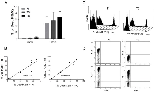
Dye exclusion tests are used to determine the number of live and dead cells. These assays are based on the principle that intact plasma membranes in live cells exclude specific dyes, whereas dead cells do not. Although widely used, the trypan blue (TB) exclusion assay has limitations. The dye can be incorporated by live cells after a short exposure time, and personal reliability, related to the expertise of the analyst, can affect the results. We propose an alternative assay for evaluating cell viability that combines the TB exclusion test and the high sensitivity of the flow cytometry technique. Previous studies have demonstrated the ability of TB to emit fluorescence when complexed with proteins. According to our results, TB/bovine serum albumin and TB/cytoplasmic protein complexes emit fluorescence at 660 nm, which is detectable by flow cytometry using a 650-nm low-pass band filter. TB at 0.002% (w/v) was defined as the optimum concentration for distinguishing unstained living cells from fluorescent dead cells, and fluorescence emission was stable for 30 min after cell treatment. Although previous studies have shown that TB promotes green fluorescence quenching, TB at 0.002% did not interfere with green fluorescence in human live T-cells stained with anti-CD3/fluorescein isothiocyanate (FITC) monoclonal antibody. We observed a high correlation between the percentage of propidium iodide+CD3/FITC+ and TB+CD3/FITC+ cells, as well as similar double-stained cell profiles in flow cytometry dot-plot graphs. Taken together, the results indicate that a TB exclusion assay by flow cytometry can be employed as an alternative tool for quick and reliable cell viability analysis.


Figures

Figure 1
Figure 1. Spectrofluorophotometry analysis of the excitation (A) and emission (B) spectra of the solutions containing 0.02% (w/v) trypan blue (TB, dotted line), bovine serum albumin (BSA, solid line) at 10% (w/v), and TB-BSA (dashed line) interaction.
Figure 2
Figure 2. Fluorescence analysis of the trypan blue (TB)-bovine serum albumin (BSA) interaction. A, Increasing BSA concentrations were added to 0.002% TB solution. The fluorimeter was configured to use fixed excitation and emission wavelengths at 488 and 660 nm, respectively. B, Human PBMC co-stained with DAPI and TB. DAPI excitation at 365 nm and the emission band-pass filter DAPI-445/50, TB excitation at 488 nm and fluorescence emission detected by LP/515.
Figure 3
Figure 3. Evaluation of the optimum concentration for trypan blue (TB) solution ranging from 0.002 to 0.4% to discriminate between live and dead peripheral blood mononuclear cells (PBMC). The analysis was performed on a FACScan cytometer (Becton & Dickinson, USA) using the long-pass filter 650/LP (FL3 detector). Staining with propidium iodide (PI) was used as a reference.
Figure 4
Figure 4. Histogram analysis of the trypan blue (TB) exclusion assay by flow cytometry to evaluate TB (at 0.002%, w/v) incorporation by live cells 0 (A), 5 (B), 10 (C), 20 (D), and 30 (E) min after dye addition.
Figure 5
Figure 5. A, Comparison of the trypan blue (TB) exclusion test using flow cytometry, propidium iodide (PI) staining and the conventional trypan blue exclusion test employing cell counting in a Neubauer chamber (NC). B, Pearson's correlation test between PI and TB or NC and TB. C, Fluorescence intensity histogram profiles of the PI and TB flow cytometry analysis. D, Fluorescence emitted by cells stained with PI and TB dyes and analyzed on FL2 (585/42) and FL3 (650 nm/LP) detectors.
Figure 6
Figure 6. Profile of T-lymphocytes stained with monoclonal antibody anti-CD3-FITC followed by treatment with propidium iodide (PI) and trypan blue (TB) at 0.002 and 0.4% (w/v) or PBS (untreated control).
Figure 7
Figure 7. A, Percentage of dead human T-lymphocytes (CD3-FITC+ cells) submitted to cell culture at temperatures (T) of 37°C (physiologic temperature) or 50°C (high-stress temperature) followed by staining with trypan blue (TB) or propidium iodide (PI). B, Dot-plot graph profile between human lymphocytes submitted to pretreatment with high-stress temperature (50°C) followed by staining with TB and PI and monoclonal antibody anti-CD3-FITC+. C, Pearson's correlation test between dead CD3+ lymphocytes using PI and TB flow cytometry assays.

Similar articles

Cited by

References

    1. Chan LL, Wilkinson AR, Paradis BD, Lai N. Rapid image-based cytometry for comparison of fluorescent viability staining methods. J Fluoresc. 2012;22:1301–1311. doi: 10.1007/s10895-012-1072-y. - DOI - PubMed
    1. Chen SF, Lu WF, Wen ZY, Li Q, Chen JH. Preparation, characterization and anticancer activity of norcantharidin-loaded poly(ethylene glycol)-poly(caprolactone) amphiphilic block copolymer micelles. Pharmazie. 2012;67:781–788. - PubMed
    1. Altiok N, Ersoz M, Koyuturk M. Estradiol induces JNK-dependent apoptosis in glioblastoma cells. Oncol Lett. 2011;2:1281–1285. - PMC - PubMed
    1. Puoci F, Morelli C, Cirillo G, Curcio M, Parisi OI, Maris P, et al. Anticancer activity of a quercetin-based polymer towards HeLa cancer cells. Anticancer Res. 2012;32:2843–2847. - PubMed
    1. Kroemer G, Galluzzi L, Vandenabeele P, Abrams J, Alnemri ES, Baehrecke EH, et al. Classification of cell death: recommendations of the Nomenclature Committee on Cell Death 2009. Cell Death Differ. 2009;16:3–11. doi: 10.1038/cdd.2008.150. - DOI - PMC - PubMed

Publication types