Review
. 2014 May;16(5):484.
doi: 10.1007/s11886-014-0484-5.

CFR and FFR assessment with PET and CTA: strengths and limitations

Affiliations
Review

CFR and FFR assessment with PET and CTA: strengths and limitations

Ryo Nakazato et al. Curr Cardiol Rep. 2014 May.

Abstract

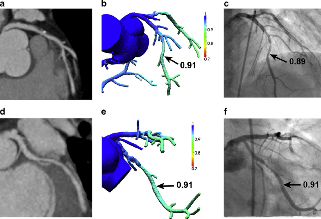
Positron emission tomography (PET) myocardial perfusion imaging (MPI) has high diagnostic accuracy and prognostic value. PET-MPI can also be used to quantitatively evaluate regional myocardial blood flow (MBF). This technique also allows the calculation of the coronary flow reserve (CFR)/myocardial flow reserve (MFR), which is the ratio of MBF at peak hyperemia to resting MBF. Coronary computed tomography angiography (CTA) is a non-invasive method for accurate detection and exclusion of high-grade coronary stenoses, when compared to an invasive coronary angiography reference standard. However, CTA assessment of coronary stenoses tends toward overestimation, and CTA cannot determine physiologic significance of lesions. Recent advances in computational fluid dynamics and image-based modeling permit calculation of non-invasive fractional flow reserve derived from CT (FFRCT), without the need for additional imaging, modification of acquisition protocols, or administration of medications. In this review, we cover the CFR/MFR assessment by PET and FFR assessment by CT.


Conflict of interest statement

Conflict of Interest Ryo Nakazato declares that he has no conflict of interest.

Ran Heo declares that he has no conflict of interest.

Jonathon Leipsic has received grant support from and been a consultant for HeartFlow. He has received payment for development of educational presentations including service on speakers’ bureaus from GE Healthcare.

James K. Min has received grant support from HeartFlow. He serves as a consultant to HeartFlow.

Figures

Fig. 1
Fig. 1
Comparison of Flurpiridaz F 18 PET MPI and SPECT MPI in (a) diagnostic certainty and (b) sensitivity and specificity. Def. = definitely; Prob. = probably, CAD = coronary artery disease; MPI = myocardial perfusion imaging; PET = positron emission tomography; SPECT = single-photon emission computed tomography (Reproduced with permission from: Berman DS, Maddahi J, Tamarappoo BK, Czernin J, Taillefer R, Udelson JE et al. Phase II safety and clinical comparison with single-photon emission computed tomography myocardial perfusion imaging for detection of coronary artery disease: flurpiridaz F 18 positron emission tomography. J Am Coll Cardiol. 2013;61(4):469–77. doi:10.1016/j. jacc.2012.11.022) [13]
Fig. 2
Fig. 2
Clinical example in which flow quantification with PET may improve diagnosis of CAD. a dipyridamole 82Rb PET MPI static images demonstrate normal relative perfusion at rest and during peak stress; b 17-segment model polar maps of rest MBF (lower left; color display scale 0 to 1.5 mL/min/g), stress MBF (upper left; scale: 0–3.0 mL/min/g), MFR (upper right; scale: 0–3.0) and MFD (lower right; scale: 0–2.0) which demonstrate global impairments, absolute values displayed in the table below; c Coronary angiogram reveals significant obstructive three-vessel CAD, relative perfusion underestimated the presence of disease (arrows point out significant stenosis). HLA, horizontal long axis; SA, short axis; VLA, vertical long axis; MBF, myocardial blood flow; MFR, myocardial flow reserve; MFD, myocardial flow difference; LAD, left anterior descending artery; LCX, left circumflex; RCA, right coronary artery; RPLS, right posterolateral branch. (Reproduced with permission from: Ziadi MC, Beanlands RS. The clinical utility of assessing myocardial blood flow using positron emission tomography. J Nucl Cardiol. 2010;17(4):571–81. doi:10.1007/s12350-010-9258-7) [60]
Fig. 3
Fig. 3
Simplified schematic of computation fluid dynamic techniques applied to CTA data for simulation of hyperemic coronary artery flow and pressure. (Reproduced with permission from: Min JK, Berman DS, Budoff MJ, Jaffer FA, Leipsic J, Leon MB et al. Rationale and design of the DeFACTO (Determination of Fractional Flow Reserve by Anatomic Computed Tomographic AngiOgraphy) study. J Cardiovasc Comput Tomogr. 2011;5(5):301–9. doi:S1934-5925(11)00317-0 [pii]) [61]
Fig. 4
Fig. 4
Representative examples of two patients from the DeFACTO study. (1) FFRCTA results for 66-year-old man with multivessel CAD but no lesion-specific ischemia (a) Coronary computed tomography angiography (CTA) demonstrating stenosis in the left anterior descending coronary artery (LAD). (b) Fractional flow reserve (FFR) derived from CTA (FFRCTA) demonstrates no ischemia in the LAD, with a computed value of 0.91. c Invasive coronary angiography (ICA) with FFR also demonstrates no ischemia in the LAD, with a measured value of 0.89. d CTA demonstrating stenosis in the left circumflex coronary (LCx) artery. e FFRCTA demonstrates no ischemia in the LCx, with a computed value of 0.91. f ICA with FFR also demonstrates no ischemia in the LCx, with a measured value of 0.91. (2) FFRCTA results for 66-year-old man with multivessel CAD and lesion-specific ischemia (a) CTA demonstrating stenosis in the LAD. b FFRCTA demonstrates ischemia in the LAD, with a computed value of 0.64. c ICA with FFR also demonstrates ischemia in the LAD, with a measured value of 0.72. d CTA demonstrating stenosis in the LCx. e FFRCTA demonstrates ischemia in the LCx, with a computed value of 0.61. f ICA with FFR also demonstrates ischemia in the LCx, with a measured value of 0.52. Abbreviations as in Fig. 4(1). (Reproduced with permission from: Taylor CA, Fonte TA, Min JK. Computational fluid dynamics applied to cardiac computed tomography for noninvasive quantification of fractional flow reserve: scientific basis. J Am Coll Cardiol. 2013;61(22):2233–41. doi:10.1016/j.jacc.2012.11.083) [48•]
Fig. 4
Fig. 4
Representative examples of two patients from the DeFACTO study. (1) FFRCTA results for 66-year-old man with multivessel CAD but no lesion-specific ischemia (a) Coronary computed tomography angiography (CTA) demonstrating stenosis in the left anterior descending coronary artery (LAD). (b) Fractional flow reserve (FFR) derived from CTA (FFRCTA) demonstrates no ischemia in the LAD, with a computed value of 0.91. c Invasive coronary angiography (ICA) with FFR also demonstrates no ischemia in the LAD, with a measured value of 0.89. d CTA demonstrating stenosis in the left circumflex coronary (LCx) artery. e FFRCTA demonstrates no ischemia in the LCx, with a computed value of 0.91. f ICA with FFR also demonstrates no ischemia in the LCx, with a measured value of 0.91. (2) FFRCTA results for 66-year-old man with multivessel CAD and lesion-specific ischemia (a) CTA demonstrating stenosis in the LAD. b FFRCTA demonstrates ischemia in the LAD, with a computed value of 0.64. c ICA with FFR also demonstrates ischemia in the LAD, with a measured value of 0.72. d CTA demonstrating stenosis in the LCx. e FFRCTA demonstrates ischemia in the LCx, with a computed value of 0.61. f ICA with FFR also demonstrates ischemia in the LCx, with a measured value of 0.52. Abbreviations as in Fig. 4(1). (Reproduced with permission from: Taylor CA, Fonte TA, Min JK. Computational fluid dynamics applied to cardiac computed tomography for noninvasive quantification of fractional flow reserve: scientific basis. J Am Coll Cardiol. 2013;61(22):2233–41. doi:10.1016/j.jacc.2012.11.083) [48•]

References

    1. Di Carli MF, Murthy VL. Cardiac PET/CT for the evaluation of known or suspected coronary artery disease. Radiographics. 2011;31(5):1239–1254. - PMC - PubMed
    1. Senthamizhchelvan S, Bravo PE, Lodge MA, Merrill J, Bengel FM, Sgouros G. Radiation dosimetry of 82Rb in humans under pharmacologic stress. J Nucl Med Off Publ Soc Nucl Med. 2011;52(3):485–491. - PMC - PubMed
    1. Stabin MG. Proposed revision to the radiation dosimetry of 82Rb. Health Phys. 2010;99(6):811–813. - PubMed
    1. Di Carli MF, Dorbala S, Meserve J, El Fakhri G, Sitek A, Moore SC. Clinical myocardial perfusion PET/CT. J Nucl Med Off Publ Soc Nucl Med. 2007;48(5):783–793. - PubMed
    1. Stabin MG. Radiopharmaceuticals for nuclear cardiology: radiation dosimetry, uncertainties, and risk. J Nucl Med Off Publ Soc Nucl Med. 2008;49(9):1555–1563. - PubMed

Publication types