. 2014 Jan 7;10(1):88-95.
doi: 10.1039/c3sm52423j.

Dynamic self-assembly of motile bacteria in liquid crystals

Affiliations

Dynamic self-assembly of motile bacteria in liquid crystals

Peter C Mushenheim et al. Soft Matter. .

Abstract

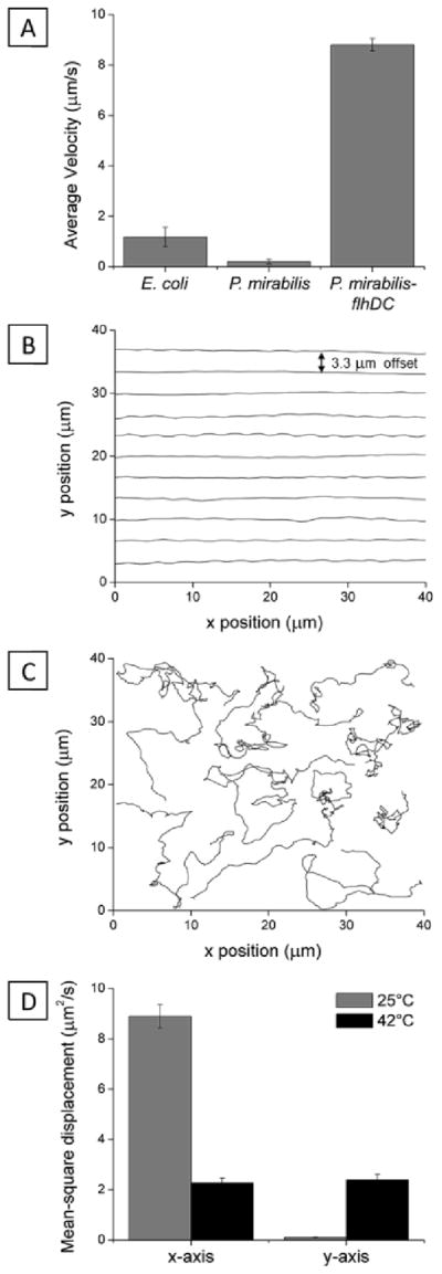
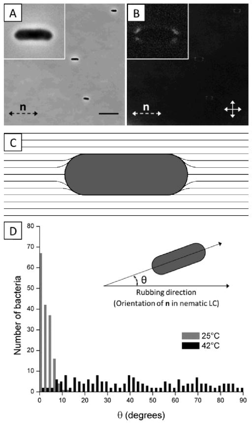
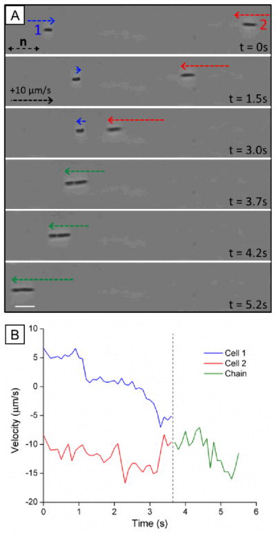
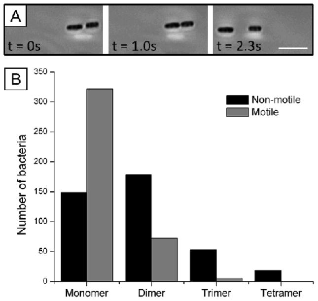
This paper reports an investigation of dynamical behaviors of motile rod-shaped bacteria within anisotropic viscoelastic environments defined by lyotropic liquid crystals (LCs). In contrast to passive microparticles (including non-motile bacteria) that associate irreversibly in LCs via elasticity-mediated forces, we report that motile Proteus mirabilis bacteria form dynamic and reversible multi-cellular assemblies when dispersed in a lyotropic LC. By measuring the velocity of the bacteria through the LC (8.8 ± 0.2 μm s(-1)) and by characterizing the ordering of the LC about the rod-shaped bacteria (tangential anchoring), we conclude that the reversibility of the inter-bacterial interaction emerges from the interplay of forces generated by the flagella of the bacteria and the elasticity of the LC, both of which are comparable in magnitude (tens of pN) for motile Proteus mirabilis cells. We also measured the dissociation process, which occurs in a direction determined by the LC, to bias the size distribution of multi-cellular bacterial complexes in a population of motile Proteus mirabilis relative to a population of non-motile cells. Overall, these observations and others reported in this paper provide insight into the fundamental dynamic behaviors of bacteria in complex anisotropic environments and suggest that motile bacteria in LCs are an exciting model system for exploration of principles for the design of active materials.


Conflict of interest statement

The authors declare the following competing financial interest(s): NLA declares a significant financial interest in Platypus Technologies LLC, a for-profit company that has developed LC-based technologies for molecular analysis.

Figures

Fig. 1
Fig. 1
Anisotropic motion of bacteria in nematic LC. (A) Average velocities of P. mirabilis-flhDC cells in nematic DSCG solutions (15 wt%) at 25°C. (B) Superimposed trajectories (offset from one another by 3.3 um) of P. mirabilis-flhDC in nematic DSCG solutions at 25°C. The LC director was oriented along the x-axis (in the direction of rubbing of the glass slides). (C) Superimposed representative trajectories of P. mirabilis-flhDC in isotropic phases of DSCG at 42°C. (D) Plot of the mean-square displacement of P. mirabilis-flhDC calculated from analysis of 139 trajectories (25°C) and 75 trajectories (42°C). The velocities of P. mirabilis-flhDC cells in DSCG solution were comparable in magnitude at 25°C (V̄ = 8.8 +/− 0.2 μm/s) and 42°C (V̄ = 8.1 +/− 0.3 μm/s). Values are reported with associated standard errors.
Fig. 2
Fig. 2
Configuration of LC around bacteria and resulting bacterial alignment. (A, B) Bright field and crossed polars images, respectively of non-motile P. mirabilis-flhDC cells dispersed in nematic DSCG solution at 25°C. The double-headed solid arrows in B indicate the positions of the polarizers while the double-headed dotted arrows depict the orientation of the LC director (n). (C) Schematic representation of the LC director profile that results from weak, tangential anchoring of the LC on the surface of P. mirabilis-flhDC cells. (D) Distribution of angles between the rubbing direction of the glass slides and the long axis of P. mirabilis-flhDC cells in DSCG solution at 25°C (nematic) and 42°C (isotropic). The scale bar in A is 10 μm. Values are reported with associated standard errors.
Fig. 3
Fig. 3
Dynamic association of motile bacteria in nematic LC. (A) Sequence of images (bright field) showing end-on-end association of two motile P. mirabilis-flhDC cells in nematic DSCG solution (15 wt%) at 25°C. Dotted arrows indicate the velocity of the bacterial cells (see calibration in t = 1.5s). (B) Plot of the velocities of the P. mirabilis-flhDC cells shown in A before and after association into the chain. The scale bar in A is 5 μm.
Fig. 4
Fig. 4
(A) Bright field and (B) crossed polars images of a non-motile P. mirabilis-flhDC multi-cellular complex (trimer) in nematic DSCG solution (15 wt%) at 25°C. The scale bar in A is 5 μm.
Fig. 5
Fig. 5
Reversible assembly of motile bacteria within LCs. (A) Sequence of images (bright field) showing both the association and dissociation of two motile bacteria in nematic DSCG solution at 25°C. (B) Populations of bacteria in multi-cellular complexes formed in nematic DSCG solution by non-motile (black) and motile (gray) bacteria. (See text for experimental details.) The total number of cells in both populations is 400. The scalebar in A is 5 μm.

References

    1. Fontes DE, Mills AL, Hornberger GM, Herman JS. Appl Environ Microbiol. 1991;57:2473–2481. - PMC - PubMed
    1. Shaw T, Winston M, Rupp CJ, Klapper I, Stoodley P. Phys Rev Lett. 2004;93:098102. - PubMed
    1. Madoff LC, Thaler SJ, Maguire JH. In: Harrison’s Principles of Internal Medicine. 16. Braunwald E, Fauci AS, Kasper DL, Hauser SL, Longo DL, Jameson JL, editors. McGraw-Hill; New York: 2005. pp. 2050–2055.
    1. Flemming HC, Wingender J. Nat Rev Microbiol. 2010;8:623–633. - PubMed
    1. Rey AD. Soft Matter. 2010;6:3402–3429.

Publication types