. 2014 Sep;25(9):1924-31.
doi: 10.1681/ASN.2013101121. Epub 2014 Mar 20.

Fluorescence microangiography for quantitative assessment of peritubular capillary changes after AKI in mice

Affiliations

Fluorescence microangiography for quantitative assessment of peritubular capillary changes after AKI in mice

Rafael Kramann et al. J Am Soc Nephrol. 2014 Sep.

Abstract

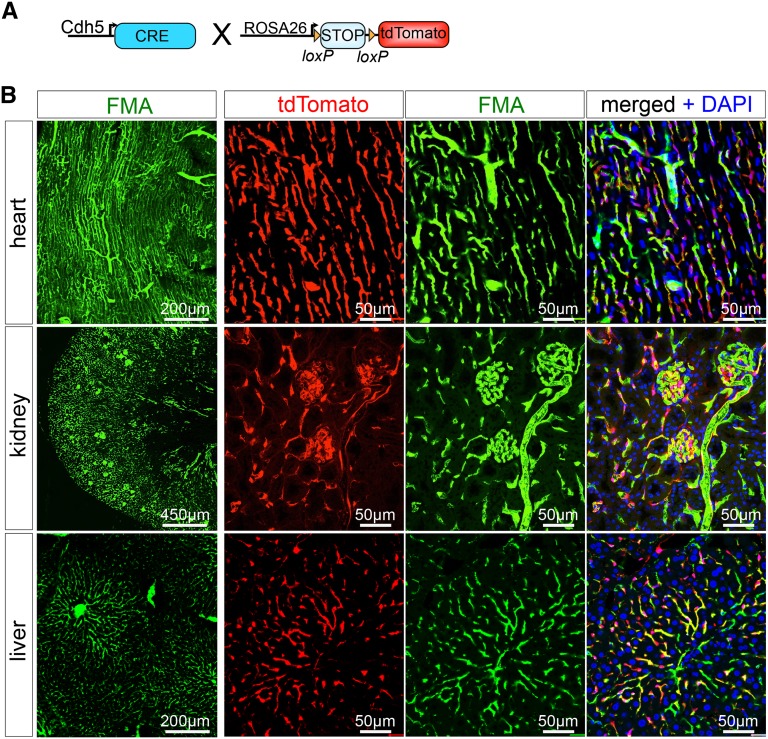
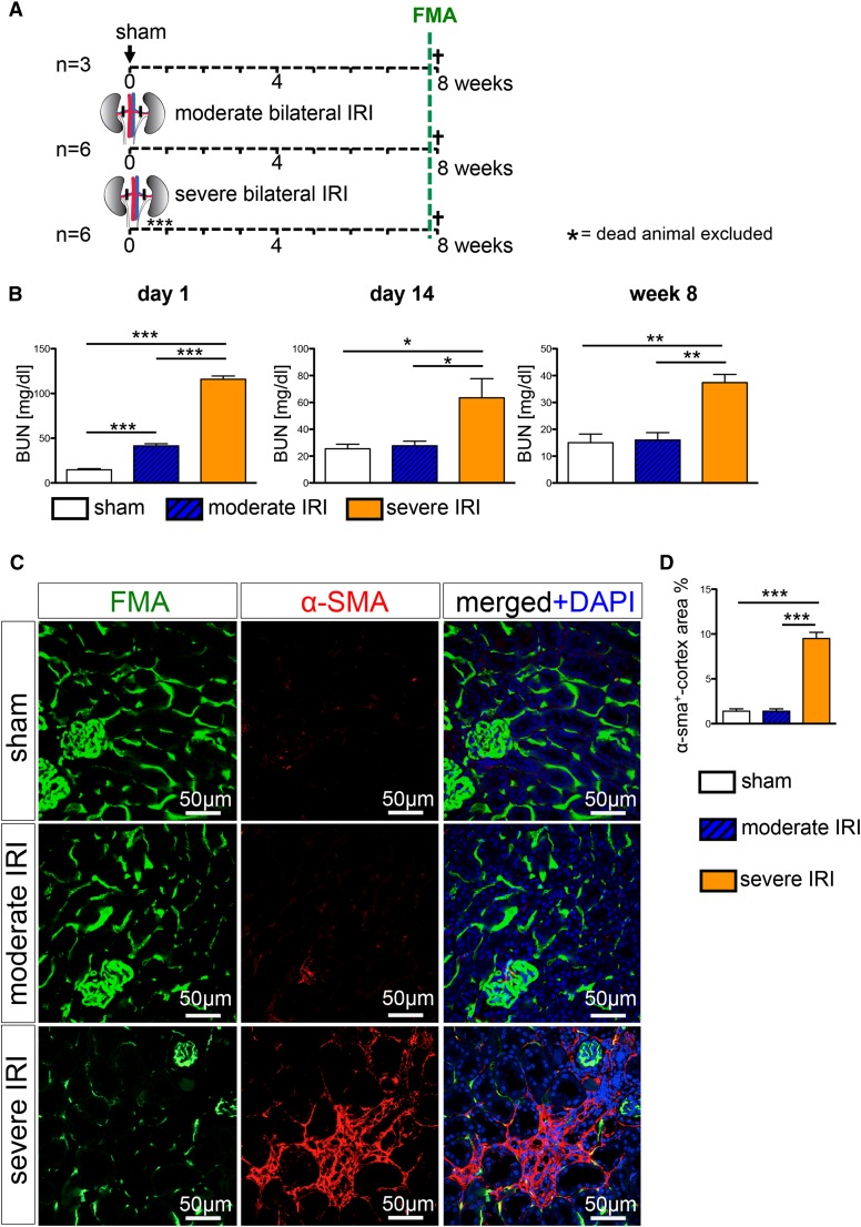
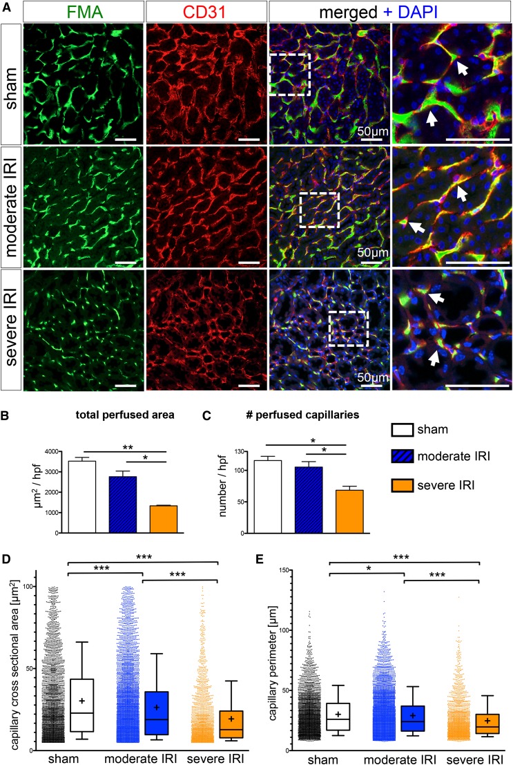
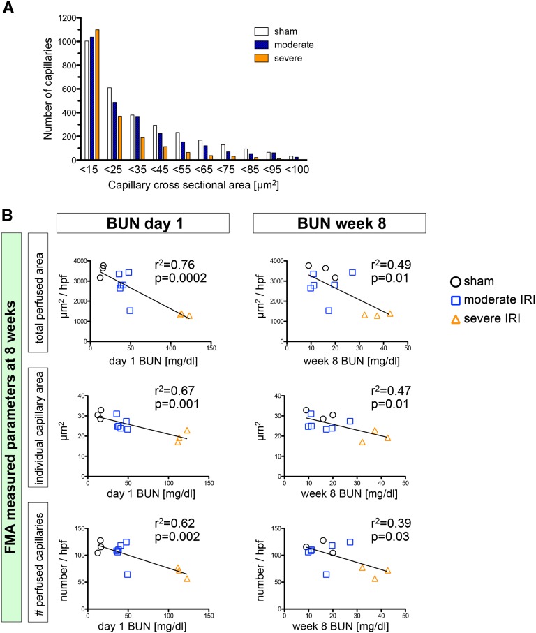
AKI predicts the future development of CKD, and one proposed mechanism for this epidemiologic link is loss of peritubular capillaries triggering chronic hypoxia. A precise definition of changes in peritubular perfusion would help test this hypothesis by more accurately correlating these changes with future loss of kidney function. Here, we have adapted and validated a fluorescence microangiography approach for use with mice to visualize, analyze, and quantitate peritubular capillary dynamics after AKI. A novel software-based approach enabled rapid and automated quantitation of capillary number, individual area, and perimeter. After validating perfusion in mice with genetically labeled endothelia, we compared peritubular capillary number and size after moderate AKI, characterized by complete renal recovery, and after severe AKI, characterized by development of interstitial fibrosis and CKD. Eight weeks after severe AKI, we measured a 40%±7.4% reduction in peritubular capillary number (P<0.05) and a 36%±4% decrease in individual capillary cross-sectional area (P<0.001) for a 62%±2.2% reduction in total peritubular perfusion (P<0.01). Whereas total peritubular perfusion and number of capillaries did not change, we detected a significant change of single capillary size following moderate AKI. The loss of peritubular capillary density and caliber at week 8 closely correlated with severity of kidney injury at day 1, suggesting irreparable microvascular damage. These findings emphasize a direct link between severity of acute injury and future loss of peritubular perfusion, demonstrate that reduced capillary caliber is an unappreciated long-term consequence of AKI, and offer a new quantitative imaging tool for understanding how AKI leads to future CKD in mouse models.

Keywords: acute renal failure; chronic kidney disease; vascular disease.


Figures

Figure 1.
Figure 1.
FMA provides highly efficient high-resolution perfusion imaging of the kidney, heart, and liver. (A) To genetically label endothelial cells in solid organ vascular beds, we bred the VE-Cadherin-Cre driver mouse against the Rosa26-tdtomato reporter line, resulting in endothelial cell–specific cre-recombination with removing of the LoxP flanked STOP codon and expression of the bright red fluorochrome tdtomato. (B) FMA was validated in Ve-CadherinCre+, R26Tomato+ mice, showing highly efficient delineation of the microvasculature in heart, kidney, and liver. DAPI, 4′,6-diamidino-2-phenylindole.
Figure 2.
Figure 2.
Severe ischemia reperfusion injury leads to CKD with interstitial fibrosis. (A) Wild-type mice were subjected to severe bilateral IRI (clamping for 28 minutes), moderate bilateral IRI (clamping for 23 minutes), or sham surgery and were euthanized following FMA at 8 weeks. (B) BUN measurement revealed significantly increased BUN values in both IRI groups at day 1 after surgery. After moderate IRI, mice showed a complete recovery, with BUN levels returned to normal values at day 14 after surgery, whereas severe IRI lead to persistently increased BUN levels at 8 weeks after surgery. (C and D) α-Smooth muscle actin staining and quantification revealed induction of interstitial fibrosis only after severe IRI. (Graphs show mean±SEM; *P<0.05; **P<0.01; ***P<0.001, one-way ANOVA with post hoc Bonferroni correction). DAPI, 4′,6-diamidino-2-phenylindole.
Figure 3.
Figure 3.
High-throughput software–based analysis of fluorescence microangiography reveals reduced capillary number and caliber after severe IRI. (A) FMA after sham surgery and moderate and severe IRI, together with CD31 immunostaining, demonstrates capillary rarefaction in response to severity of injury (arrows indicate capillaries with red CD31+endothelial cells surrounding the green FMA solution; all scale bars are 50 µm). (B and C) Severe IRI results in a significant reduction of the total cortical cross-sectional capillary area per high-power field [×400, inner cortex] (B) and a significant reduction in capillary number (C). (D and E) The cortical individual capillary cross-sectional area (D) (mean±SEM, sham: 30.31±0.42 µm2; moderate IRI: 26.29±0.28 µm2; severe IRI: 19.34±0.38 µm2) and perimeter (E) (sham: 30.19±0.31 µm; moderate IRI: 29.13±0.22 µm; severe IRI: 24.77±0.35 µm) was significantly reduced after both moderate and severe IRI. (Of note, data represent n=3 mice in the sham group and severe IRI group and n=6 mice in the moderate IRI group; mean±SEM in B and C; box and whiskers with 10th–90th percentiles in D and E; + indicates mean in D and E. *P<0.05; **P<0.01; ***P<0.001, one-way ANOVA with post hoc Bonferroni correction). DAPI, 4′,6-diamidino-2-phenylindole.
Figure 4.
Figure 4.
Severity of initial injury determines shift in capillary size and extent of capillary rarefaction. (A) Distribution of capillary size demonstrates a loss of larger capillaries (>15 µm2) after moderate and severe IRI, with a slight increase in counted small capillaries (<15 µm2) (data represent three mice per group). (B) FMA assessed total capillary cross-sectional area (total perfused area, µm2/high-power field), individual capillary cross-sectional area (µm2), and capillary number (number/high-power field) shows highly significant correlation with day 1 BUN and lower correlation with week 8 BUN.

References

    1. Coca SG, Singanamala S, Parikh CR: Chronic kidney disease after acute kidney injury: A systematic review and meta-analysis. Kidney Int 81: 442–448, 2012 - PMC - PubMed
    1. Chawla LS, Kimmel PL: Acute kidney injury and chronic kidney disease: An integrated clinical syndrome. Kidney Int 82: 516–524, 2012 - PubMed
    1. Kusaba T, Lalli M, Kramann R, Kobayashi A, Humphreys BD: Differentiated kidney epithelial cells repair injured proximal tubule. Proc Natl Acad Sci U S A 28: 1527–1532, 2014 - PMC - PubMed
    1. Humphreys BD, Valerius MT, Kobayashi A, Mugford JW, Soeung S, Duffield JS, McMahon AP, Bonventre JV: Intrinsic epithelial cells repair the kidney after injury. Cell Stem Cell 2: 284–291, 2008 - PubMed
    1. Venkatachalam MA, Griffin KA, Lan R, Geng H, Saikumar P, Bidani AK: Acute kidney injury: A springboard for progression in chronic kidney disease. Am J Physiol Renal Physiol 298: F1078–F1094, 2010 - PMC - PubMed

Publication types

MeSH terms