. 2014 Mar 21;343(6177):1353-6.
doi: 10.1126/science.1248373.

β-Catenin activation regulates tissue growth non-cell autonomously in the hair stem cell niche

Affiliations

β-Catenin activation regulates tissue growth non-cell autonomously in the hair stem cell niche

Elizabeth R Deschene et al. Science. .

Abstract

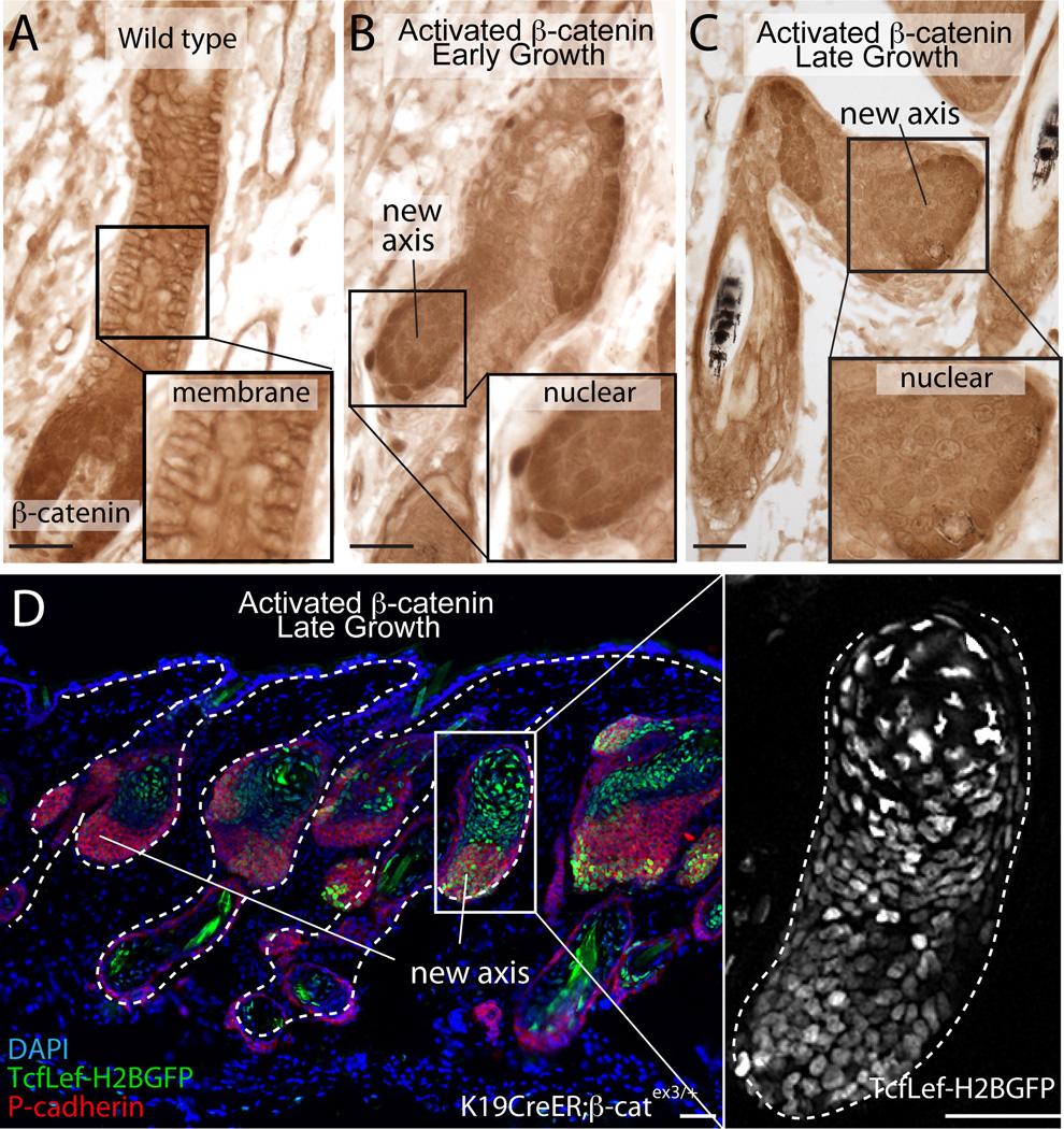
Wnt/β-catenin signaling is critical for tissue regeneration. However, it is unclear how β-catenin controls stem cell behaviors to coordinate organized growth. Using live imaging, we show that activation of β-catenin specifically within mouse hair follicle stem cells generates new hair growth through oriented cell divisions and cellular displacement. β-Catenin activation is sufficient to induce hair growth independently of mesenchymal dermal papilla niche signals normally required for hair regeneration. Wild-type cells are co-opted into new hair growths by β-catenin mutant cells, which non-cell autonomously activate Wnt signaling within the neighboring wild-type cells via Wnt ligands. This study demonstrates a mechanism by which Wnt/β-catenin signaling controls stem cell-dependent tissue growth non-cell autonomously and advances our understanding of the mechanisms that drive coordinated regeneration.


Conflict of interest statement

We declare no conflict of interest.

Figures

Fig. 1
Fig. 1. Activated β-catenin-induced cellular mechanisms that promote new hair growths
(A) Hair growths in Tamoxifen-treated K19CreER;β-catnflox(Ex3)/+. New hair growths are P-cadherin+ (green) and proliferative (Ki67, red). Nuclei (DAPI, blue). (B) Optical sections of z-stacks of a K19CreER;β-catnflox(Ex3)/+;K14-H2BGFP HF depict upward nuclear movement (Movie S2). Epithelial nuclei (K14-H2BGFP, white). Arrowheads indicate points of reference of original nuclei positions. Colored circles represent moving nuclei, black circles represent stationary nuclei. (C) Formation (c’ inset) and expansion (cc’) of nuclear clusters in two separate K19CreER;β-catnflox(Ex3)/+;K14-H2BGFP mice. Nuclei are pseudo-colored (Movie S3–S4). (D) K19CreER;β-catnflox(Ex3)/+;K14-H2BGFP HFs just after DP ablations (Day 0, yellow arrows). DPs marked by Lef1-RFP (red), epithelial nuclei by K14-H2BGFP (green). (E) Same HFs after 6 days of Tamoxifen treatment (Day 7). (F–G) Revisit of the same HFs (Day 12–19). Asterisk denotes HF out of plane of view. Scale bars=25µm.
Fig. 2
Fig. 2. β-catenin activation triggers new axes composed of both mutant and wild-type cells
(A–B) Optical sections of z-stacks of K19CreER;β-catnflox(Ex3)/+;K14-H2BGFP;tdTom mutant HFs show early and late hair growths (K14-H2BGFP, green; tdTom Cre-reporter, red). (C) Genotype of FACS-isolated populations from the new growths based on Pcadherin- enrichment (top gel) (n=2 mice) or TcfLef-H2BGFP Wnt reporter (bottom gel) (n=2 mice). (D) Both tdTom+ and tdTom cells dividing within a new growth. Schematic depicts representative divisions within both tdTom+ and tdTom populations over a time-lapse recording (see Movie S5). Scale bars=50µm.
Fig. 3
Fig. 3. β-catenin-mutant cells activate Wnt signaling in neighboring wild-type cells
(A) Immunohistochemistry of β-catenin in growing wild-type HF. Inset highlights membrane bound staining. (B-C) Nuclear β-catenin in new hair growths in Tamoxifen-induced K19CreER;β-catnflox(Ex3)/+ mice at early and late growth stages. Insets highlight nuclear staining. (D) Wnt reporter expression (TcfLef-H2BGFP, green) in late growths (P-cadherin, red). Scale bars=50µm.
Fig. 4
Fig. 4. β-catenin acts non cell-autonomously via Wnt ligands
(A) Representative qRT-PCR analysis of Wnt ligands and target genes comparing sorted PcadHi;tdTom+ versus PcadHi;tdTom populations from K19CreER;β-catnflox(Ex3)/+;tdTom mice (n=2; p<0.005). (B) Model of non-cell autonomous β-catenin signaling. (C) Wntless staining (red) in K19CreER;β-catnflox(Ex3)/+ mice. (D) Following Tamoxifen induction, H&E of K19CreER;β-catnflox(Ex3)/+;Wlsfl/fl mice show anagen-like stage HFs with growths of varying sizes versus (G) K19CreER;β-catnflox(Ex3)/+;Wlsfl/+ mutant controls which show growths with a higher frequency and larger size (n=3 experimental litters, n=7 K19CreER;β-catnflox(Ex3)/+;Wlsfl/fl mice). (E) Nuclear β-catenin within growths of K19CreER;β-catnflox(Ex3)/+;Wlsfl/fl and controls (H). (F) Wntless (red) is absent in most of the growths of K19CreER;β-catnflox(Ex3)/+;Wlsfl/fl compared to mutant controls (I). Nuclei (DAPI, blue). Scale bars=50µm.

References

    1. Morrison SJ, Spradling AC. Cell. 2008;132:598–611. - PMC - PubMed
    1. Rompolas P, Greco V. Semin Cell Dev Biol. 2013 - PMC - PubMed
    1. Cotsarelis G, Sun TT, Lavker RM. Cell. 1990;61:1329–1337. - PubMed
    1. Greco V, et al. Cell stem cell. 2009;4:155–169. - PMC - PubMed
    1. Blanpain C, Lowry WE, Geoghegan A, Polak L, Fuchs E. Cell. 2004;118:635–648. - PubMed

Publication types