. 2014 Mar 21;343(6177):1363-6.
doi: 10.1126/science.1248725.

Structure of the mitochondrial translocator protein in complex with a diagnostic ligand

Affiliations

Structure of the mitochondrial translocator protein in complex with a diagnostic ligand

Lukasz Jaremko et al. Science. .

Abstract

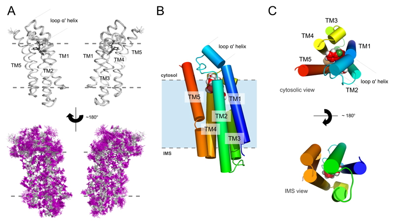
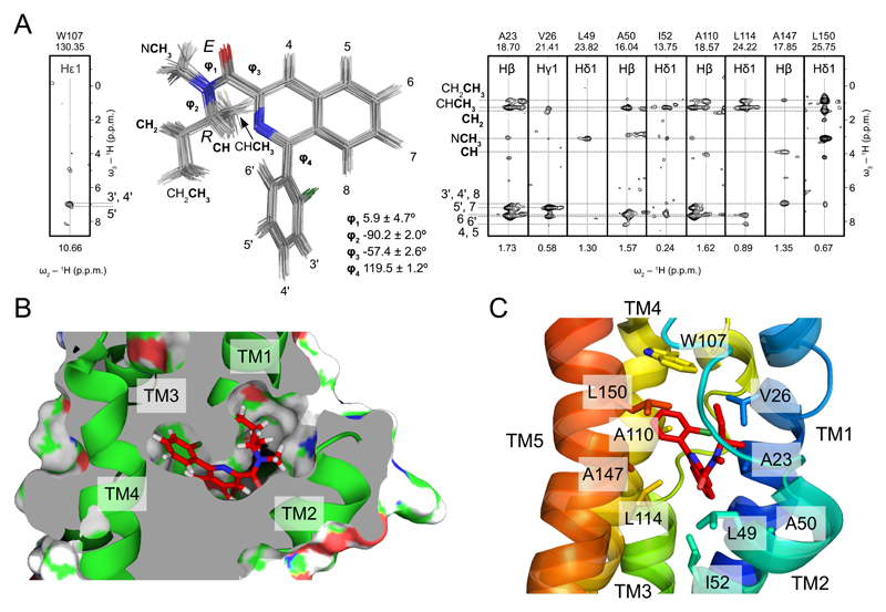
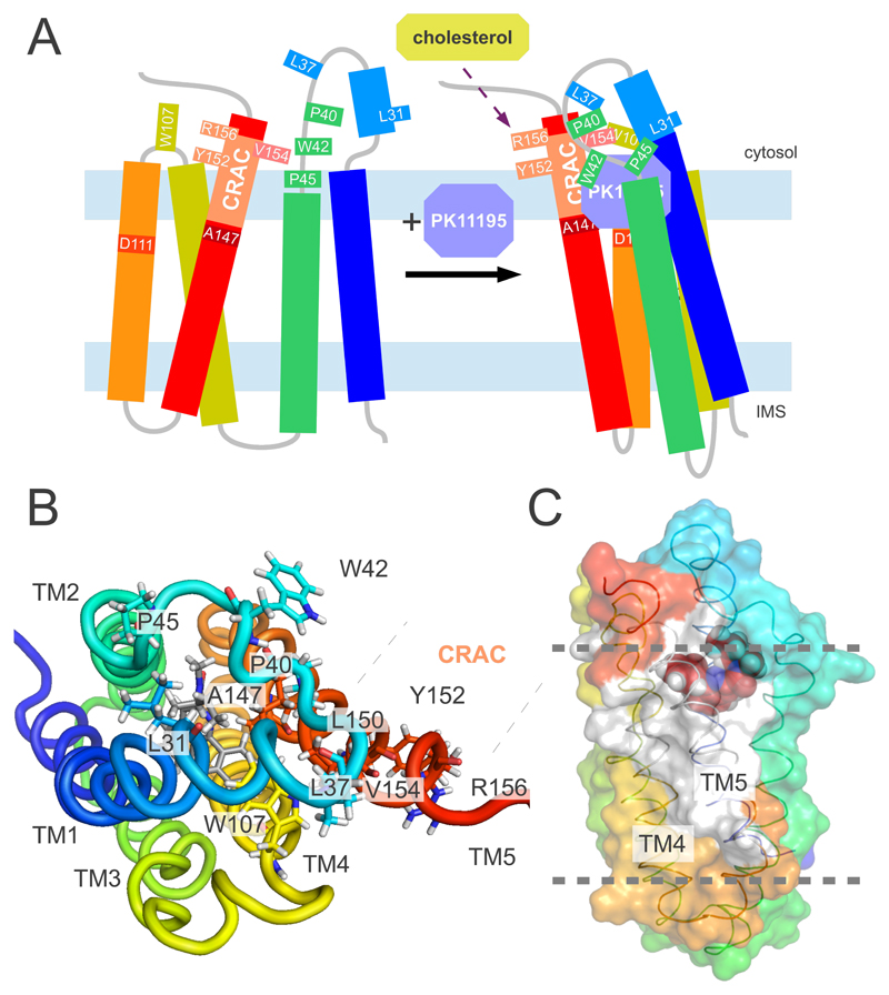
The 18-kilodalton translocator protein TSPO is found in mitochondrial membranes and mediates the import of cholesterol and porphyrins into mitochondria. In line with the role of TSPO in mitochondrial function, TSPO ligands are used for a variety of diagnostic and therapeutic applications in animals and humans. We present the three-dimensional high-resolution structure of mammalian TSPO reconstituted in detergent micelles in complex with its high-affinity ligand PK11195. The TSPO-PK11195 structure is described by a tight bundle of five transmembrane α helices that form a hydrophobic pocket accepting PK11195. Ligand-induced stabilization of the structure of TSPO suggests a molecular mechanism for the stimulation of cholesterol transport into mitochondria.


Figures

Fig. 1
Fig. 1. High-resolution solution structure of the mTSPO-PK11195 complex.
(A) Backbone ribbon trace (upper panel) and all atom view (lower panel) of the 20 lowest-energy structures determined by NMR spectroscopy. The backbone and the side chains are shown in silver and magenta, respectively. The ligand is black. (B) Cylindrical representation of the lowest-energy complex structure with the ligand atoms shown as spheres. (C) Cytosolic and IMS views of the mTSPO-PK11195 complex. Gray dotted lines in (A) and (B) indicate approximate membrane boundaries.
Fig. 2
Fig. 2. Binding of PK11195 to TSPO.
(A) Superposition of the 20 lowest-energy (R)-PK11195 conformations as found in the TSPO-PK11195 complex structure. Strips from 3D F1-13C,15N-filtered/edited-NOESY-1H-13C-HSQC (right) and 3D F1-13C,15N-filtered/edited-NOESY-1H-15N-HSQC (left) experiments show intermolecular NOEs between PK11195 and selected TSPO residues. Ligand atoms are labeled. Dihedral angles φ1, φ2, φ3, and φ4 are the average values found in the 20 lowest-energy structures ± st.dev. (B) Detailed view of the PK11195 binding cavity. Transmembrane helices are shown as green ribbons, the ligand in a stick representation. (C) TSPO residues that form the ligand binding pocket. The same residues as in (A) are shown in a stick representation. Transmembrane helices are color coded.
Fig. 3
Fig. 3. Model for the ligand-induced modulation of the TSPO structure.
(A) Cartoon representation of the conformation of TSPO in the absence (left) and presence (right) of PK11195. The representation of the ligand-bound state is based on the 3D structure of the mTSPO-PK11195 complex shown in Fig. 1. In agreement with circular dichroism (17), the secondary motifs in the ligand-free state have been assumed to be similar to the one in the mTSPO-PK11195 complex. The 2D correlation spectrum of mTSPO in DPC micelles displayed narrow signal dispersion (Fig. S1), indicating increased mobility and decreased helix packing of the ligand-free structure. The cholesterol recognition amino acid consensus, CRAC, is labeled. Upon ligand binding, the cytosolic entrance to the channel is covered by the lid comprising the TM1-TM2 loop. (B) Atomic resolution view onto the TM1-TM2 lid. The CRAC residues Y152 and R156, which are essential for cholesterol binding (19, 27), are located on the outside of the TSPO structure and are not involved in PK11195 binding. (C) Surface representation of the TSPO-PK11195 complex highlighting the residues that were most protected after 28 h of hydrogen/deuterium exchange (marked in white). The rest of the protein is colored as in (A). Gray dotted lines indicate the approximate membrane boundaries.

References

    1. Papadopoulos V, et al. Translocator protein (18kDa): new nomenclature for the peripheral-type benzodiazepine receptor based on its structure and molecular function. Trends Pharmacol Sci. 2006;27:402. - PubMed
    1. Rupprecht R, et al. Translocator protein (18 kDa) (TSPO) as a therapeutic target for neurological and psychiatric disorders. Nat Rev Drug Discov. 2010;9:971. - PubMed
    1. Fan J, Lindemann P, Feuilloley MG, Papadopoulos V. Structural and functional evolution of the translocator protein (18 kDa) Curr Mol Med. 2012;12:369. - PubMed
    1. Braestrup C, Squires RF. Specific benzodiazepine receptors in rat brain characterized by high-affinity (3H)diazepam binding. Proc Natl Acad Sci U S A. 1977;74:3805. - PMC - PubMed
    1. Veenman L, Papadopoulos V, Gavish M. Channel-like functions of the 18-kDa translocator protein (TSPO): regulation of apoptosis and steroidogenesis as part of the host-defense response. Curr Pharm Des. 2007;13:2385. - PubMed

Publication types

MeSH terms

Associated data