

Microsurgical excision of symptomatic sacral perineurial cyst with sacral recapping laminectomy : a case report in technical aspects
- PMID: 24653808
- PMCID: PMC3958574
- DOI: 10.3340/jkns.2014.55.2.110
Microsurgical excision of symptomatic sacral perineurial cyst with sacral recapping laminectomy : a case report in technical aspects
Abstract
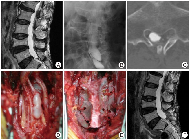
Perineurial cysts (Tarlov cysts) are lesions of the nerve root that are often observed in the sacral area. There is debate about whether symptomatic perineurial cysts should be treated surgically. We presented three patients with symptomatic perineurial cyst who were treated surgically, and introduced sacral recapping laminectomy. Patients complained of low back pain and hypesthesia on lower extremities. We performed operations with sacral recapping technique for all three. The outcome measure was baseline visual analogue score and post operative follow up magnetic resonance images. All patients were completely relieved of symptoms after operation. Although not sufficient to address controversies, this small case series introduces successful use of a particular surgical technique to treat sacral perineural cyst, with resolution of most symptoms and no sequelae.
Keywords: Laminectomy; Tarlov cysts.
Figures


Similar articles
-
Surgical excision of symptomatic sacral perineurial Tarlov cyst: case series and review of the literature.Eur Spine J. 2016 Nov;25(11):3385-3392. doi: 10.1007/s00586-016-4584-3. Epub 2016 May 6. Eur Spine J. 2016. PMID: 27154168 Review.
-
Symptomatic sacral perineurial (Tarlov) cysts.Coll Antropol. 2009 Dec;33(4):1401-3. Coll Antropol. 2009. PMID: 20102100
-
Sacral laminoplasty and cystic fenestration in the treatment of symptomatic sacral perineural (Tarlov) cysts: Technical case report.Surg Neurol Int. 2011;2:129. doi: 10.4103/2152-7806.85469. Epub 2011 Sep 27. Surg Neurol Int. 2011. PMID: 22059124 Free PMC article.
-
Microsurgical treatment of sacral perineural (Tarlov) cysts: case series and review of the literature.J Neurosurg Spine. 2016 May;24(5):700-7. doi: 10.3171/2015.9.SPINE153. Epub 2016 Jan 8. J Neurosurg Spine. 2016. PMID: 26745352 Review.
-
Tarlov cyst: Case report and review of literature.Indian J Orthop. 2007 Oct;41(4):401-3. doi: 10.4103/0019-5413.37007. Indian J Orthop. 2007. PMID: 21139800 Free PMC article.
Cited by
-
Preventive effect of dexamethasone gelatin sponge on the lumbosacral epidural adhesion.Int J Clin Exp Med. 2015 Apr 15;8(4):5478-84. eCollection 2015. Int J Clin Exp Med. 2015. PMID: 26131126 Free PMC article.
-
Tarlov cysts: long-term follow-up after microsurgical inverted plication and sacroplasty.Eur Spine J. 2016 Nov;25(11):3403-3410. doi: 10.1007/s00586-016-4744-5. Epub 2016 Aug 23. Eur Spine J. 2016. PMID: 27554352
-
Surgical management of symptomatic Tarlov cysts: cyst fenestration and nerve root imbrication-a single institutional experience.J Spine Surg. 2019 Dec;5(4):496-503. doi: 10.21037/jss.2019.11.11. J Spine Surg. 2019. PMID: 32043000 Free PMC article.
-
Management of perineural (Tarlov) cysts: a population-based cohort study and algorithm for the selection of surgical candidates.Acta Neurochir (Wien). 2019 Sep;161(9):1909-1915. doi: 10.1007/s00701-019-04000-5. Epub 2019 Jul 3. Acta Neurochir (Wien). 2019. PMID: 31270612 Free PMC article.
-
A Hidden Condition: Multiple Tarlov Cysts Unveiled in a Young Woman Seeking Primary Care for Debilitating Low Back Pain.Am J Case Rep. 2023 Jul 30;24:e940600. doi: 10.12659/AJCR.940600. Am J Case Rep. 2023. PMID: 37516905 Free PMC article.
References
-
- Bartels RH, van Overbeeke JJ. Lumbar cerebrospinal fluid drainage for symptomatic sacral nerve root cysts : an adjuvant diagnostic procedure and/or alternative treatment? Technical case report. Neurosurgery. 1997;40:861–864. discussion 864-865. - PubMed
-
- Caspar W, Papavero L, Nabhan A, Loew C, Ahlhelm F. Microsurgical excision of symptomatic sacral perineurial cysts: a study of 15 cases. Surg Neurol. 2003;59:101–105. discussion 105-106. - PubMed
-
- Cattaneo L, Pavesi G, Mancia D. Sural nerve abnormalities in sacral perineural (Tarlov) cysts. J Neurol. 2001;248:623–624. - PubMed
-
- Davis DH, Wilkinson JT, Teaford AK, Smigiel MR. Sciatica produced by a sacral perineurial cyst. Tex Med. 1987;83:55–56. - PubMed
Publication types
LinkOut - more resources
Full Text Sources
Other Literature Sources
