Superior canal dehiscence patients have smaller mastoid volume than age- and sex-matched otosclerosis and temporal bone fracture patients
- PMID: 24653885
- PMCID: PMC3936659
- DOI: 10.7874/kja.2012.16.3.120
Superior canal dehiscence patients have smaller mastoid volume than age- and sex-matched otosclerosis and temporal bone fracture patients
Abstract
Background and objectives: The purpose of the study was to compare the mastoid air-cell volume of the patients with superior semicircular canal dehiscence syndrome (SCDS) and that of the control patients with otosclerosis and temporal bone (TB) fracture.
Subjects and methods: Ten patients with SCDS were enrolled and 10 patients with bilateral otosclerosis and TB fracture were selected as control groups by age and sex matching. To measure the mastoid air-cell volume, 3D reconstruction software was used.
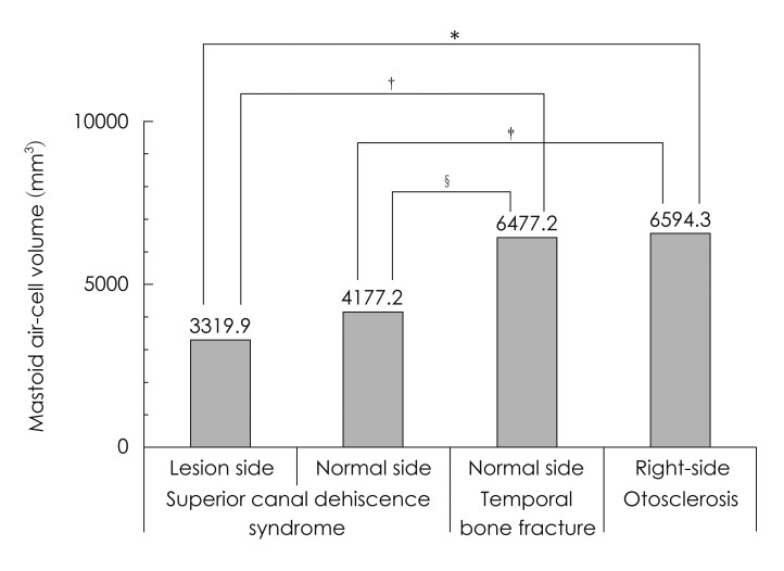
Results: In 10 patients with SCDS, the mean age was 44.5 years, ranging from 16 to 79 years (M : F=4 : 6). Mean mastoid air-cell volume in the SCDS side was 3319.9 mm(3), whereas 4177.2 mm(3) in the normal side (p=0.022). Mean mastoid air-cell volume in the right side of otosclerosis patients was 6594.3 mm(3) and it was not different from 6380.5 mm(3) in the left side (p=0.445). Mean mastoid air-cell volume in normal side of TB fracture was 6477.2 mm(3). The mastoid air-cell volume in the SCDS side was significantly smaller than that of otosclerosis and TB fracture patients (p=0.009, p=0.002, respectively). The mastoid air-cell volume in the normal side of SCDS was significantly smaller than that of TB fracture (p=0.019), but not significant with that of otosclerosis (p=0.063).
Conclusions: Our findings revealed that the mastoid air-cell volume in the SCDS side was significantly smaller than control group, which suggest that the decreased mastoid pneumatization is closely related to the generation of SCDS.
Keywords: Computed tomography; Dehiscence; Mastoid; Radiology; Semicircular canals.
Figures

Similar articles
-
Surgical decision-making in superior canal dehiscence syndrome with concomitant otosclerosis.Eur Arch Otorhinolaryngol. 2024 Jul;281(7):3859-3865. doi: 10.1007/s00405-024-08679-w. Epub 2024 May 23. Eur Arch Otorhinolaryngol. 2024. PMID: 38780629 Free PMC article.
-
Relationship Between Clinical Features and the Arc and Length of Dehiscence in SCDS: A Single Center Review of 42 Cases.Otol Neurotol. 2022 Feb 1;43(2):236-243. doi: 10.1097/MAO.0000000000003398. Otol Neurotol. 2022. PMID: 34699403
-
Superior semicircular canal dehiscence syndrome as assessed by oVEMP and temporal bone computed tomography imaging.Eur Arch Otorhinolaryngol. 2012 May;269(5):1545-9. doi: 10.1007/s00405-011-1893-3. Epub 2011 Dec 23. Eur Arch Otorhinolaryngol. 2012. PMID: 22193872
-
Superior Canal Dehiscence Syndrome Affecting 3 Families.JAMA Otolaryngol Head Neck Surg. 2017 Jul 1;143(7):656-662. doi: 10.1001/jamaoto.2016.4743. JAMA Otolaryngol Head Neck Surg. 2017. PMID: 28384775 Free PMC article.
-
Latent superior canal dehiscence syndrome unmasked by stapedotomy for otosclerosis.J Laryngol Otol. 2010 Apr;124(4):428-30. doi: 10.1017/S0022215109991654. Epub 2009 Oct 20. J Laryngol Otol. 2010. PMID: 19840429 Review.
Cited by
-
Arcuate eminence distance to temporal bone outer table in the middle fossa repair of superior canal dehiscence.Eur Arch Otorhinolaryngol. 2025 Apr;282(4):1801-1808. doi: 10.1007/s00405-024-09067-0. Epub 2024 Dec 7. Eur Arch Otorhinolaryngol. 2025. PMID: 39643813 Free PMC article.
-
Superior Semicircular Canal Dehiscence : Covering Defects in Understanding from Clinical to Radiologic Evaluation.Clin Neuroradiol. 2021 Dec;31(4):933-941. doi: 10.1007/s00062-021-01037-x. Epub 2021 Jun 7. Clin Neuroradiol. 2021. PMID: 34097081 Review.
-
Temporal bone anatomy characteristics in superior semicircular canal dehiscence.J Otol. 2017 Dec;12(4):185-191. doi: 10.1016/j.joto.2017.08.003. Epub 2017 Aug 10. J Otol. 2017. PMID: 29937854 Free PMC article.
-
Third Window Syndrome: An Up-to-Date Systematic Review of Causes, Diagnosis, and Treatment.J Audiol Otol. 2025 Apr;29(2):86-94. doi: 10.7874/jao.2024.00696. Epub 2025 Apr 18. J Audiol Otol. 2025. PMID: 40296471 Free PMC article. Review.
-
Tympanic Resonance Hypothesis.Front Neurol. 2020 Jan 30;11:14. doi: 10.3389/fneur.2020.00014. eCollection 2020. Front Neurol. 2020. PMID: 32117001 Free PMC article.
References
-
- Minor LB, Solomon D, Zinreich JS, Zee DS. Sound- and/or pressure-induced vertigo due to bone dehiscence of the superior semicircular canal. Arch Otolaryngol Head Neck Surg. 1998;124:249–258. - PubMed
-
- Minor LB. Clinical manifestations of superior semicircular canal dehiscence. Laryngoscope. 2005;115:1717–1727. - PubMed
-
- Mikulec AA, McKenna MJ, Ramsey MJ, Rosowski JJ, Herrmann BS, Rauch SD, et al. Superior semicircular canal dehiscence presenting as conductive hearing loss without vertigo. Otol Neurotol. 2004;25:121–129. - PubMed
-
- Cremer PD, Minor LB, Carey JP, Della Santina CC. Eye movements in patients with superior canal dehiscence syndrome align with the abnormal canal. Neurology. 2000;55:1833–1841. - PubMed
-
- Friedland DR, Michel MA. Cranial thickness in superior canal dehiscence syndrome: implications for canal resurfacing surgery. Otol Neurotol. 2006;27:346–354. - PubMed
LinkOut - more resources
Full Text Sources