Pulsatile tinnitus with a dural arterio-venous fistula diagnosed by computed tomography-angiography
- PMID: 24653921
- PMCID: PMC3936554
- DOI: 10.7874/kja.2013.17.3.133
Pulsatile tinnitus with a dural arterio-venous fistula diagnosed by computed tomography-angiography
Abstract
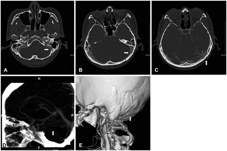
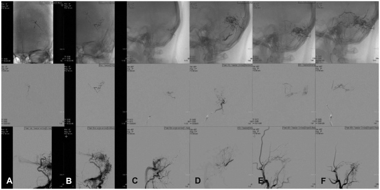
A 43 year-old female patient suffered the sudden onset of pulsatile tinnitus in the left ear 2 months ago. The tinnitus did not subside spontaneously and remained unchanged. The patient had no history of head trauma or surgery of the head and neck. The character of the tinnitus was pulsatile, and it was synchronous with the heart beat. Audiologic examinations were performed and all of the results were normal. Computed tomography with angiography was performed and evidence of an arterio-venous fistula (AVF) was found. 4-vessel angiography was performed to confirm the dural AVF between the external carotid artery and sigmoid sinus. Embolization of the feeder-vessels was done under a fluoroscope and 70% of the fistula flow was controlled after embolization and the tinnitus totally subsided during the embolization.
Keywords: Angiography; Arteriovenous fistula; Embolization; Pulsatile tinnitus; Therapeutic.
Figures
Similar articles
-
Cranial dural arteriovenous fistula as a rare cause of tinnitus - case report.Pol J Radiol. 2013 Oct;78(4):65-9. doi: 10.12659/PJR.889701. Epub 2013 Nov 19. Pol J Radiol. 2013. PMID: 24505226 Free PMC article.
-
[Use of embolization in the treatment of pulsatile tinnitus secondary to dural arteriovenous fistula].Acta Otorrinolaringol Esp. 1996 Nov-Dec;47(6):449-52. Acta Otorrinolaringol Esp. 1996. PMID: 9044584 Spanish.
-
Selective transvenous liquid embolization of a Type 1 dural arteriovenous fistula at the junction of the transverse and sigmoid sinuses. Case report.J Neurosurg. 2000 Jun;92(6):1045-9. doi: 10.3171/jns.2000.92.6.1045. J Neurosurg. 2000. PMID: 10839269
-
Spontaneous closure of transverse sinus dural arteriovenous fistula: case report.Neurol Med Chir (Tokyo). 2008 Dec;48(12):564-8. doi: 10.2176/nmc.48.564. Neurol Med Chir (Tokyo). 2008. PMID: 19106495 Review.
-
[Dural arteriovenous fistula of the transverse sigmoid sinus after transvenous embolization of the carotid cavernous fistula].No To Shinkei. 1999 Dec;51(12):1065-9. No To Shinkei. 1999. PMID: 10654304 Review. Japanese.
Cited by
-
Ear ischemia induced by endovascular therapy for arteriovenous fistula of the sigmoid sinus: A case report.World J Clin Cases. 2021 Dec 26;9(36):11443-11447. doi: 10.12998/wjcc.v9.i36.11443. World J Clin Cases. 2021. PMID: 35071576 Free PMC article.
-
Multiple Dural Arteriovenous Fistulas Presenting as Pulsatile Tinnitus Treated with External Manual Compression.J Audiol Otol. 2017 Sep;21(3):156-159. doi: 10.7874/jao.2017.00115. Epub 2017 Sep 19. J Audiol Otol. 2017. PMID: 28942629 Free PMC article.
References
-
- Park SN. Tinnitus: recent treatment. Res Vestib Sci. 2009;8:108–116.
-
- Madani G, Connor SE. Imaging in pulsatile tinnitus. Clin Radiol. 2009;64:319–328. - PubMed
-
- Sismanis A. Pulsatile tinnitus. A 15-year experience. Am J Otol. 1998;19:472–477. - PubMed
-
- Waldvogel D, Mattle HP, Sturzenegger M, Schroth G. Pulsatile tinnitus--a review of 84 patients. J Neurol. 1998;245:137–142. - PubMed
-
- Malek AM, Halbach VV, Higashida RT, Phatouros CC, Meyers PM, Dowd CF. Treatment of dural arteriovenous malformations and fistulas. Neurosurg Clin N Am. 2000;11:147–166. ix. - PubMed
Publication types
LinkOut - more resources
Full Text Sources
Other Literature Sources