Review
. 2014:32:609-34.
doi: 10.1146/annurev-immunol-032713-120236.

Metabolic regulation of immune responses

Affiliations
Review

Metabolic regulation of immune responses

Kirthana Ganeshan et al. Annu Rev Immunol. 2014.

Abstract

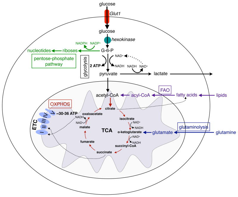
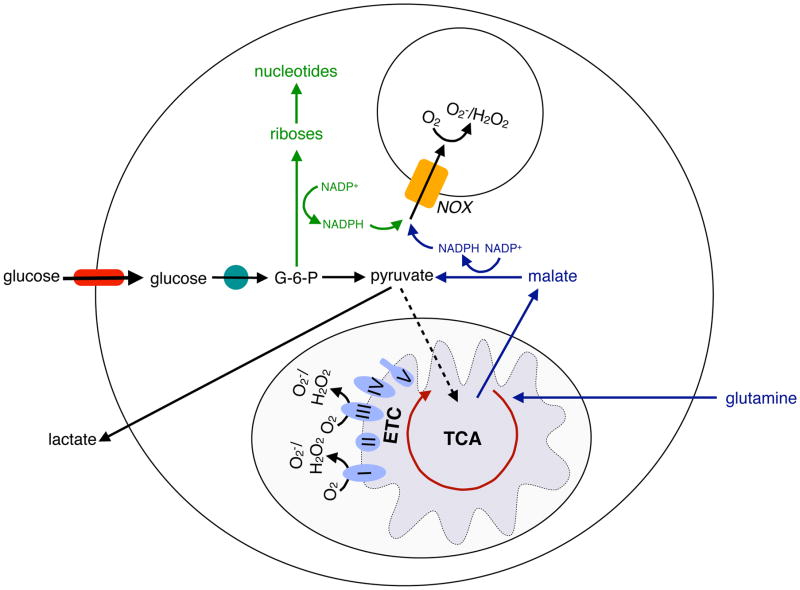
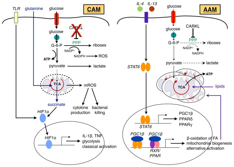
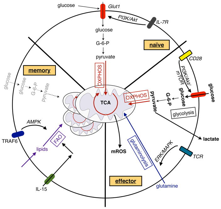
The immune system defends against pathogens and maintains tissue homeostasis for the life of the organism. These diverse functions are bioenergetically expensive, requiring precise control of cellular metabolic pathways. Although initial observations in this area were made almost a century ago, studies over the past decade have elucidated the molecular basis for how extracellular signals control the uptake and catabolism of nutrients in quiescent and activated immune cells. Collectively, these studies have revealed that the metabolic pathways of oxidative metabolism, glycolysis, and glutaminolysis preferentially fuel the cell fate decisions and effector functions of immune cells. Here, we discuss these findings and provide a general framework for understanding how metabolism fuels and regulates the maturation of immune responses. A better understanding of the metabolic checkpoints that control these transitions might provide new insights for modulating immunity in infection, cancer, or inflammatory disorders.


Conflict of interest statement

Authorship

The authors declare that they have no competing financial interests.

Figures

Figure 1
Figure 1. Major metabolic pathways of immune cells
Glucose enters the cells through the glucose transporter, GLUT1 and is phosphorylated to glucose-6-phosphate (G-6-P) by hexokinases. During glycolysis, G-6-P is metabolized to pyruvate, reducing NAD+ to NADH, and generating 2 ATP. In hypoxia, pyruvate is oxidized to lactate, restoring NAD+ levels in the cell. In normoxia, pyruvate is metabolized to acetyl-CoA, which is oxidized in the TCA cycle to generate NADH. In the redox reactions of OXPHOS, electrons are sequentially transferred to generate a H+ gradient across the inner mitochondrial membrane, which drives the synthesis of ATP. In contrast to glycolysis, mitochondrial OXPHOS is a highly efficient form of generating ATP, yielding ~30–36 ATP per molecule of glucose. Immune cells utilize three additional pathways, the pentose phosphate shunt (PPP), glutaminolysis, and fatty acid oxidation, to meet their metabolic and functional demands. G-6-P is the entry point for PPP, which generate riboses for nucleotide synthesis. During this process, NADP+ is reduced to NADPH, forming the critical co-factor required for ROS production via the NADPH oxidase system in neutrophils and macrophages. During glutaminolysis, glutamine is metabolized to glutamate and subsequently toα -ketoglutarate, which then enters the TCA cycle. The fate of glutamine is dependent on the activation state of the immune cell; it can either be oxidized completely to generate ATP or used to replenish the metabolic intermediates of TCA cycles, which are diverted for macromolecule biosynthesis. The β-oxidation of fatty acids yields acetyl-CoA, which enter the TCA cycle and OXPHOS pathways to generate ATP.
Figure 2
Figure 2. Metabolic pathways supporting ROS generation in macrophages and neutrophils
Macrophages and neutrophils rely on the production of reactive oxygen species (ROS) for their bactericidal actions. NADPH, which is required for the redox reactions of NADPH oxidase (NOX) system, is generated in the PPP branch of glycolysis or from the oxidation of glutamine-derived malate to pyruvate. In this manner, the metabolic pathways of both glycolysis and glutaminolysis contribute to the microbicidal activity of macrophages and neutrophils. In addition, mitochondrial ROS, which is derived from the inefficient flow of electrons through complex I and III of the electron transport chain (ETC), is a significant source of microbicidal ROS in macrophages.
Figure 3
Figure 3. CAMs and AAMs use distinct metabolic programs to fuel their functions
Stimulation of TLRs by PAMPs, such as LPS, drives the expression of glycolytic genes and inflammatory cytokines, a transcriptional program that is primarily coordinated by HIF-1α in classically activated macrophages (CAMs). This transition to glycolysis allows CAMs to generate NADPH through the PPP to support their respiratory burst. Succinate, derived from glutamine-dependent anaplerosis, furthers stabilizes HIF-1α protein, resulting in increased transcription and release of IL-1β. Moreover, downstream of TLR signaling, mitochondrial ROS (mROS) can also support the microbicidal functions of CAMs. In contrast to the glycolytic program of CAMs, alternatively activated macrophages (AAMs) preferentially rely on β-oxidation of fatty acids and mitochondrial respiration for their sustenance and functional activation. Type 2 cytokines, such as IL-4 and IL-13, signal through their cognate receptors to activate the latent STAT6 transcription factor. STAT6 promotes the metabolic transition to oxidative metabolism by inducting genes important in fatty acid oxidation (FAO) and mitochondrial biogenesis. In addition, STAT6 transcriptionally induces PGC1β, PPARγ, and PPARδ, which synergize with STAT6 to enhance expression of alternative activation markers and stabilize the metabolic switch to oxidative metabolism. Induction of CARKL in AAMs results in inhibition of PPP and glycolysis, thereby favoring the oxidative program of AAMs.
Figure 4
Figure 4. Metabolism of naïve, activated and memory T cells
Signaling via the IL-7 receptor (IL-7R) maintains Glut1 expression, glucose uptake, and mitochondrial OXPHOS in naïve CD4+ T cells. Upon activation, signaling via CD28 activates the PI3K/Akt/mTOR pathway to induce glycolysis in activated T cells, whereas TCR-driven ERK/MAPK signaling initiates glutaminolysis to support T cell proliferation. In addition, glycolysis promotes the translation of cytokines in activated T cells. In metabolically restrictive environments, OXPHOS can promote the proliferation of activated T cells, whereas mROS can enhance antigen-specific T cell activation. In contrast to the activation of T cells, memory formation of CD8+ T cells is dependent on TRAF6 and IL-15 driven FAO and mitochondrial biogenesis; the former is, in part, supported by activation of the cellular energy sensor AMPK.

References

    1. Taniguchi C, Emanuelli B, Kahn C. Critical nodes in signalling pathways: insights into insulin action. Nature reviews. Molecular cell biology. 2006;7:85–96. - PubMed
    1. Levene P, Meyer G. The action of leucocytes on glucose. Journal of Biological Chemistry 1912
    1. Ardawi M, Newsholme E. Maximum activities of some enzymes of glycolysis, the tricarboxylic acid cycle and ketone-body and glutamine utilization pathways in lymphocytes of the rat. The Biochemical journal. 1982;208:743–8. - PMC - PubMed
    1. Newsholme E, Crabtree B, Ardawi M. Glutamine metabolism in lymphocytes: its biochemical, physiological and clinical importance. Quarterly journal of experimental physiology (Cambridge, England) 1985;70:473–89. - PubMed
    1. Wellen K, Hatzivassiliou G, Sachdeva U, Bui T, Cross J, Thompson C. ATP-citrate lyase links cellular metabolism to histone acetylation. Science (New York, NY ) 2009;324:1076–80. - PMC - PubMed

Publication types