. 2014 May;18(5):780-9.
doi: 10.1111/jcmm.12259. Epub 2014 Mar 21.

Intramyocardial transplantation of cardiac telocytes decreases myocardial infarction and improves post-infarcted cardiac function in rats

Affiliations

Intramyocardial transplantation of cardiac telocytes decreases myocardial infarction and improves post-infarcted cardiac function in rats

Baoyin Zhao et al. J Cell Mol Med. 2014 May.

Abstract

The midterm effects of cardiac telocytes (CTs) transplantation on myocardial infarction (MI) and the cellular mechanisms involved in the beneficial effects of CTs transplantation are not understood. In the present study, we have revealed that transplantation of CTs was able to significantly decrease the infarct size and improved cardiac function 14 weeks after MI. It has established that CT transplantation exerted a protective effect on the myocardium and this was maintained for at least 14 weeks. The cellular mechanism behind this beneficial effect on MI was partially attributed to increased cardiac angiogenesis, improved reconstruction of the CT network and decreased myocardial fibrosis. These combined effects decreased the infarct size, improved the reconstruction of the LV and enhanced myocardial function in MI. Our findings suggest that CTs could be considered as a potential cell source for therapeutic use to improve cardiac repair and function following MI, used either alone or in tandem with stem cells.

Keywords: cardiac telocytes; myocardial infarction; post-infarction cardiac function.


Figures

Fig. 1
Fig. 1
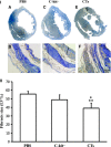
Transplantation of cardiac telocytes decreased the infarct size in myocardial infarction (MI). CTs (106) were simultaneously injected into the infarcted area and the border zone after LAD ligation MI. The infarction size was identified by Masson*s trichrome staining and analysed 14 weeks after LAD ligation. The infarction size (%LV) in the CT-treated group was significantly smaller (P < 0.05) than the c-kit cell-treated group and PBS-treated group. N = 4–5 for each group. *P < 0.05, CT-treated group versus c-kit cell-treated group and PBS-treated group.
Fig. 2
Fig. 2
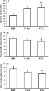
Transplantation of cardiac telocytes improved myocardial function in myocardial infarction (MI). Myocardial function of MI hearts was analysed by echocardiography 14-week post-LAD ligation showing (A) the ejection fraction of the CT-treated group was significantly higher than the PBS-treated group (P < 0.01). (B) the final diastolic diameters of the CT-treated group was significantly lower than the PBS-treated group (P < 0.05). (C) the final systolic diameters of the CT-treated group was significantly lower than the PBS-treated group (P < 0.01). N = 4–5 for each group. *P < 0.05, CT-treated group versusPBS-treated group; **P < 0.01, CT-treated group versusPBS-treated group.
Fig. 3
Fig. 3
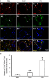
Transplantation of CTs increased c-kit+/CD34+ in 14-week-old infarct site. Double immunofluorescent staining for c-kit+/CD34+ cells was examined 14 weeks after transplantation. (I) Representative morphology of c-kit+/CD34+ CTs. (A1-4) PBS-treated group. (B1-4) c-kit cell-treated group; (C1-4) CT-treated group. C-kit+/CD34+ CTs were found near and surrounding the blood vessels in CT-treated group, c-kit cell-treated group and PBS-treated group. Open arrow: Cell body and/or nucleus of CT. Arrow: Telopode of CT; bar = 20 μm. (II) Semiquantitative image analysis of c-kit+/CD34+ CTs revealed that the density of c-kit+/CD34+ CTs in the CT-treated group was significantly higher than in the c-kit cell-treated group and PBS-treated group (P < 0.001). In addition, the density of c-kit+/CD34+ CTs in the c-kit cell-treated group was significantly higher than the PBS-treated group (P < 0.05), while the density of c-kit+/CD34+ CTs in CT-treated group was approximately fourfold higher than the c-kit cell-treated group (P < 0.001). *P < 0.001; **P < 0.05; N = 3.
Fig. 4
Fig. 4
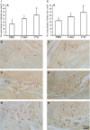
Transplantation of cardiac telocytes enhanced angiogenesis in myocardial infarction (MI). The density of vessels in MI hearts 14-week post-LAD ligation was determined by comparing the presence of vWF+ vessels in the infarct zone and border zone. (I) In the infarcted area, the vessel density of the CT-treated group was significantly larger than the PBS-treated group (P < 0.05). The vessel density of the c-kit cell-treated group appeared larger than the PBS-treated group, but was not statistically significant (P > 0.05). (A) Showing the semi-quantification analysis of vessel density. The representative morphologies of the PBS-treated group (B), the c-kit cell-treated group (C) and the CT-treated group (D). (II): In the border zone, the vessel density of CT-treated group was significantly larger than the PBS-treated group (P < 0.05). (E) showing the semi-quantification analysis of vessel density. The representative morphologies of the PBS-treated group (F), c-kit cell-treated group (G) and CT-treated group (H). Arrows indicate the vWF+ vessels; bar = 40 μm. N = 4–5 for each group. *P < 0.05, CT-treated group versusPBS-treated group.
Fig. 5
Fig. 5
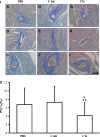
Transplantation of cardiac telocytes decreased collagen surrounding the peripheral vessels in non-infarcted zone. The perivascular collagen volume area (PVCA) of non-infarcted zone, 14 weeks after LAD ligation, was determined by analysis of collagen fibre staining. The PVCA of the CT-treated group was significantly reduced compared with the c-kit cell-treated group (P < 0.01) and PBS-treated group (P < 0.05). (IA-C) Representative morphology of PVCA in different size of vessel for the PBS-treated group; (ID-F) representative morphology of PVCA in different size of vessel for the c-kit cell-treated group; (IG-I) representative morphology of PVCA in different size of vessel for the CT-treated group. (II) Semi-quantification analysis of PVCA; bar = 60 μm. N = 4–5 for each group. **P < 0.01, CT-treated group versus c-kit cell-treated group; *P < 0.05, CT-treated group versusPBS-treated group.
Fig. 6
Fig. 6
Transplantation of cardiac telocytes decreased collagen coverage in the infarcted zone. The collagen covered area of the infarcted zone (CAIZ), 14 weeks after LAD ligation, was determined and compared. The CAIZ of the CT-treated group was found to be significantly lower than the c-kit cell-treated group (P < 0.05) and PBS-treated group (P < 0.01). (IA) Representative cross dimension with collagen fibre staining for the PBS-treated group; (IB) representative morphology of CAIZ in infracted zone under higher power (20 × ) for the PBS-treated group; (IC) representative cross dimension with collagen fibre staining for the c-kit cell-treated group; (ID) representative morphology of CAIZ in infracted zone under higher power (20 × ) for the c-kit cell-treated group; (IE) representative cross dimension with collagen fibre staining for the CT-treated group; (IF) representative morphology of CAIZ in infracted zone under higher power (20 × ) for the CT-treated group. (II) Semi-quantification analysis of the CAIZ; bar = 200 μm. N = 4–5 for each group. **P < 0.01, CT-treated group versusPBS-treated group; *P < 0.05, CT-treated group versus c-kit cell-treated group.
Fig. 7
Fig. 7
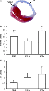
Transplantation of cardiac telocytes increased the thickness of infarcted myocardium and border zone wall. The thickness of the infarcted myocardium (TIM) and wall thickness of border zone (WTBZ) in the LV, 14-week post-LAD ligation, were determined by Masson*s trichrome staining. (A) Schematic drawing of the TIM and WTBZ. (B) The WTBZ of the CT-treated group was significantly higher than the c-kit cell-treated group and PBS-treated group (P < 0.01). The WTBZ of the c-kit cell-treated group was significantly lower than the PBS-treated group (P < 0.01). N = 4–5 for each group. *P < 0.01, CT-treated group versus c-kit cell-treated group and PBS-treated group. **P < 0.01 PBS-treated group versus c-kit cell-treated group. (C) The TIM of the CT-treated group was significantly wider than the PBS-treated group (P < 0.05). N = 4–5 for each group. *P < 0.05 CT-treated group versusPBS-treated group.

References

    1. Bernstein HS, Srivastava D. Stem cell therapy for cardiac disease. Pediatr Res. 2012;71:491–9. - PubMed
    1. Ptaszek LM, Mansour M, Ruskin JN, et al. Towards regenerative therapy for cardiac disease. Lancet. 2012;10–16:933–42. - PubMed
    1. Segers VF, Lee RT. Stem-cell therapy for cardiac disease. Nature. 2008;451:937–42. - PubMed
    1. Ieda M, Tsuchihashi T, Ivey KN, et al. Cardiac fibroblasts regulate myocardial proliferation through beta1 integrin signaling. Dev Cell. 2009;16:233–44. - PMC - PubMed
    1. Assmus B, Schachinger V, Teupe C, et al. Transplantation of progenitor cells and regeneration enhancement in acute myocardial infarction (TOPCARE-AMI) Circulation. 2002;106:3009–17. - PubMed

Publication types

MeSH terms