

Predictive value of neonatal MRI showing no or minor degrees of brain injury after hypothermia
- PMID: 24656462
- PMCID: PMC4006931
- DOI: 10.1016/j.pediatrneurol.2014.01.013
Predictive value of neonatal MRI showing no or minor degrees of brain injury after hypothermia
Abstract
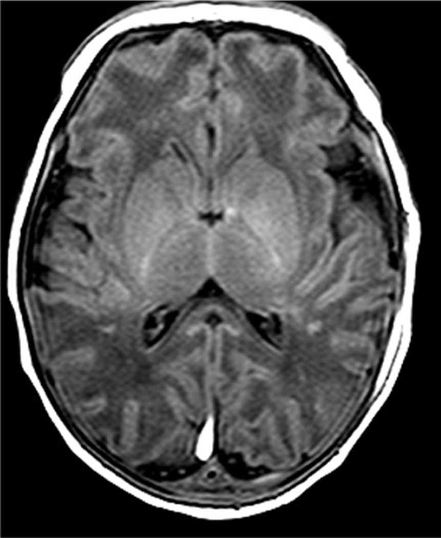
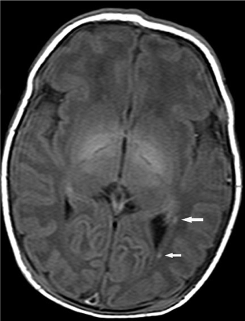
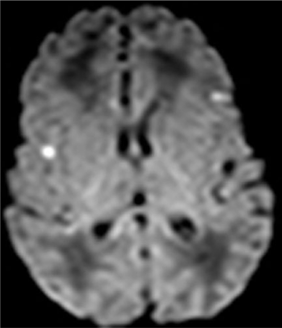
Background: Magnetic resonance imaging is a surrogate biomarker for major neurodevelopmental disabilities in survivors of perinatal hypoxic-ischemic encephalopathy because injury to the basal ganglia/thalami is highly predictive of major neuromotor and cognitive problems. Major disabilities and the appearance of neonatal magnetic resonance imaging are improved with therapeutic hypothermia. We evaluated neurodevelopmental outcomes when conventional magnetic resonance imaging showed minimal or no brain injury.
Methods: Institutional review board-approved series of 62 infants (≥36 weeks; ≥1800 g; 34 boys/28 girls) cooled for hypoxic-ischemic encephalopathy between 2005 and 2011 who underwent neonatal magnetic resonance imaging and Bayley Scales of Infant and Toddler Development-III at 22 ± 7 months of age. Magnetic resonance imaging at 5-14 (mean 8) days was scored as normal (score = 0), showing focal gray or white matter injury only (score = 1), or basal ganglia/thalamic and/or watershed lesions with or without more extensive hemispheric injury (score = 2). Sensitivity, specificity, and positive and negative predictive values for magnetic resonance scores 0 and 1 and statistical interaction between magnetic resonance imaging score and age at magnetic resonance imaging were determined.
Results: Magnetic resonance score = 0 was seen in 35/62 patients; 26/35 (74%) were typically developing, seven (20%) had moderate and two (6%) had severe delay. Magnetic resonance score = 1 was seen in 17/62 (27%) patients; 5/17 (29%) were normal, 11/17 (65%) had moderate delay, and 1/17 (6%) had severe neurodevelopmental delay. Of the 52 patients with magnetic resonance scores of 0 and 1, 40% were abnormal. The negative predictive value of a normal magnetic resonance imaging was 74%. For score 1, sensitivity was 95% (confidence interval 63%-83%), specificity 84% (confidence interval 70%-90%), positive predictive value 84% (confidence interval 71%-93%), and negative predictive value 74% (confidence interval 62%-82%).
Conclusions: Caution is warranted when prognosticating about neurodevelopmental status in early childhood after hypoxic ischemic encephalopathy with cooling, and longer follow-up studies are needed to determine the prognostic significance of a neonatal magnetic resonance imaging showing no or minor degrees of brain injury.
Keywords: MRI; hypothermia; hypoxic ischemic encephalopathy; infant; newborn; prognosis.
Copyright © 2014 Elsevier Inc. All rights reserved.
Figures
Similar articles
-
Prognostic utility of magnetic resonance imaging in neonatal hypoxic-ischemic encephalopathy: substudy of a randomized trial.Arch Pediatr Adolesc Med. 2012 Jul 1;166(7):634-40. doi: 10.1001/archpediatrics.2012.284. Arch Pediatr Adolesc Med. 2012. PMID: 22751877 Clinical Trial.
-
Therapeutic hypothermia for neonatal hypoxic-ischemic encephalopathy: magnetic resonance imaging findings and neurological outcomes in a Brazilian cohort.J Matern Fetal Neonatal Med. 2019 Aug;32(16):2727-2734. doi: 10.1080/14767058.2018.1448773. Epub 2018 Mar 13. J Matern Fetal Neonatal Med. 2019. PMID: 29504433
-
MRI findings in infants with infantile spasms after neonatal hypoxic-ischemic encephalopathy.Pediatr Neurol. 2013 Dec;49(6):401-5. doi: 10.1016/j.pediatrneurol.2013.08.007. Epub 2013 Oct 2. Pediatr Neurol. 2013. PMID: 24095571 Free PMC article.
-
Prognostic Value of Brain Magnetic Resonance Imaging in Neonatal Hypoxic-Ischemic Encephalopathy: A Meta-analysis.J Child Neurol. 2017 Nov;32(13):1065-1073. doi: 10.1177/0883073817726681. Epub 2017 Sep 19. J Child Neurol. 2017. PMID: 28925315 Review.
-
MRI for neurodevelopmental prognostication in the high-risk term infant.Semin Perinatol. 2015 Mar;39(2):159-67. doi: 10.1053/j.semperi.2015.01.009. Epub 2015 Feb 21. Semin Perinatol. 2015. PMID: 25712162 Review.
Cited by
-
Wavelet coherence analysis of dynamic cerebral autoregulation in neonatal hypoxic-ischemic encephalopathy.Neuroimage Clin. 2016 Jan 25;11:124-132. doi: 10.1016/j.nicl.2016.01.020. eCollection 2016. Neuroimage Clin. 2016. PMID: 26937380 Free PMC article.
-
Management and investigation of neonatal encephalopathy: 2017 update.Arch Dis Child Fetal Neonatal Ed. 2017 Jul;102(4):F346-F358. doi: 10.1136/archdischild-2015-309639. Epub 2017 Apr 6. Arch Dis Child Fetal Neonatal Ed. 2017. PMID: 28389438 Free PMC article.
-
Prospective research on infants with mild encephalopathy: the PRIME study.J Perinatol. 2018 Jan;38(1):80-85. doi: 10.1038/jp.2017.164. Epub 2017 Nov 2. J Perinatol. 2018. PMID: 29095433 Free PMC article.
-
Derivation of Novel Imaging Biomarkers of Neonatal Brain Injury Using Bedside Diffuse Optical Tomography: Protocol for a Prospective Feasibility Study.NeuroSci. 2025 Jun 30;6(3):60. doi: 10.3390/neurosci6030060. NeuroSci. 2025. PMID: 40700125 Free PMC article.
-
MR spectroscopy in children: protocols and pitfalls in non-tumorous brain pathology.Pediatr Radiol. 2016 Jun;46(7):963-82. doi: 10.1007/s00247-014-3270-z. Epub 2016 May 27. Pediatr Radiol. 2016. PMID: 27233789 Review.
References
-
- Shankaran S, Laptook AR, Ehrenkranz RA, Tyson JE, McDonald SA, Donovan EF, et al. Wholebodyhypothermia for neonates with hypoxic-ischemic encephalopathy. N Engl J Med. 2005 Oct;353(15):1574–1584. - PubMed
-
- Azzopardi DV, Strohm B, Edwards AD, Dyet L, Halliday HL, Juszczak E, et al. Moderatehypothermia to treat perinatal asphyxia encephalopathy. N Engl J Med. 2009 Oct;361(14):1349–1358. - PubMed
-
- Zhou WH, Cheng GQ, Shao XM, et al. Selective head cooling with mild systemic hypothermia afterneonatal hypoxic-ischemic encephalopathy: a multicenter randomized controlled trial in China. J Pediatr. 2010 Sep;157(3):367–372. 372, e361–e363. - PubMed
MeSH terms
Grants and funding
LinkOut - more resources
Full Text Sources
Other Literature Sources
Medical
