. 2014 Apr 10;7(1):19-26.
doi: 10.1016/j.celrep.2014.03.013. Epub 2014 Mar 20.

Phosphorylation-dependent PIH1D1 interactions define substrate specificity of the R2TP cochaperone complex

Affiliations

Phosphorylation-dependent PIH1D1 interactions define substrate specificity of the R2TP cochaperone complex

Zuzana Hořejší et al. Cell Rep. .

Abstract

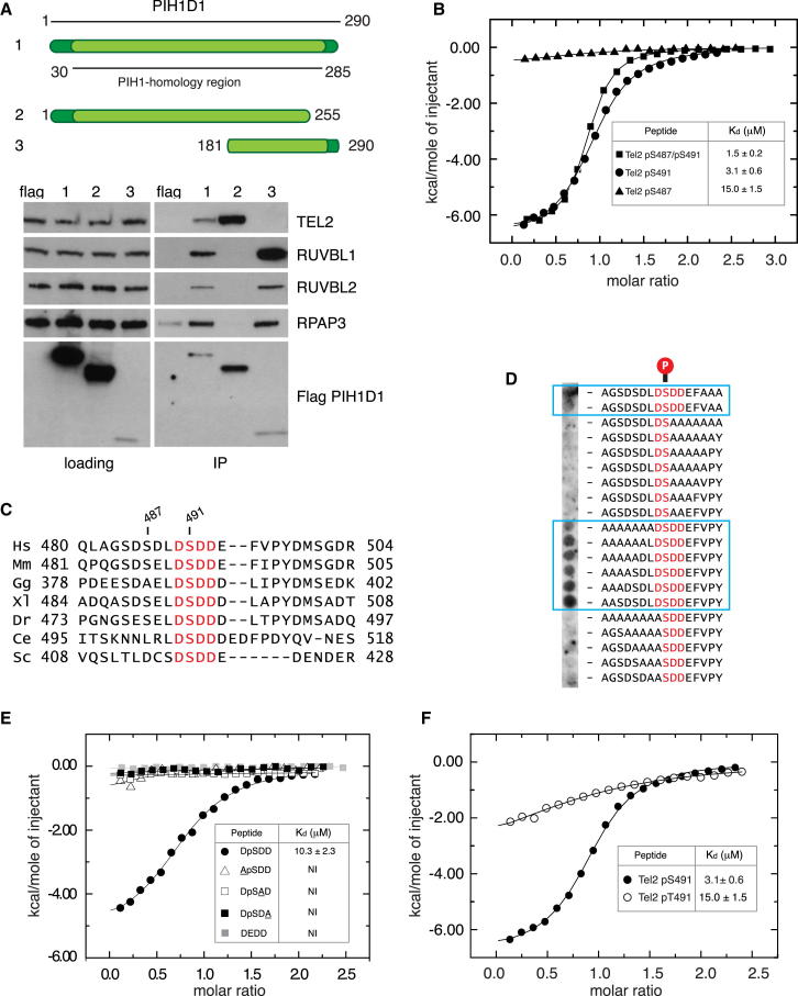
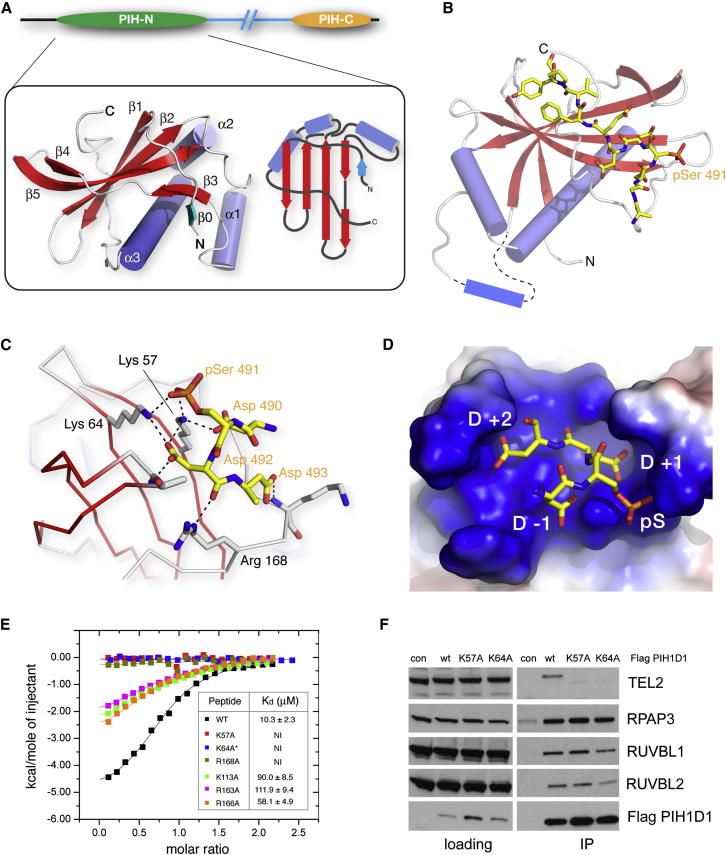
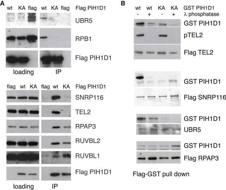
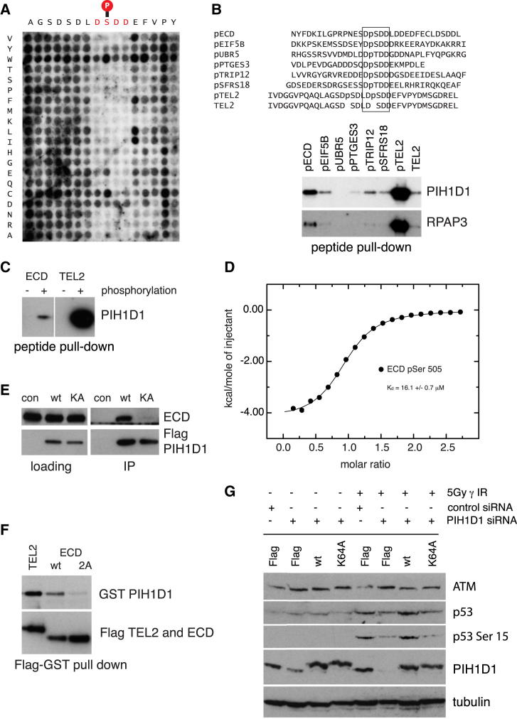
The R2TP cochaperone complex plays a critical role in the assembly of multisubunit machines, including small nucleolar ribonucleoproteins (snoRNPs), RNA polymerase II, and the mTORC1 and SMG1 kinase complexes, but the molecular basis of substrate recognition remains unclear. Here, we describe a phosphopeptide binding domain (PIH-N) in the PIH1D1 subunit of the R2TP complex that preferentially binds to highly acidic phosphorylated proteins. A cocrystal structure of a PIH-N domain/TEL2 phosphopeptide complex reveals a highly specific phosphopeptide recognition mechanism in which Lys57 and 64 in PIH1D1, along with a conserved DpSDD phosphopeptide motif within TEL2, are essential and sufficient for binding. Proteomic analysis of PIH1D1 interactors identified R2TP complex substrates that are recruited by the PIH-N domain in a sequence-specific and phosphorylation-dependent manner suggestive of a common mechanism of substrate recognition. We propose that protein complexes assembled by the R2TP complex are defined by phosphorylation of a specific motif and recognition by the PIH1D1 subunit.


Figures

None
Graphical abstract
Figure 1
Figure 1
PIH-N Domain Is Responsible for Binding of PIH1D1 to Phosphorylated TEL2 but Not Components of R2TP Complex (A) Top, schematic representation of the WT and truncated PIH1D1 proteins. The region of highest homology with budding yeast PIH1 is highlighted in light green. Bottom, interaction of PIH1D1 with components of R2TP complex and TEL2. RUVBL1, RUVBL2, RPAP3, and TEL2 were immunoprecipitated from human embryonic kidney 293T (HEK293T) cells transiently transfected with FLAG-tagged PIH1D1 proteins or empty vector expressing FLAG. (B) Isothermal titration calorimetry (ITC) analysis of PIH1D1 1–180 binding to singly and doubly phosphorylated TEL2 peptides. (C) A core DSDD motif is absolutely conserved in TEL2 orthologs from yeast humans (Hs, Homo sapiens; Mm, Mus musculus; Gg, Gallus gallus; Dr, Danio rerio; Ce, Caenorhabditis elegans; Sc, Saccharomyces cerevisiae). (D) 1D pep-spot array containing TEL2 phosphopeptides in which polyalanine tracts were substituted for regions flanking the core DpS491DD motif. (E) ITC analysis of PIH1D1 51–180 binding to WT and mutant 8-mer peptides encompassing the core conserved DpSDD TEL2 motif. NI, no quantifiable interaction. (F) ITC analysis of WT TEL2 peptide and a variant in which pSer491 was substituted with pThr.
Figure 2
Figure 2
PIH-N Domain Structure and Tel2 Interactions (A) Left, ribbons representation of the uncomplexed PIH1D1 PIH-N domain. Right, schematic representation of the overall α + β topology. β0 (cyan) contains four residues of vector encoded sequence that most likely mimics the conformation of native sequence that was removed in construction of the crystallizable fragment. (B) Structure of the PIH-N domain of PIH1D1 bound to a TEL2 phosphopeptide. The protein is shown as ribbons and the phosphopeptide as a stick representation. The loop containing α1 is disordered in the phosphopeptide complex structure and is shown schematically. (C) The core DpSDD motif in TEL2 is secured via a network of salt bridge and hydrogen bonding interactions with three basic residues: Lys57, Lys64, and Arg168. (D) Aspartates in the pSer +1 and +2 positions (and, to a lesser extent the −1 position) within the TEL2 peptide pack tightly against the protein surface. (E) ITC isotherms show that mutation of the core-motif-interacting side chains from Lys57, Lys64, and Arg168 are most deleterious to TEL2 binding. The asterisk shows that the titration with K64A was carried out at higher concentration/molar ratio, but, nonetheless, no interaction was detectable. (F) Lys57 and Lys64, but not R2TP, are essential for TEL2 binding components in vivo. RUVBL1, RUVBL2, RPAP3, and TEL2 were immunoprecipitated from HEK293T cells transiently transfected with FLAG-tagged WT PIH1D1 or PIH1D1 with K64A and K57A mutations.
Figure 3
Figure 3
Identification of Phosphopeptide-Specific Interacting Partners of PIH1D1 (A) FLAG-tagged WT PIH1D1 and K64A mutant immunoprecipitates with RPAP3, RUVBL1, and RUVBL2 subunits of the R2TP complex from HEK293T cells. FLAG-tagged WT PIH1D1, but not the K64A phosphopeptide binding mutant, immunoprecipitates UBR5, RPB1, SNRP116, and TEL2. Proteins were immunoprecipitated from HEK293T cells transiently transfected with FLAG-tagged PIH1D1 proteins or empty vector expressing FLAG. (B) FLAG-tagged TEL2, SNRP116, and UBR5 untreated with lambda phosphatase bind to recombinant GST-tagged WT PIH1D1 but not K64A mutant. Interactions with GST-tagged WT PIH1D1 are disrupted by lambda phosphatase treatment. FLAG-tagged RPAP3 untreated or treated with lambda phosphatase binds to GST-tagged WT PIH1D1 and K64A mutant.
Figure 4
Figure 4
A Minimal Consensus Sequence for PIH1 Domain Binding Reveals Potential R2TP Substrates (A) A pep-spot array was synthesized in which each residue in the TEL2 phosphopolypeptide (16 amino acids) was substituted with all other amino acids. Peptides were spotted on a peptide array and incubated with purified 6× His-tagged PIH1D1 fragment 1–180. (B) Peptide pull-down of PIH1D1 from HEK293T whole-cell extract. PIH1D1 and RPAP3 were pulled down from whole-cell extract by biotinylated peptides containing the phosphorylated consensus PIH-N domain binding sequence. Negative control (line 8) was unphosphorylated TEL2 peptide. (C) ECD peptide binds PIH1D1 in a phosphopeptide-specific manner. PIH1D1 was pulled down from HEK293T whole-cell extract by biotinylated ECD peptide containing phosphorylated Ser505 but not with nonphosphorylated ECD peptide. (D) ITC analysis of ECD binding (RPNESDpS505DDLDDY) to PIH1D1 1–180. (E) FLAG-tagged WT PIH1D1, but not K64A mutant, immunoprecipitates ECD from HEK293T whole-cell extract. Proteins were immunoprecipitated from HEK293T cells transiently transfected with FLAG-tagged PIH1D1 proteins or empty vector expression FLAG. (F) Mutation of ECD Ser505 and Ser518 to Ala disrupt binding of FLAG-tagged ECD to GST-tagged WT PIH1D1. (G) Mutation of PIH1D1 leads to decreased levels of p53 after DNA damage. Retinal pigment epithelium cells were stably transfected with FLAG-tagged WT PIH1D1, PIH1D1 K64A, or empty vector expressing FLAG. The cells were treated with control siRNA (si con) or siRNA targeting 5′ untranslated region of PIH1D1 cDNA and irradiated with 5Gy. Samples were collected 1 hr after irradiation.

References

    1. Ahn S., Kim J., Hwang J. CK2-mediated TEL2 phosphorylation augments nonsense-mediated mRNA decay (NMD) by increase of SMG1 stability. Biochim. Biophys. Acta. 2013;1829:1047–1055. - PubMed
    1. Back R., Dominguez C., Rothé B., Bobo C., Beaufils C., Moréra S., Meyer P., Charpentier B., Branlant C., Allain F.H., Manival X. High-resolution structural analysis shows how Tah1 tethers Hsp90 to the R2TP complex. Structure. 2013;21:1834–1847. - PubMed
    1. Boulon S., Marmier-Gourrier N., Pradet-Balade B., Wurth L., Verheggen C., Jády B.E., Rothé B., Pescia C., Robert M.C., Kiss T. The Hsp90 chaperone controls the biogenesis of L7Ae RNPs through conserved machinery. J. Cell Biol. 2008;180:579–595. - PMC - PubMed
    1. Boulon S., Pradet-Balade B., Verheggen C., Molle D., Boireau S., Georgieva M., Azzag K., Robert M.C., Ahmad Y., Neel H. HSP90 and its R2TP/Prefoldin-like cochaperone are involved in the cytoplasmic assembly of RNA polymerase II. Mol. Cell. 2010;39:912–924. - PMC - PubMed
    1. Clapperton J.A., Manke I.A., Lowery D.M., Ho T., Haire L.F., Yaffe M.B., Smerdon S.J. Structure and mechanism of BRCA1 BRCT domain recognition of phosphorylated BACH1 with implications for cancer. Nat. Struct. Mol. Biol. 2004;11:512–518. - PubMed

Publication types

MeSH terms