. 2014 Apr 10;54(1):193-202.
doi: 10.1016/j.molcel.2014.02.016. Epub 2014 Mar 20.

BRD7, a tumor suppressor, interacts with p85α and regulates PI3K activity

Affiliations

BRD7, a tumor suppressor, interacts with p85α and regulates PI3K activity

Yu-Hsin Chiu et al. Mol Cell. .

Abstract

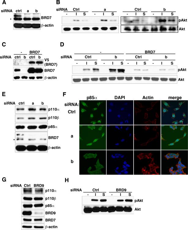
Phosphoinositide 3-kinase (PI3K) activity is important for regulating cell growth, survival, and motility. We report here the identification of bromodomain-containing protein 7 (BRD7) as a p85α-interacting protein that negatively regulates PI3K signaling. BRD7 binds to the inter-SH2 (iSH2) domain of p85 through an evolutionarily conserved region located at the C terminus of BRD7. Via this interaction, BRD7 facilitates nuclear translocation of p85α. The BRD7-dependent depletion of p85 from the cytosol impairs formation of p85/p110 complexes in the cytosol, leading to a decrease in p110 proteins and in PI3K pathway signaling. In contrast, silencing of endogenous BRD7 expression by RNAi increases the steady-state level of p110 proteins and enhances Akt phosphorylation after stimulation. These data suggest that BRD7 and p110 compete for the interaction to p85. The unbound p110 protein is unstable, leading to the attenuation of PI3K activity, which suggests how BRD7 could function as a tumor suppressor.


Figures

Figure 1
Figure 1. BRD7 binds to p85α
(A) HEK293T cells were co-transfected with HA-tagged p85α and GST protein, GST-tagged wild-type BRD7 or GST-tagged NLS deleted BRD7 as indicated, followed by GST pull-down (GST-PD) and immunoblotting analysis with antibodies against GST or HA. (B) HEK293T cells were co-transfected with Flag-tagged p85α and various GST-tagged BRD7 fragments, followed by immunoprecipitation with anti-FLAG M2-agarose and immunoblot analysis with antibodies against GST or Flag. (C) Schematic illustration of domain structures of BRD7 (upper panel). The lower panel shows the alignment of the p85-binding region of BRD7 protein sequences from human (H. sapiens), mouse (M. musculus), cow (B. taurus), chicken (G. gallus), zebrafish (D. rerio), fruit fly (D. melanogaster) and worm (C. elegans). (D) GST-tagged BRD7 and various Flag-tagged p85α fragments were transfected into HEK293T cells and then subsequently analyzed by GST pull-down and immunoblot assay with anti-GST or Flag antibodies. (E) Schematic illustration of domain structures of p85α (upper panel). Alignment of the BRD7-binding region of p85α protein sequences (lower panel). See also Figure S1.
Figure 2
Figure 2. BRD7 induces the nuclear translocation of p85α
(A) COS7 cells were transfected with GFP-p85 and GST-tagged wild-type or mutant (ΔNLS or Δ543-604) BRD7. Anti-GST antibody was used to detect BRD7 in the immunofluorescence analysis. Cells were stained with DAPI and imaged with a fluorescent microscope. (B) Crude lysates from HeLa cells transfected with Flag-tagged p85α and increasing amounts of myc-tagged BRD7 (0, 1, 2 or 4 μg) were fractionated into cytosolic and nuclear fractions as described in Experimental Procedures. Proteins in these fractions were analyzed by immunoblotting with antibodies against p85α, myc, lamin B1 and α-tubulin. (C) COS7 cells were transfected with mCherry-BRD7 and then stained with anti-p85α antibody and DAPI. The localization of p85α and BRD7 were analyzed by immunofluorescence. (D) HEK293T cells were transfected with GST protein alone, GST-tagged wild-type or Δ543-604 BRD7, followed by GST pull-down and immunoblotting analysis with antibodies against p85α or GST. (E) Endogenous BRD7 was immunoprecipitated with anti-BRD7 antibodies from mononuclear cell lysates. Rabbit IgG was used as a control. Cell lysates and immunoprecipitates were analyzed by immunoblotting with antibodies against p85α and BRD7. (F) CHO-K1 cells were co-transfected with GFP-p85α and HA-tagged p110α together with GST-tagged wild-type or ΔNLS BRD7. The cells were immunostained with anti-GST and anti-HA antibodies, and imaged with a fluorescent microscope. (G) Cell lysates from HeLa cells transfected with GST-tagged BRD7 and HA-tagged p85α were separated by Superdex 200 using the FPLC system. Every other fractions were subjected to immunoblotting analysis with antibodies against GST, HA, p110α, p110β or ARID2. The fractions contained 670 or 200 kDa proteins were indicated. (H) Fraction 14 from (G) was subjected to GST-pull down assay (GSTPD) and immunoprecipitation (IP) using rabbit IgG, anti-p85α, p110α or p110β antibodies. Samples were further analyzed by immunoblotting using antibodies against GST, p85α, p110α, p110β and ARID2. See also Figure S2 and S3.
Figure 3
Figure 3. BRD7 reduces PI3K signaling and p110 expression
(A) HeLa cells transfected with myc-tagged BRD7 or a control plasmid were under serum starvation overnight and then stimulated with 100μM insulin (I) or 15% serum in DMEM (S). Cell lysates were separated by SDS-PAGE and analyzed with antibodies against phospho-Akt (S473), total Akt, and myc. (B) HeLa cells transfected with increasing amount of myc-tagged BRD7 and immunoblotted with antibodies against p110α, p110β, p85α, myc and β-actin. (C) Stable cells expressing V5-tagged wild-type or ΔNLS BRD7 were confirmed by immunoblotting with anti-V5 antibody. The amount of β-actin was used as a loading control (lower panel). Stable cells were stimulated with insulin or serum, followed by immunoblot analysis as metioned in (A). (D) HeLa cells expressing wild-type or ΔNLS BRD7 were analyzed as indicated in (B). (E and F) HeLa cells were transfected with the indicated amount of BRD7 DNA. Total RNAs were extracted from those cells and analyzed with real-time RT-PCR using primers specifically designed for detecting p110α, p110β, p85α, BRD7 and GAPDH. The mRNA level of p110α, p110β, p85α (E), or BRD7 (F) was normalized with the level of the housekeeping gene, GAPDH. ns: non-significant difference. The values are the average of triplicates ± SD.
Figure 4
Figure 4. BRD7 destabilizes p110 and negatively regulates PI3K signaling
(A) HeLa cells were transfected with two different pairs of siRNA oligos (a and b) against BRD7 or a control oligo. High-salt cell lysates were immunoblotted with antibodies against BRD7 (for RNAi efficiency) or β-actin (for loading control). An asterisk (*) indicates a non-specific band detected by anti-BRD7 antibody. (B) Cells from (A) were serum-starved for overnight and subsequently treated with 100μM insulin (I) or 15% serum in DMEM (S). Cell lysates were analyzed by immunoblotting with an antibody against phospho-Akt (S473) or total Akt. (C) BRD7 siRNA-resistant cells and the parental HeLa cells were transfected with control siRNA or siRNA against BRD7 (oligos b) and then immunoblotted with anti-V5, BRD7 or β-actin. (D) Cells from (C) were analyzed as described in (B). (E) NCI-H520 cells were transfected with control siRNA and siRNA oligos against BRD7 (a and b). Cell lysates were immunoblotted with antibodies against p110α, p110β, p85α, BRD7 or β-actin. An asterisk (*) indicates a non-specific band detected by anti-BRD7 antibody. (F) Cells from (E) were examined with immunofluorescence using anti-p85α antibody, followed by Alexa fluor 568 phalloidin and DAPI staining and imaged with a fluorescent microscope. (G) HeLa cells were transfected with siRNA oligos against BRD9 or a control oligo. High-salt cell lysates from those cells were immunoblotted with antibodies against p110α, p110β, p85α, BRD9, BRD7 or β-actin. (H) Cells from (G) were serum-starved for overnight and then treated with 100μM insulin (I) or 15% serum in DMEM (S). Cell lysates were analyzed by immunoblotting with an antibody against phospho-Akt (S473) or total Akt. See also Figure S4.

References

    1. Barretina J, Caponigro G, Stransky N, Venkatesan K, Margolin AA, Kim S, Wilson CJ, Lehar J, Kryukov GV, Sonkin D, et al. The Cancer Cell Line Encyclopedia enables predictive modelling of anticancer drug sensitivity. Nature. 2012;483:603–607. - PMC - PubMed
    1. Bavelloni A, Santi S, Sirri A, Riccio M, Faenza I, Zini N, Cecchi S, Ferri A, Auron P, Maraldi NM, et al. Phosphatidylinositol 3-kinase translocation to the nucleus is induced by interleukin 1 and prevented by mutation of interleukin 1 receptor in human osteosarcoma Saos-2 cells. Journal of cell science. 1999;112(Pt 5):631–640. - PubMed
    1. Burrows AE, Smogorzewska A, Elledge SJ. Polybromo-associated BRG1- associated factor components BRD7 and BAF180 are critical regulators of p53 required for induction of replicative senescence. Proceedings of the National Academy of Sciences of the United States of America. 2010;107:14280–14285. - PMC - PubMed
    1. Carpenter CL, Duckworth BC, Auger KR, Cohen B, Schaffhausen BS, Cantley LC. Purification and characterization of phosphoinositide 3-kinase from rat liver. The Journal of biological chemistry. 1990;265:19704–19711. - PubMed
    1. Chagpar RB, Links PH, Pastor MC, Furber LA, Hawrysh AD, Chamberlain MD, Anderson DH. Direct positive regulation of PTEN by the p85 subunit of phosphatidylinositol 3-kinase. Proceedings of the National Academy of Sciences of the United States of America. 2010;107:5471–5476. - PMC - PubMed

Publication types

MeSH terms

Substances