. 2014 Apr 10;54(1):17-29.
doi: 10.1016/j.molcel.2014.02.018. Epub 2014 Mar 20.

Molecular basis for specific recognition of bacterial ligands by NAIP/NLRC4 inflammasomes

Affiliations

Molecular basis for specific recognition of bacterial ligands by NAIP/NLRC4 inflammasomes

Jeannette L Tenthorey et al. Mol Cell. .

Abstract

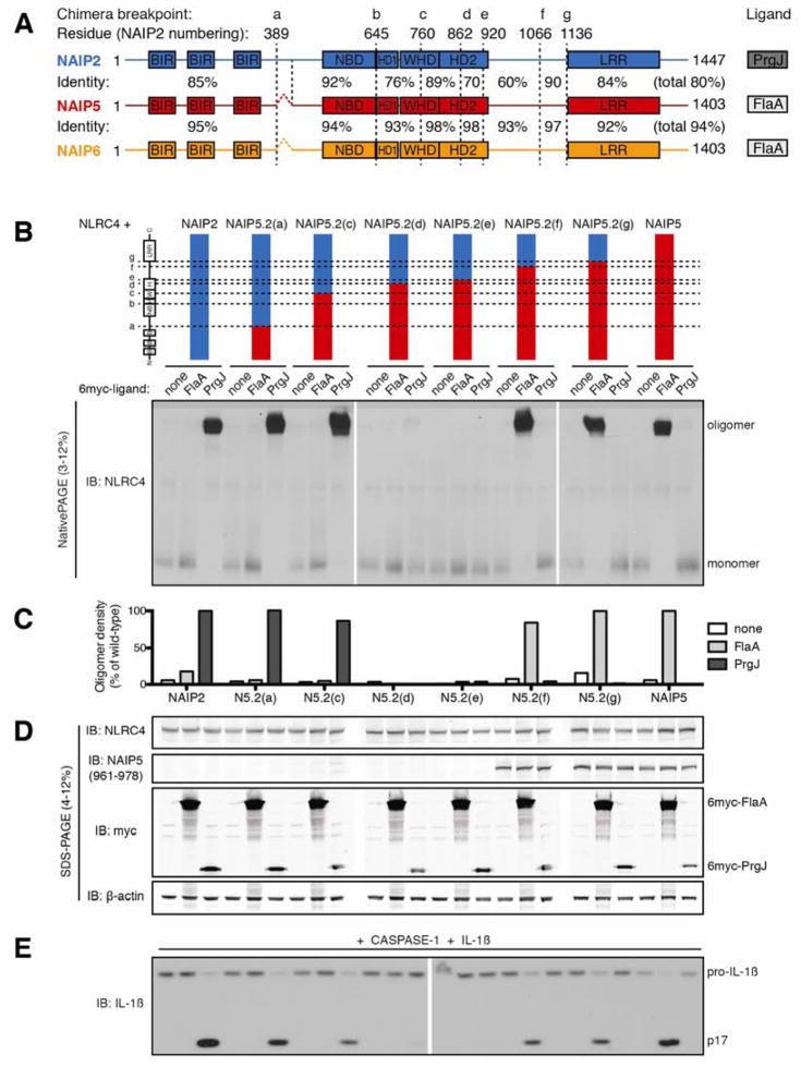
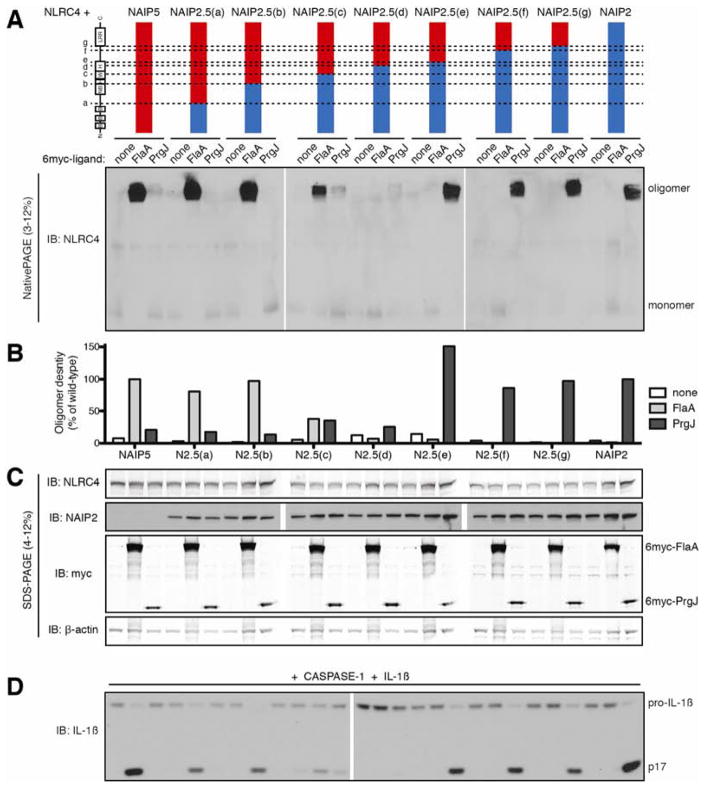
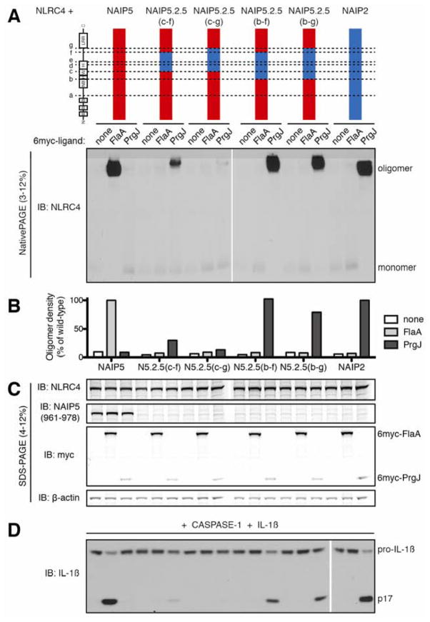
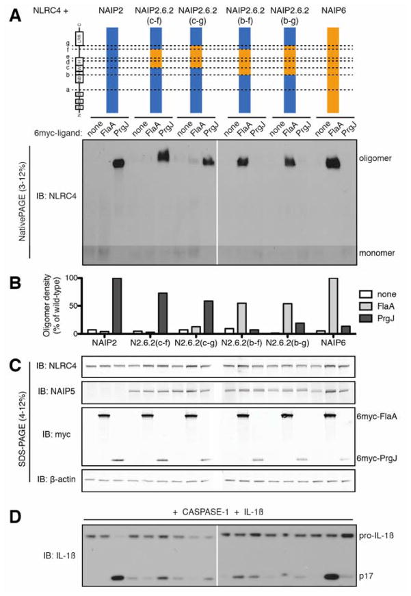
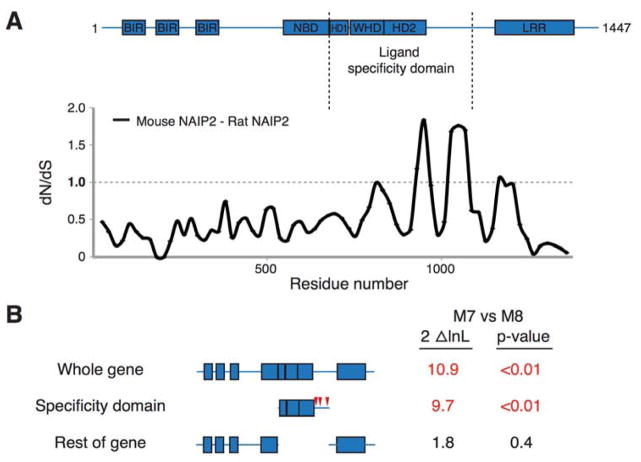
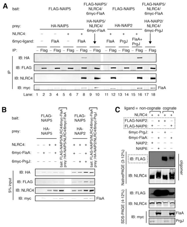
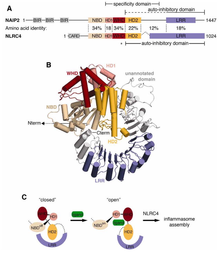
NLR (nucleotide-binding domain [NBD]- and leucine-rich repeat [LRR]-containing) proteins mediate innate immune sensing of pathogens in mammals and plants. How NLRs detect their cognate stimuli remains poorly understood. Here, we analyzed ligand recognition by NLR apoptosis inhibitory protein (NAIP) inflammasomes. Mice express multiple highly related NAIP paralogs that recognize distinct bacterial proteins. We analyzed a panel of 43 chimeric NAIPs, allowing us to map the NAIP domain responsible for specific ligand detection. Surprisingly, ligand specificity was mediated not by the LRR domain, but by an internal region encompassing several NBD-associated α-helical domains. Interestingly, we find that the ligand specificity domain has evolved under positive selection in both rodents and primates. We further show that ligand binding is required for the subsequent co-oligomerization of NAIPs with the downstream signaling adaptor NLR family, CARD-containing 4 (NLRC4). These data provide a molecular basis for how NLRs detect ligands and assemble into inflammasomes.


Figures

Figure 1
Figure 1. NAIP5 LRR is dispensable for recognition of flagellin
(A) Schematic of NAIP predicted domains drawn to scale. Baculovirus inhibitor of apoptosis repeat (BIR) and leucine-rich repeat (LRR) domains were identified by amino acid query against the NCBI Conserved Domain Database. The nucleotide binding domain (NBD), helix domain 1 (HD1), winged helix domain (WHD), and helix domain 2 (HD2) were annotated by homology modeling to NLRC4 (PDB 4KXF). Chimera breakpoints are identified by lowercase letters (a–g) and by dashed lines. Amino acid identity between NAIP2 and NAIP5 or NAIP5 and NAIP6 is indicated for each segment and across total protein length. (B) Oligomerization assay to test the specificity of NAIP5.2 chimeras. HEK293T cells were transfected with NLRC4 and the indicated NAIP chimera and 6myc-tagged ligand. After 48 hours, cell lysates were harvested, normalized for total protein, subjected to blue native PAGE, and immunoblotted (IB) for NLRC4, as previously described (Kofoed and Vance, 2011). Results shown are representative of at least 3 independent trials. See also Figure S1. (C) NAIP chimera responses to each ligand in (B) were quantified by densitometry of the oligomer species and normalized to wild-type NAIP2 or NAIP5 response to PrgJ or FlaA, respectively. (D) Lysates from (B) were subjected to denaturing SDS-PAGE and immunoblotted for NLRC4, NAIP5, myc, and β-actin to control for equal transfection and loading. (E) NAIP2.5 inflammasomes are functional and induce IL-1β cleavage. HEK293T cells were transfected as in (B) but with the addition of CASP-1 and pro-IL-1β. Cell lysates were harvested at 24 hours and immunoblotted for IL-1β; full-length (pro) and cleaved (p17) forms are indicated. Results shown are representative of 2 independent trials.
Figure 2
Figure 2. NAIP2 LRR is dispensable for recognition of PrgJ
(A) Oligomerization assay to test the specificity of NAIP2.5 chimeras. HEK293T cells were transfected with NLRC4 and the indicated NAIP chimera and 6myc-tagged ligand. Cell lysates were subjected to blue native PAGE as in Figure 1. Results shown are representative of at least 3 independent trials. See also Figure S2. (B) NAIP chimera responses to each ligand in (A) were quantified by densitometry of the oligomer species and normalized to wild-type NAIP2 or NAIP5 response to PrgJ or FlaA, respectively. (C) Lysates from (A) were subjected to denaturing SDS-PAGE and immunoblotted for NLRC4, NAIP2, myc, and β-actin. (D) NAIP5.2 inflammasomes are functional. HEK293T cells were transfected as in (A) but with the addition of CASP-1 and pro-IL-1β. Cell lysates were analyzed for IL-1β cleavage after 24 hours. Results shown are representative of 2 independent trials.
Figure 3
Figure 3. Central NBD-associated domains of NAIP2, including HD1, WHD, and HD2, are sufficient for recognition of PrgJ
(A) Oligomerization assay to test the specificity of NAIP5.2.5 chimeras. HEK293T cells were transfected with NLRC4 and the indicated NAIP chimera and 6myc-tagged ligand. Cell lysates were subjected to blue native PAGE as in Figure 1. Results shown are representative of at least 3 independent trials. See also Figure S3. (B) NAIP chimera responses to each ligand in (A) were quantified by densitometry of the oligomer species and normalized to wild-type NAIP2 or NAIP5 response to PrgJ or FlaA, respectively. (C) Lysates from (A) were subjected to denaturing SDS-PAGE and immunoblotted for NLRC4, NAIP5, myc, and β-actin. (D) NAIP5.2.5 inflammasomes are functional. HEK293T cells were transfected as in (A) but with the addition of CASP-1 and pro-IL-1β. Cell lysates were analyzed for IL-1β cleavage after 24 hours. Results shown are representative of 2 independent trials.
Figure 4
Figure 4. Central NBD-associated domains of NAIP6, including HD1, WHD and HD2, are sufficient for recognition of flagellin
(A) Oligomerization assay to test the specificity of NAIP2.6.2 chimeras. HEK293T cells were transfected with NLRC4 and the indicated NAIP chimera and 6myc-tagged ligand. Cell lysates were subjected to blue native PAGE as in Figure 1. See also Figure S4. (B) NAIP chimera responses to each ligand in (A) were quantified by densitometry of the oligomer species and normalized to wild-type NAIP2 or NAIP6 response to PrgJ or FlaA, respectively. (C) Lysates from (A) were subjected to denaturing SDS-PAGE and immunoblotted for NLRC4, NAIP5, myc, and β-actin. The NAIP5 antibody also detects NAIP6. (D) NAIP2.6.2 chimeras exhibit basal IL-1β cleavage. HEK293T cells were transfected as in (A) but with the addition of CASP-1 and pro-IL-1β. Cell lysates were analyzed for IL-1β cleavage after 24 hours. All results shown are representative of 3 independent trials.
Figure 5
Figure 5. The ligand specificity domain has evolved under positive selection
(A) Sliding window comparison of the dN/dS ratio between mouse and rat Naip2 genes. dN/dS ratios were calculated every 20 codons with a window size of 50 codons. Shown above is the domain structure of mouse NAIP2. (B) Results of PAML analyses on the entire primate NAIP gene, the ligand specificity domain alone, and the entire gene outside the ligand specificity domain. The left column shows two times the log likelihood difference between a model that allows (M8) or disallows (M7) positive selection. The right column shows the statistical significance of support for the gene, or domain, having evolved under positive selection. Values in red indicate strong support for positive selection. Red triangles indicate primate NAIP codons (corresponding to mouse NAIP2 residues 941, 965 and 1049) identified as having evolved under recurrent positive selection with a posterior probability of >0.95. See also Figure S5.
Figure 6
Figure 6. NAIPs require cognate ligand to assemble into an inflammasome
(A) HA-NAIP2 associates with FLAG-NAIP5 only when both cognate ligands are present. HEK293T cells were co-transfected with multiple NAIP constructs, distinguishable by FLAG or HA tags, and NLRC4 and 6myc-tagged ligands as indicated. After 48 hours, cell lysates were divided equally, and assembled inflammasomes were immunoprecipitated (IP) with anti-FLAG antibody (Flag) or normal mouse IgG (−) as a negative control. To control for non-specific inter-oligomer association, separately assembled inflammasomes were mixed in lysate (denoted by arrows). Immunoprecipitates were separated by SDS-PAGE and immunoblotted for HA, FLAG, NLRC4 and myc. Results are representative of at least 4 independent trials. (B) Prior to immunoprecipitations in (A), 5% of lysate volume was removed as input, separated by SDS-PAGE, and immunoblotted in parallel with IP samples. (C) FLAG-NAIPs are detectable in assembled inflammasomes only when provided with their cognate ligand. HEK293T cells were co-transfected with NLRC4 and the indicated 6myc-tagged ligand and NAIP construct(s). After 48 hours, cell lysates were harvested, separated in parallel by blue native PAGE and SDS-PAGE, and immunoblotted for FLAG followed by NLRC4 and myc. See also Figure S6.
Figure 7
Figure 7. Model of NAIP autoinhibition relief by ligand binding
(A) Comparison of NLRC4 (PDB 4KXF) and predicted NAIP2 domain architecture. Solid lines indicate the NAIP ligand specificity domain identified in this study and autoinhibitory domains previously identified for NAIP (Kofoed and Vance, 2011) and NLRC4 (Hu et al., 2013). The dashed line denotes a potential extension of the autoinhibitory domain based on comparison with NLRC4; the asterisk marks the NLRC4 autoinhibitory residue H443 that is not conserved in NAIPs. Amino acid identity between NAIP2 and NLRC4 is indicated for each segment. See also Figure S7. (B) Homology model of NAIP2 by multiple template threading using the Phyre2 server. NAIP2 predicted domains are colored as follows: NBD (tan), HD1 (pink), WHD (red), HD2 (orange), and LRR (blue). The N-terminal BIR domains of NAIP2 are not shown. Regions outside of the predicted domains are depicted in gray; predicted structure of the unannotated domain (especially residues 987–1040) is based on low-confidence ab initio modeling. (C) Model for NAIP activation by ligand binding. Regions of low-confidence structural modeling have been replaced with dashed lines. Ligand, depicted as a green oval, is predicted to bind primarily within the NAIP ligand specificity domain. Ligand binding may sterically occlude autoinhibitory interactions and/or allosterically induce rotation of autoinhibitory domains away from the NBD in a manner similar to apoptosome assembly (Reubold et al., 2011). Exposure of NBD oligomerization surfaces triggers assembly of the NAIP/NLRC4 inflammasome.

Comment in

References

    1. Bergsbaken T, Fink SL, Cookson BT. Pyroptosis: host cell death and inflammation. Nat Rev Microbiol. 2009;7:99–109. - PMC - PubMed
    1. Bonardi V, Cherkis K, Nishimura MT, Dangl JL. A new eye on NLR proteins: focused on clarity or diffused by complexity? Curr Opin Immunol. 2012;24:41–50. - PMC - PubMed
    1. Chavarria-Smith J, Vance RE. Direct proteolytic cleavage of NLRP1B is necessary and sufficient for inflammasome activation by anthrax lethal factor. PLoS pathogens. 2013;9:e1003452. - PMC - PubMed
    1. Chisholm ST, Coaker G, Day B, Staskawicz BJ. Host-microbe interactions: shaping the evolution of the plant immune response. Cell. 2006;124:803–814. - PubMed
    1. Comeron JM. K-Estimator: calculation of the number of nucleotide substitutions per site and the confidence intervals. Bioinformatics. 1999;15:763–764. - PubMed

Publication types

MeSH terms