. 2014 Apr 10;54(1):119-132.
doi: 10.1016/j.molcel.2014.02.020. Epub 2014 Mar 20.

Inositol pyrophosphates mediate the DNA-PK/ATM-p53 cell death pathway by regulating CK2 phosphorylation of Tti1/Tel2

Affiliations

Inositol pyrophosphates mediate the DNA-PK/ATM-p53 cell death pathway by regulating CK2 phosphorylation of Tti1/Tel2

Feng Rao et al. Mol Cell. .

Erratum in

Abstract

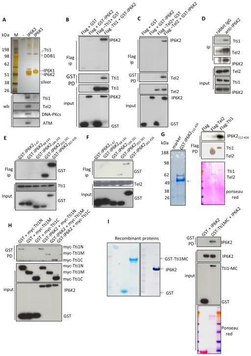
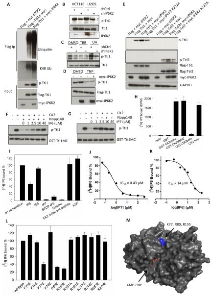
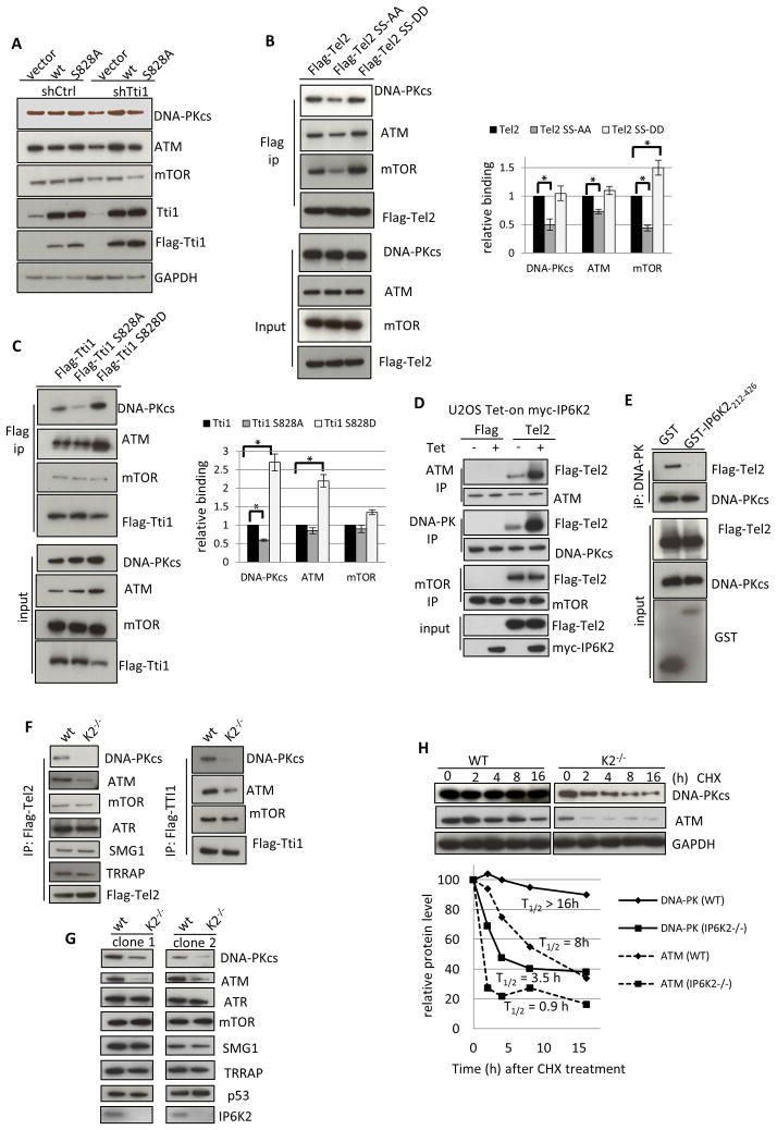
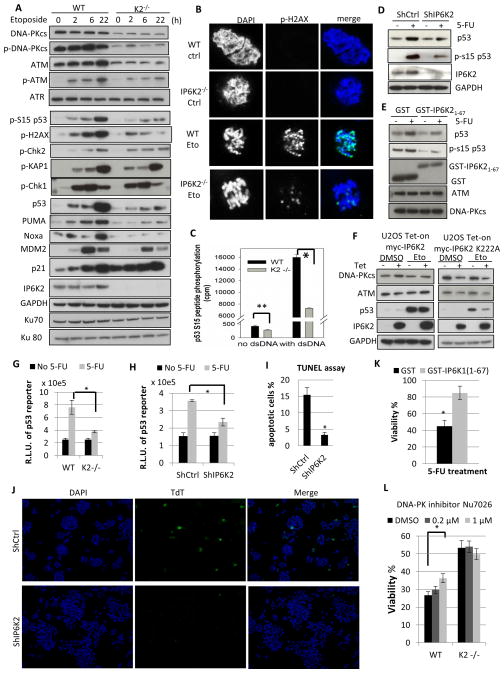
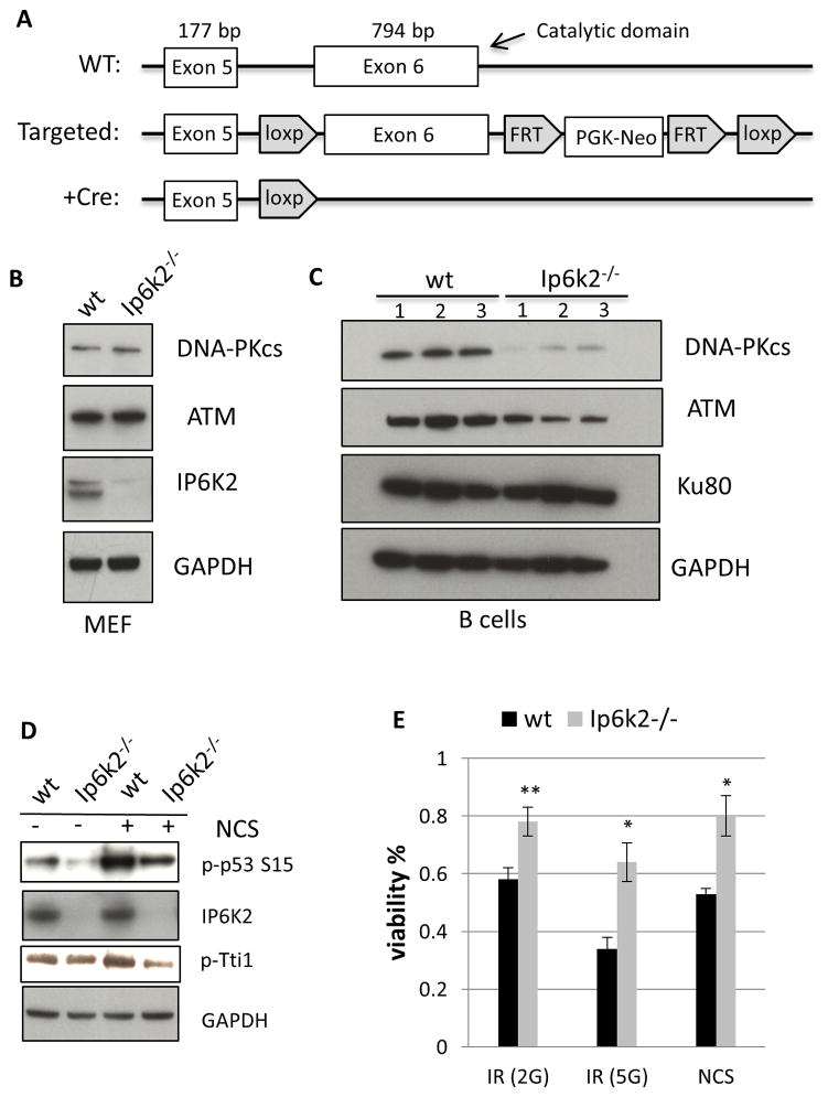
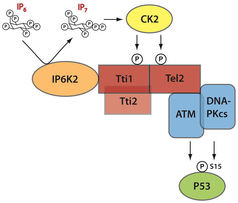
The apoptotic actions of p53 require its phosphorylation by a family of phosphoinositide-3-kinase-related-kinases (PIKKs), which include DNA-PKcs and ATM. These kinases are stabilized by the TTT (Tel2, Tti1, Tti2) cochaperone family, whose actions are mediated by CK2 phosphorylation. The inositol pyrophosphates, such as 5-diphosphoinositol pentakisphosphate (IP7), are generated by a family of inositol hexakisphosphate kinases (IP6Ks), of which IP6K2 has been implicated in p53-associated cell death. In the present study we report an apoptotic signaling cascade linking CK2, TTT, the PIKKs, and p53. We demonstrate that IP7, formed by IP6K2, binds CK2 to enhance its phosphorylation of the TTT complex, thereby stabilizing DNA-PKcs and ATM. This process stimulates p53 phosphorylation at serine 15 to activate the cell death program in human cancer cells and in murine B cells.


Figures

Figure 1
Figure 1. IP6K2 binds to the TTT complex
(A) SDS-PAGE of tandem-affinity purified IP6Ks, followed by silver-staining. Lower panel, western-blot of tandem-affinity purified IP6Ks identifies binding partner. (B) Co-IP between GST-IP6K2 and Flag-Tti1 expressed in HEK293 cells. (C) Co-IP between GST-IP6K2 and Flag-Tel2 expressed in HEK293 cells. (D) Endogenous Co-IP between IP6K2 and Tel2/Tti1 in HCT116 cells. (E) Co-IP between GST-IP6K2 fragments and Flag-Tti1. (F) Co-IP between GST-IP6K2 fragments and Flag-Tel2. (G) Direct binding between recombinant GST-IP6K2212-426 and purified Flag-Tti1 in vitro. (H) Co-IP between myc-Tti1 N (aa1-460), M (aa 461-825), and C (aa 826-1089) fragments and GST-IP6K2. (I) Direct binding between purified recombinant full-length IP6K2 and recombinant GST-Tti1MC (aa 461-1089) in vitro. see also Figure S1.
Figure 2
Figure 2. IP7 binds to CK2 and enhances CK2-mediated phosphorylation of Tti1/Tel2
(A) Coexpression of IP6K2 increases the phosphorylation (S828) and ubiqutination of Tti1. Cells were harvested 28 h after transfection. The amount of Tti1 plasmids used for transfection was adjusted to achieve equal expression levels. (B) Lentiviral shRNA knockdown of IP6K2 diminishes endogenous Tti1 phosphorylation. (C) Effect of CK2 inhibitor TBB (20 μM, 4 h) and protein phosphatase inhibitor okadaic acid (OA) (50 nM, 1 h) on Tti1 phosphorylation. U2OS cells were transfected with the indicated lentiviral shRNA constructs and selected with puromycin (1 μg/ml) prior to drug treatment. (D) Effect of TNP treatment (10 μM, 1.5 h) on the phosphorylation of Tti1, with/without IP6K2 co-expression. (E) Co-expression of IP6K2 wildtype, but not the K222A mutant, increases phospho-Tti1 (S828) and phospho-Tel2 (S487/S491). (F–G) Concentration-dependent effects of IP6 (F) and IP7 (G) on reversing hNopp140 (500 nM) inhibition of GST-Tti1MC phosphorylation by CK2. Reaction conditions were: 20 mM MgCl2 (pH 7.5), 50 mM KCl, 10 mM MgCl2, 200 μM ATP, 30 °C, 15 min. (H) [3H]IP6 binding to various CK2 preparations. Twenty μg purified recombinant CK2 proteins on glutathione beads were incubated with 10 μl [3H]IP6 (65 μCi/ml) overnight at 4 °C. Controls included buffer or GST alone. (I–J) Concentration-dependent competition of [3H]IP6 binding to CK2α by unlabeled IP6 (I) and IP7 (J). (K) [3H]IP6 binding to CK2α in the presence of various competitors. The concentrations of small molecules used were: ATP (100 μM), the rest (25 μM). (L) [3H]IP6 binding to CK2α mutants. (M) Location of K77, R80 and R155 in the 3D structure of CK2α displayed in surface mode (PDB id: 2PVR). The ATP analog AMPPNP is located in the catalytic active site. Residues K77, R80 and R155 are highlighted in blue. The figure was generated by Pymol. see also Figure S2.
Figure 3
Figure 3
IP6K2 is required for the binding of the TTT complex to DNA-PKcs/ATM and for the stability of DNA-Pkcs/ATM. (A) Protein levels of DNA-PKcs, ATM and mTOR in HCT116 cells that were transfected with shTti1 for 3 d, and with siRNA-resistant flag-Tti1 or flag-Tti1-S828A for 2 d. (B) Immunoprecipitation of Tel2 wildtype, S478A/491A, and S478D/S491D mutant proteins, followed by western-blot analysis. The bar graph represents average data from three different experiments. Relative binding was determined as the amount of co-immunoprecipitated PIKKs divided by the levels of Flag-Tti1 variants. (C) Immunoprecipitation of Tel2 wildtype, S828A, and S828D mutant proteins, followed by western-blot analysis. (D) Overexpression of myc-IP6K2 in U2OS cells increases the co-IP between Flag-Tel2 and ATM/DNA-PKcs, but not that between Flag-Tel2 and mTOR. Four h after cells were transfected with Flag-Tel2 (lipofectamine), tetracycline (1 μM) was added for another 20 h. (E) Overexpression of GST-IP6K2212-426 abolishes the co-IP between Flag-Tel2 and DNA-PKcs. (F) Flag-Tel2 and Flag-Tti1 immunoprecipitation from wildtype and isogenic IP6K2−/− HCT116 cells, followed by western-blot analysis. (G). Western-blot analysis of the expression levels of PIKKs in two clones of IP6K2 wildtype and null cell lines. (H) Cycloheximide treatment (50 μM) decreases DNA-PKcs and ATM protein levels more rapidly in IP6K2−/− than wild-type cells. see also Figure S3.
Figure 4
Figure 4
IP6K2 influence the p53 cell-death pathway by promoting p53 S15-phosphorylation by DNA-PKcs/ATM. (A) Western blots of DNA-PKcs, ATM, their active phosphorylated forms, and their downstream targets after treatment with etoposide (20 μM) for 2, 6, and 22 h, respectively. (B) Immunofluorescence staining for p-H2AX in WT and IP6K2−/− HCT116 cells with or without etoposide treatment. (C) [32P]ATP-dependent phosphorylation of a p53 S15-peptide by wild-type and IP6K2−/− cell lysate in vitro, in the presence or absence of dsDNA. For experimental details see Materials and Methods. (D) Knockdown of IP6K2 in HCT116 cells by shRNA (lipofectamine) abolishes p53 induction upon DNA damage. (E) Effect of GST-IP6K21-67 expression on induction of p53 and its S15 phosphorylation upon 5-FU treatment. (F) Kinase dead IP6K2 destabilizes DNA-PKcs/ATM and diminishes p53 induction. IP6K2 wildtype and K222A mutant were overexpressed (16 h) in a Tet-on U2OS cell system, followed by etoposide treatment (20 μM, 16 h). (G) Luciferase activity from the p53 consensus promoter driven reporter PG13-luc. R.L.U.: relative luciferase unit. (H) Knockdown of IP6K2 attenuates p53 transcriptional activation of the PG13-luc luciferase reporter. (I–J) Knockdown of IP6K2 increases cell viability as measured by TUNEL assay. Forty-eight h after transfection (lipofectamine) of shRNA constructs, cells were treated with etoposide (40 μM, 24 h). Cells were stained using the Terminal deoxynucleotidyl Transferase (TdT)-mediated dUTP-biotin Nick End Labeling (TUNEL) assay system. Fragmented apoptotic cell nuclei were visualized by TUNEL (TdT, green), and the nucleus was stained with DAPI (blue). (K). Viability of HCT116 cells with or without overexpression of IP6K21-67 followed by 5-FU treatment. (L) Viability of HCT116 wildtype and IP6K2 null cells upon treatment with cytotoxic concentrations of 5-FU (400 μM) with or without pre-treatment with the DNA-PK inhibitor Nu7026 for 1 h. see also Figure S4.
Figure 5
Figure 5
IP6K2 mediates the DNA-PKcs/ATM-p53 pathway in murine B cells. (A) Schematic depiction of the targeting strategy used to generate Ip6k2−/− mice. Exon 6, containing the majority of the catalytic domain and the 3′ UTR, was flanked by loxP sites to generate the Flox/Flox mice. Breading with a Cre driver mice leads to targeted deletion of exon 6 and loss of IP6K2 expression. (B) Western-blot analysis of primary MEFs prepared from littermate wildtype and Ip6k2−/− mice. (C) Western-blot analysis of resting B cells prepared from littermate wildtype and Ip6k2−/− mice. (D) Western-blot analysis of resting B cells treated with or without NCS (20 μM, 1 h). (E) Viability of B cells after treatment with ionizing radiation (2G, 5 G) or NCS (40 μM). see also Figure S5.
Figure 6
Figure 6
Schematic depiction of IP6K2’s apoptotic actions involving the DNA-PKcs/ATM-p53 pathway. IP7 generated by IP6K2 enhances CK2 phosphorylation of Tti1 and Tel2, leading to their binding of DNA-PKcs/ATM. This binding augments the stability and catalytic activities of DNA-PKcs and ATM, leading to the activating S-15 phosphorylation of p53. S-15 phosphorylation stabilizes p53 and promotes the transcription of apoptotic effectors.

References

    1. Abraham RT. PI 3-kinase related kinases: ‘big’ players in stress-induced signaling pathways. DNA Repair (Amst) 2004;3:883–887. - PubMed
    1. Achari Y, Lees-Miller SP. Detection of DNA-dependent protein kinase in extracts from human and rodent cells. Methods Mol Biol. 2000;99:85–97. - PubMed
    1. Albert C, Safrany ST, Bembenek ME, Reddy KM, Reddy K, Falck J, Brocker M, Shears SB, Mayr GW. Biological variability in the structures of diphosphoinositol polyphosphates in Dictyostelium discoideum and mammalian cells. Biochem J. 1997;327(Pt 2):553–560. - PMC - PubMed
    1. Banin S, Moyal L, Shieh S, Taya Y, Anderson CW, Chessa L, Smorodinsky NI, Prives C, Reiss Y, Shiloh Y, et al. Enhanced phosphorylation of p53 by ATM in response to DNA damage. Science. 1998;281:1674–1677. - PubMed
    1. Barker CJ, Illies C, Gaboardi GC, Berggren PO. Inositol pyrophosphates: structure, enzymology and function. Cell Mol Life Sci. 2009;66:3851–3871. - PMC - PubMed

Publication types

MeSH terms

Substances

LinkOut - more resources