

Altered vascular smooth muscle function in the ApoE knockout mouse during the progression of atherosclerosis
- PMID: 24657385
- PMCID: PMC3997800
- DOI: 10.1016/j.atherosclerosis.2014.02.014
Altered vascular smooth muscle function in the ApoE knockout mouse during the progression of atherosclerosis
Abstract
Objectives: Relaxation of vascular smooth muscle (VSM) requires re-uptake of cytosolic Ca(2+) into the sarcoplasmic reticulum (SR) via the Sarco/Endoplasmic Reticulum Ca(2+) ATPase (SERCA), or extrusion via the Plasma Membrane Ca(2+) ATPase (PMCA) or sodium Ca(2+) exchanger (NCX). Peroxynitrite, a reactive species formed in vascular inflammatory diseases, upregulates SERCA activity to induce relaxation but, chronically, can contribute to atherogenesis and altered vascular function by escalating endoplasmic reticulum stress. Our objectives were to determine if peroxynitrite-induced relaxation and Ca(2+) handling processes within vascular smooth muscle cells were altered as atherosclerosis develops.
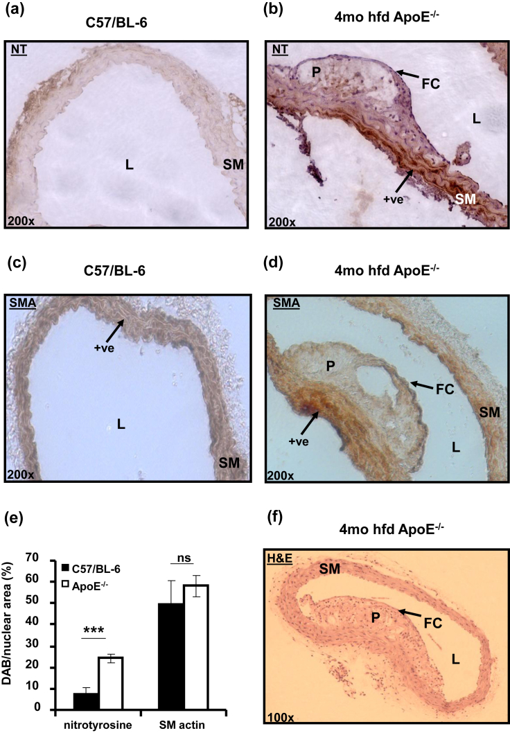
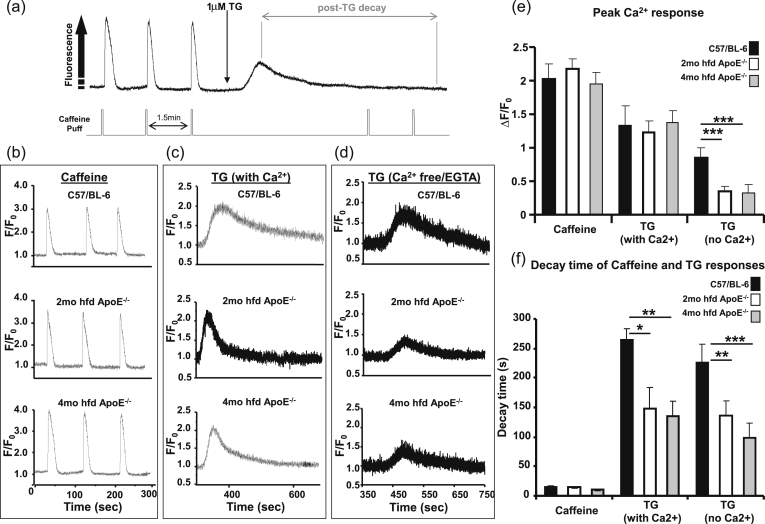
Methods: Aortae from control and ApoE(-/-) mice were studied histologically, functionally and for protein expression levels of SERCA and PMCA. Ca(2+) responses were assessed in dissociated aortic smooth muscle cells in the presence and absence of extracellular Ca(2+).
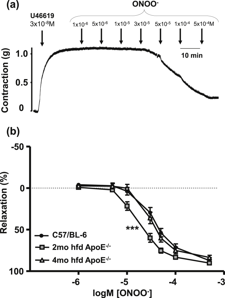
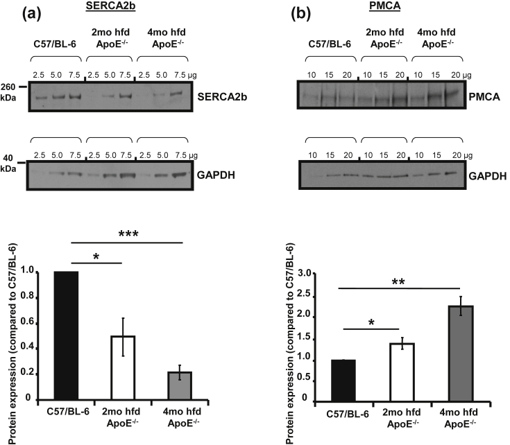
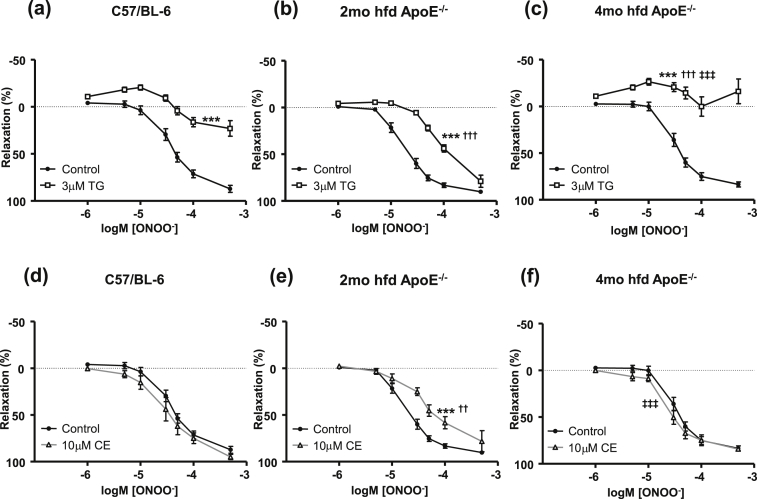
Results: Relaxation to peroxynitrite was concentration-dependent and endothelium-independent. The abilities of the SERCA blocker thapsigargin and the PMCA inhibitor carboxyeosin to block this relaxation were altered during fat feeding and plaque progression. SERCA levels were progressively reduced, while PMCA expression was upregulated. In ApoE(-/-) VSM cells, increases in cytosolic Ca(2+) [Ca(2+)]c in response to SERCA blockade were reduced, while SERCA-independent Ca(2+) clearance was faster compared to control.
Conclusion: As atherosclerosis develops in the ApoE(-/-) mouse, expression and function of Ca(2+) handling proteins are altered. Up-regulation of Ca(2+) removal via PMCA may offer a potential compensatory mechanism to help normalise the dysfunctional relaxation observed during disease progression.
Keywords: Atherosclerosis; Ca(2+); PMCA; Peroxynitrite; SERCA.
Copyright © 2014 The Authors. Published by Elsevier Ireland Ltd.. All rights reserved.
Figures
Similar articles
-
Changes in IP3 Receptor Expression and Function in Aortic Smooth Muscle of Atherosclerotic Mice.J Vasc Res. 2017;54(2):68-78. doi: 10.1159/000461581. Epub 2017 Apr 1. J Vasc Res. 2017. PMID: 28365690 Free PMC article.
-
Peroxynitrite resistance of sarco/endoplasmic reticulum Ca2+ pump in pig coronary artery endothelium and smooth muscle.Cell Calcium. 2004 Jul;36(1):77-82. doi: 10.1016/j.ceca.2003.12.002. Cell Calcium. 2004. PMID: 15126058
-
Role of plasma membrane Ca2+-ATPase in contraction-relaxation processes of the bladder: evidence from PMCA gene-ablated mice.Am J Physiol Cell Physiol. 2006 Apr;290(4):C1239-47. doi: 10.1152/ajpcell.00440.2005. Epub 2005 Nov 16. Am J Physiol Cell Physiol. 2006. PMID: 16291816
-
Modulation of vascular sarco/endoplasmic reticulum calcium ATPase in cardiovascular pathophysiology.Adv Pharmacol. 2010;59:165-95. doi: 10.1016/S1054-3589(10)59006-9. Adv Pharmacol. 2010. PMID: 20933202 Review.
-
Calcium transporters and signalling in smooth muscles.Cell Calcium. 2007 Oct-Nov;42(4-5):467-76. doi: 10.1016/j.ceca.2007.05.011. Epub 2007 Jul 10. Cell Calcium. 2007. PMID: 17624426 Review.
Cited by
-
Atherosclerosis differentially affects calcium signalling in endothelial cells from aortic arch and thoracic aorta in Apolipoprotein E knockout mice.Physiol Rep. 2014 Oct 24;2(10):e12171. doi: 10.14814/phy2.12171. Print 2014 Oct 1. Physiol Rep. 2014. PMID: 25344475 Free PMC article.
-
The relationship between estrogen-induced phenotypic transformation and proliferation of vascular smooth muscle and hypertensive intracerebral hemorrhage.Ann Transl Med. 2020 Jun;8(12):762. doi: 10.21037/atm-20-4567. Ann Transl Med. 2020. PMID: 32647687 Free PMC article.
-
Pulmonary vascular dysfunction in metabolic syndrome.J Physiol. 2019 Feb;597(4):1121-1141. doi: 10.1113/JP275856. Epub 2018 Sep 12. J Physiol. 2019. PMID: 30125956 Free PMC article. Review.
-
Changes in IP3 Receptor Expression and Function in Aortic Smooth Muscle of Atherosclerotic Mice.J Vasc Res. 2017;54(2):68-78. doi: 10.1159/000461581. Epub 2017 Apr 1. J Vasc Res. 2017. PMID: 28365690 Free PMC article.
-
The hypotensive effect of acute and chronic AMP-activated protein kinase activation in normal and hyperlipidemic mice.Vascul Pharmacol. 2015 Nov;74:93-102. doi: 10.1016/j.vph.2015.07.010. Epub 2015 Jul 18. Vascul Pharmacol. 2015. PMID: 26196300 Free PMC article.
References
-
- Touyz R.M., Briones A.M. Reactive oxygen species and vascular biology: Implications in human hypertension. Hypertens Res. 2011;34:5–14. - PubMed
-
- Touyz R.M., Schiffrin E.L. Reactive oxygen species in vascular biology: Implications in hypertension. Histochem Cell Biol. 2004;122:339–352. - PubMed
-
- Park J.G., Oh G.T. The role of peroxidases in the pathogenesis of atherosclerosis. BMB Rep. 2011;44:497–505. - PubMed
-
- Taniyama Y., Griendling K.K. Reactive oxygen species in the vasculature: molecular and cellular mechanisms. Hypertension. 2003;42:1075–1081. - PubMed
Publication types
MeSH terms
Substances
Grants and funding
LinkOut - more resources
Full Text Sources
Other Literature Sources
Medical
Research Materials
Miscellaneous
