. 2014 Jun:312:69-80.
doi: 10.1016/j.heares.2014.03.002. Epub 2014 Mar 20.

Viscoelastic properties of the human tympanic membrane studied with stroboscopic holography and finite element modeling

Affiliations

Viscoelastic properties of the human tympanic membrane studied with stroboscopic holography and finite element modeling

Daniel De Greef et al. Hear Res. 2014 Jun.

Abstract

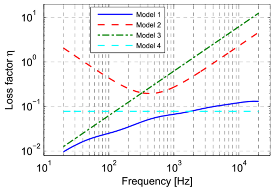
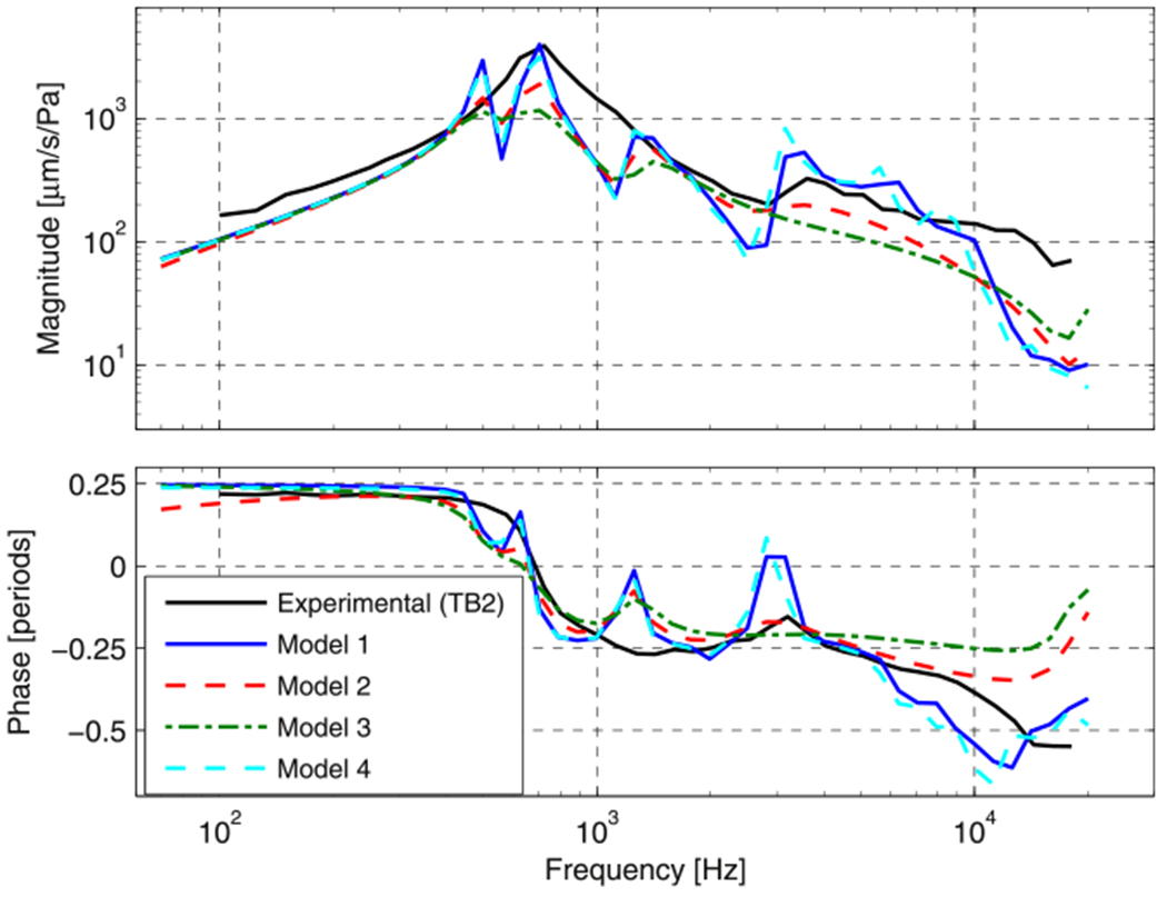
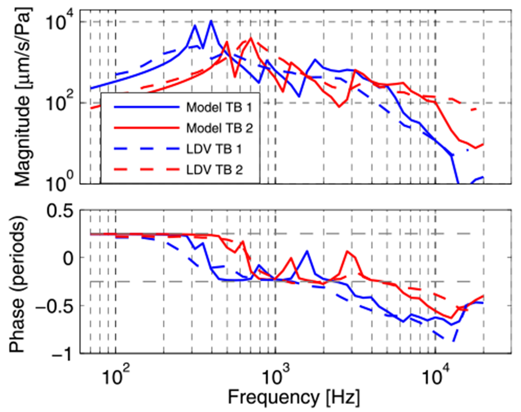
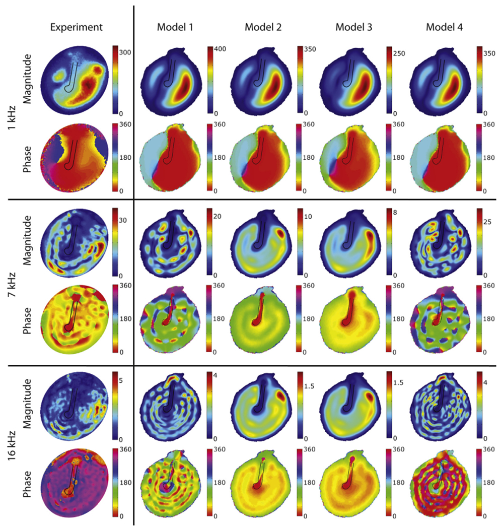
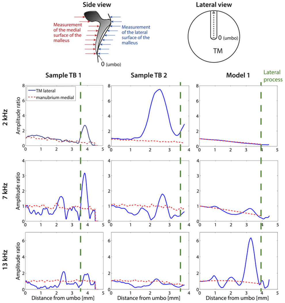
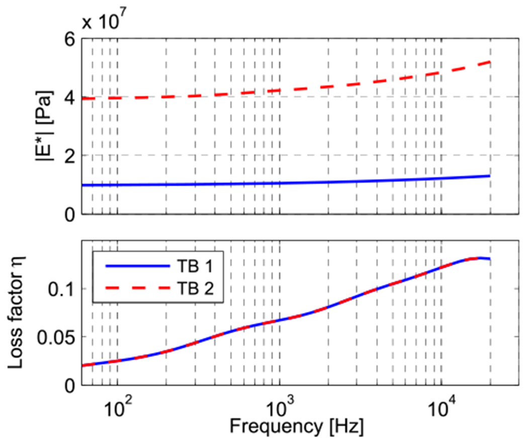
A new anatomically-accurate Finite Element (FE) model of the tympanic membrane (TM) and malleus was combined with measurements of the sound-induced motion of the TM surface and the bony manubrium, in an isolated TM-malleus preparation. Using the results, we were able to address two issues related to how sound is coupled to the ossicular chain: (i) Estimate the viscous damping within the tympanic membrane itself, the presence of which may help smooth the broadband response of a potentially highly resonant TM, and (ii) Investigate the function of a peculiar feature of human middle-ear anatomy, the thin mucosal epithelial fold that couples the mid part of the human manubrium to the TM. Sound induced motions of the surface of ex vivo human eardrums and mallei were measured with stroboscopic holography, which yields maps of the amplitude and phase of the displacement of the entire membrane surface at selected frequencies. The results of these measurements were similar, but not identical to measurements made in intact ears. The holography measurements were complemented by laser-Doppler vibrometer measurements of sound-induced umbo velocity, which were made with fine-frequency resolution. Comparisons of these measurements to predictions from a new anatomically accurate FE model with varied membrane characteristics suggest the TM contains viscous elements, which provide relatively low damping, and that the epithelial fold that connects the central section of the human manubrium to the TM only loosely couples the TM to the manubrium. The laser-Doppler measurements in two preparations also suggested the presence of significant variation in the complex modulus of the TM between specimens. Some animations illustrating the model results are available at our website (www.uantwerp.be/en/rg/bimef/downloads/tympanic-membrane-motion).


Figures

Fig. 1.
Fig. 1.
A photograph and sketch of the medial surface of a prepared sample (TB1): (a) pars tensa of the TM, (b) umbo of the malleus, (c) mallear head, (d) pars flaccida of the TM, and (e) the anterior malleal spine and fold. Six plastic reflective beads (each marked by an *) are visible along the manubrial arm and neck of the malleus. The six beads were used in measurements reported in Horwitz et al. (2012). In this report we only report measurements made at the umbo.
Fig. 2.
Fig. 2.
Schematics of the measurement setups. In each case a sound source is coupled to the lateral (external) surface of the TM via a clear artificial ear canal, and a probe microphone determines the sound pressure at the boney tympanic ring. (A) Holographic measurement of the lateral TM surface: The blue light is the dispersed laser illumination beam. The red Optoelectronic Holograph box contains the CCD camera and its optics. (B) Holographic measurements of the medial surface while acoustically stimulating the lateral surface. The foam yellow plug sealed the lateral end of the artificial ear canal. (C) Laser-Doppler measurements of the umbo of the malleus during sound stimulation of the TM lateral surface. (For interpretation of the references to colour in this figure legend, the reader is referred to the web version of this article.)
Fig. 3.
Fig. 3.
Finite element model with imported middle ear structures of a right human ear. The 5 mm and 2 mm scale bars are located in the model’s coordinate planes.
Fig. 4.
Fig. 4.
The thickness of the TM coded in the finite element model composed from segmentation of CT-images of a right human ear. The segmented TM opposite the manubrium of the malleus is significantly thicker than most other locations.
Fig. 5.
Fig. 5.
Histological sections collected from a single human right ear: at the lateral process (left), at a point midway between the lateral process and the umbo (middle), and at the umbo (right). Images from www.temporalboneconsortium.org.
Fig. 6.
Fig. 6.
Loss factor curves of the four different damping cases, defined in Section 2.2.3.
Fig. 7.
Fig. 7.
Normalized umbo velocity magnitude and angle for the four damping cases (see Section 2.2.3 and Fig. 6) and experimental data from TB2 (black line).
Fig. 8.
Fig. 8.
Experimental (laser Doppler Vibrometry) and model umbo velocity response for the two samples.
Fig. 9.
Fig. 9.
Displacement magnitudes [nm/Pa] and phase [deg] of the lateral surface of the TM for selected tonal stimuli of 1, 7 and 16 kHz (the phase is relative to the umbo position). The loss-factors used in the models are defined in Section 2.2.3 and illustrated in Fig. 6.
Fig. 10.
Fig. 10.
Comparison of lateral displacements of the TM along the length of its attachment to the manubrium and medial displacements of the manubrium and malleus neck and head, for frequencies of 2, 7 and 13 kHz. The schematic on the upper right represents the view of the lateral surface of the manubrium from the ear canal. The schematic on the upper left illustrates the motion components that were measured by the holography system when it viewed either the lateral or medial surface of the TM. The data are normalized to the umbo. Left and middle column: Holographically measured displacements from sample TB1 and TB2. Right column: model prediction of the motion of the lateral surface of the TM opposite to the manubrium and the medial surface of the manubrium. The vertical dashed lines on the plots indicate the location of the lateral process. Locations on the lateral surface of the ear canal superior to the lateral process represent measurements on the surface of the pars flaccida of the TM. (For interpretation of the references to colour in this figure legend, the reader is referred to the web version of this article.)
Fig. 11.
Fig. 11.
Complex modulus magnitude and phase values of the TM used for the two model data sets. Both magnitude curves differ merely by a multiplication factor, while the phase data is equal for both models and represents model 1 defined in Section 2.2.3 and depicted in Fig. 6.
Fig. 12.
Fig. 12.
Model representation for the generalized Maxwell model.

References

    1. Aernouts J, Aerts JRM, Dirckx JJJ, 2012. Mechanical properties of human tympanic membrane in the quasi-static regime from in situ point indentation measurements. Hear. Res 290 (1–2), 45–54. 10.1016/j.heares.2012.05.001. - DOI - PubMed
    1. Cheng JT, Aarnisalo AA, Harrington E, Hernandez-Montes MDS, Furlong C, Merchant SN, Rosowski JJ, 2010. Motion of the surface of the human tympanic membrane measured with stroboscopic holography. Hear. Res 263 (1–2), 66–77. 10.1016/j.heares.2009.12.024. - DOI - PMC - PubMed
    1. Cheng JT, Hamade M, Merchant SN, Rosowski JJ, Harrington E, Furlong C, 2013. Wave motion on the surface of the human tympanic membrane: holographic measurement and modeling analysis. J. Acoust. Soc. Am 133 (2), 918–937. 10.1121/1.4773263. - DOI - PMC - PubMed
    1. De La Rochefoucauld O, Olson ES, 2010. A sum of simple and complex motions on the eardrum and manubrium in gerbil. Hear. Res 263, 9–15. 10.1016/j.heares.2009.10.014. - DOI - PMC - PubMed
    1. Decraemer W, Khanna S, 2004. Measurement, visualization and quantitative analysis of complete three-dimensional kinematical data sets of human and cat middle ear. In: Wada H (Ed.), Proceedings of the 3rd Symposium on Middle Ear Mechanics in Research and Otology, Singapore, pp. 3–10.

Publication types