. 2014 May;7(3):517-25.
doi: 10.1161/CIRCIMAGING.113.000305. Epub 2014 Mar 21.

Near-infrared II fluorescence for imaging hindlimb vessel regeneration with dynamic tissue perfusion measurement

Affiliations

Near-infrared II fluorescence for imaging hindlimb vessel regeneration with dynamic tissue perfusion measurement

Guosong Hong et al. Circ Cardiovasc Imaging. 2014 May.

Abstract

Background: Real-time vascular imaging that provides both anatomic and hemodynamic information could greatly facilitate the diagnosis of vascular diseases and provide accurate assessment of therapeutic effects. Here, we have developed a novel fluorescence-based all-optical method, named near-infrared II (NIR-II) fluorescence imaging, to image murine hindlimb vasculature and blood flow in an experimental model of peripheral arterial disease, by exploiting fluorescence in the NIR-II region (1000-1400 nm) of photon wavelengths.

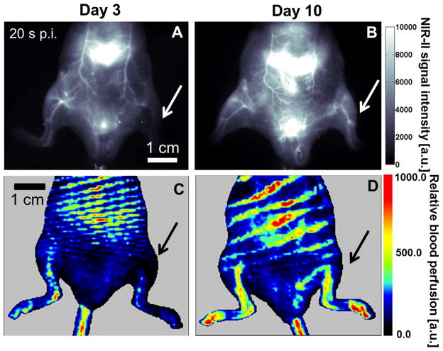
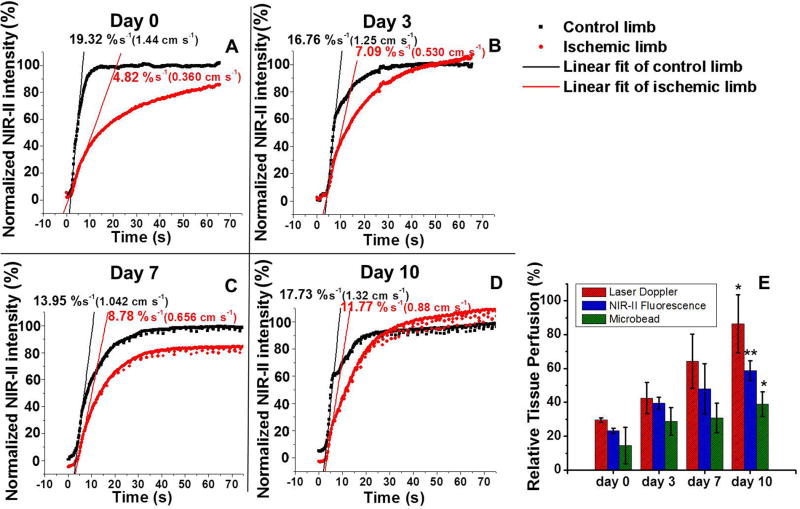
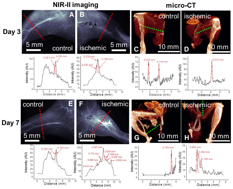
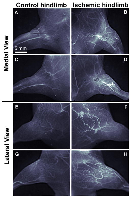
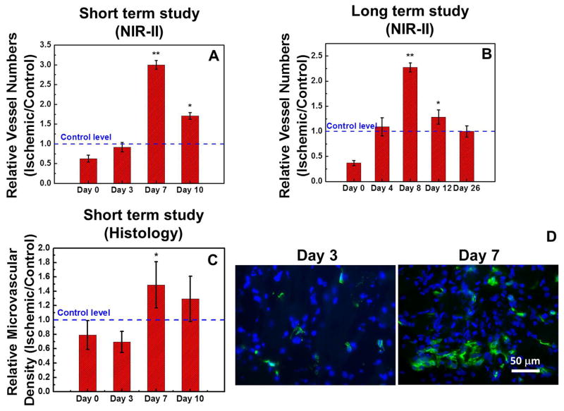
Methods and results: Because of the reduced photon scattering of NIR-II fluorescence compared with traditional NIR fluorescence imaging and thus much deeper penetration depth into the body, we demonstrated that the mouse hindlimb vasculature could be imaged with higher spatial resolution than in vivo microscopic computed tomography. Furthermore, imaging during 26 days revealed a significant increase in hindlimb microvascular density in response to experimentally induced ischemia within the first 8 days of the surgery (P<0.005), which was confirmed by histological analysis of microvascular density. Moreover, the tissue perfusion in the ischemic hindlimb could be quantitatively measured by the dynamic NIR-II method, revealing the temporal kinetics of blood flow recovery that resembled microbead-based blood flowmetry and laser Doppler blood spectroscopy.

Conclusions: The penetration depth of millimeters, high spatial resolution, and fast acquisition rate of NIR-II imaging make it a useful imaging tool for murine models of vascular disease.

Keywords: angiography; hemodynamics; nanotubes, carbon.


Figures

Figure 1
Figure 1
Corresponding NIR-II (A&B) and laser Doppler perfusion images (C&D) on day 3 (A&C) and day 10 (B&D) after surgery-induced hindlimb ischemia, where the arrow denotes the ischemic hindlimb. Note that the NIR-II images in A&B are extracted from Videos 1&2 that dynamically record the tissue perfusion labeled by NIR-II contrast, and that these two images both correspond to the time point of 20 s p.i.
Figure 2
Figure 2
Comparison of quantitative tissue perfusion after induction of hindlimb ischemia. (A–D) Normalized NIR-II fluorescence intensity in control and ischemic hindlimbs as a function of time p.i. on post-operative days 0, 3, 7 and 10. (E) A bar chart showing the relative tissue perfusion values (ischemic/control) on day 0, 3, 7 and 10 derived from laser Doppler technique (red bars), NIR-II fluorescence method (blue bars) and microbead measurements (green bars). Statistically significant differences of perfusion are found between days 0 and 10 for all methods (*P < 0.05 and **P < 0.005).
Figure 3
Figure 3
Comparison of NIR-II fluorescence (A, B, E, F) and μCT images (C, D, G, H) of mouse hindlimb vasculature (top) and cross-sectional vessel width measurements (bottom) on day 3 (A–D) and day 7 (E–H) after surgery-induced hindlimb ischemia.
Figure 4
Figure 4
High magnification NIR-II fluorescence images of mouse hindlimb vasculature of two different mice (one mouse is shown in A,B,E,F and the other in C,D,G,H) at day 7 after surgery-induced hindlimb ischemia. Both medial (A–D) and lateral (E–H) aspects of control (A,C,E,G) and ischemic (B,D,F,H) hindlimbs are shown.
Figure 5
Figure 5
Quantification of collateral vessel formation in the ischemic limb during revascularization. NIR-II fluorescence based results of short-term (A) and long-term (B) revascularization studies are expressed relative to the vessel numbers in the control limb, and histology based results of a short-term study (C) are expressed in microvascular density. (D) Fluorescence microscopy images of ischemic hindlimb tissues resected on day 3 (left) and day 7 (right) after the induction of acute hindlimb ischemia with CD31 staining (green) and nucleus staining (blue). Statistically significant difference in comparison to day 0 is shown as *P < 0.05 and **P < 0.005.

References

    1. Rosamond W, Flegal K, Furie K, Go A, Greenlund K, Haase N, Hailpern SM, Ho M, Howard V, Kissela B, Kittner S, Lloyd-Jones D, McDermott M, Meigs J, Moy C, Nichol G, O’Donnell C, Roger V, Sorlie P, Steinberger J, Thom T, Wilson M, Hong Y, Stroke AHASC. Heart disease and stroke statistics - 2008 update - a report from the american heart association statistics committee and stroke statistics subcommittee. Circulation. 2008;117:E25–E146. - PubMed
    1. Hong G, Lee JC, Robinson JT, Raaz U, Xie L, Huang NF, Cooke JP, Dai H. Multifunctional in vivo vascular imaging using near-infrared ii fluorescence. Nat Med. 2012;18:1841–1846. - PMC - PubMed
    1. Iijima S, Ichihashi T. Single-shell carbon nanotubes of 1-nm diameter. Nature. 1993;363:603–605.
    1. Welsher K, Liu Z, Sherlock SP, Robinson JT, Chen Z, Daranciang D, Dai HJ. A route to brightly fluorescent carbon nanotubes for near-infrared imaging in mice. Nat Nanotechnol. 2009;4:773–780. - PMC - PubMed
    1. Robinson JT, Hong GS, Liang YY, Zhang B, Yaghi OK, Dai HJ. In vivo fluorescence imaging in the second near-infrared window with long circulating carbon nanotubes capable of ultrahigh tumor uptake. J Am Chem Soc. 2012;134:10664–10669. - PMC - PubMed

Publication types