. 2014 Jun 10:183:1-8.
doi: 10.1016/j.jconrel.2014.03.025. Epub 2014 Mar 21.

Ultrasound-assisted siRNA delivery via arginine-grafted bioreducible polymer and microbubbles targeting VEGF for ovarian cancer treatment

Affiliations

Ultrasound-assisted siRNA delivery via arginine-grafted bioreducible polymer and microbubbles targeting VEGF for ovarian cancer treatment

Stelios Florinas et al. J Control Release. .

Abstract

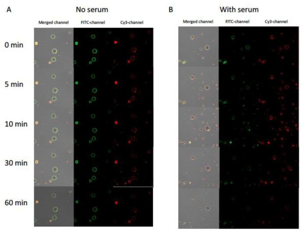
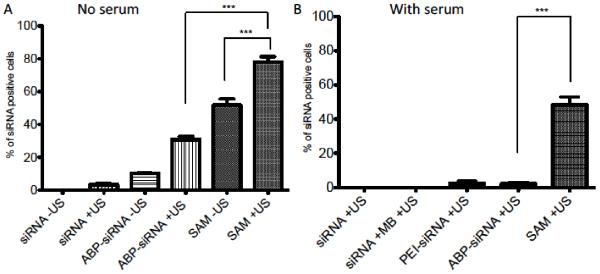
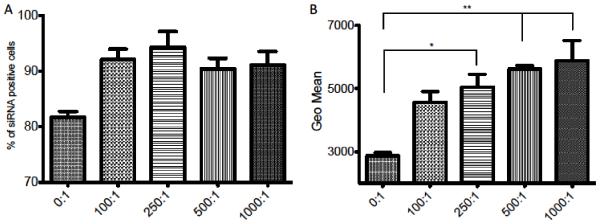
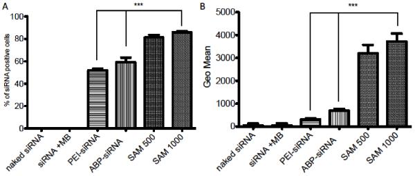
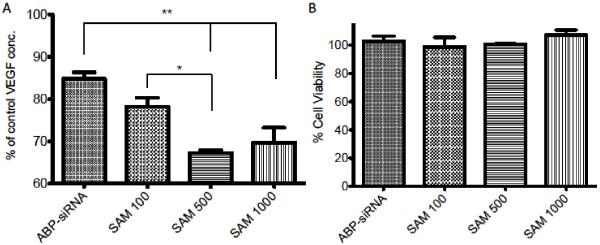
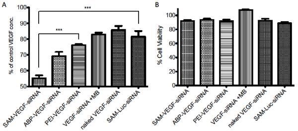
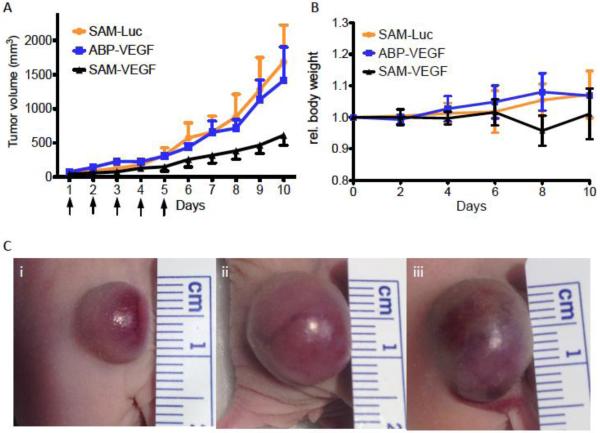
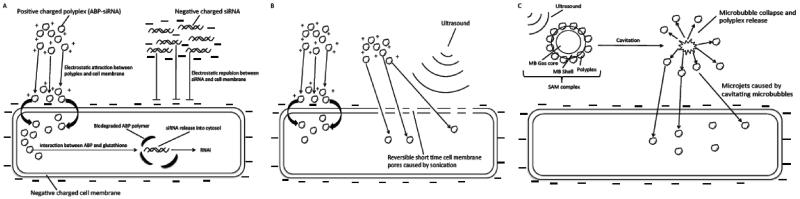
The major drawback hampering siRNA therapies from being more widely accepted in clinical practice is its insufficient accumulation at the target site mainly due to poor cellular uptake and rapid degradation in serum. Therefore, we designed a novel polymeric siRNA carrier system, which would withstand serum-containing environments and tested its performance in vitro as well as in vivo. Delivering siRNA with a system combining an arginine-grafted bioreducible polymer (ABP), microbubbles (MBs), and ultrasound technology (US) we were able to synergize the advantages each delivery system owns individually, and created our innovative siRNA-ABP-MB (SAM) complexes. SAM complexes show significantly higher siRNA uptake and VEGF protein knockdown in vitro with serum-containing media when compared to naked siRNA, and 25k-branched-polyethylenimine (bPEI) representing the current standard in nonviral gene therapy. SAM complexes activated by US are also able to improve siRNA uptake in tumor tissue resulting in decelerating tumor growth in vivo.

Keywords: Cancer; Microbubbles; RNAi; Ultrasound; VEGF; siRNA.


Figures

Figure 1
Figure 1
Confocal microscopy of SAM complexes incubated for 60 minutes in serum free (A) and serum containing (10% FBS) (B) cell culture media. SAM complexes with FITC labeled human serum albumin (HSA) microbubble shell (FITC-channel) and Cy3 labeled polyplexes siRNA-Cy3-ABP (Cy3-channel). Merged channel represents FITC channel plus Cy3-channel.
Figure 2
Figure 2
Comparison of cellular uptake delivering siRNA in serum free and serum containing media determined by FACS analysis in A2780 cell line. (A) Cellular uptake in serum free media with 5 nM siRNA complexed in polyplexes (ABP-siRNA), SAM complexes or as naked siRNA using 1 MHz US conditions with 0.5 Watt/cm2 and 50% duty or no US treatment. (B) Intracellular delivery in serum containing (10% FBS) cell culture media with with 5 nM siRNA complexed in polyplexes (ABP-siRNA, PEI-siRNA), SAM complexes, combined with MBs or as naked siRNA using 1 MHz US conditions with 0.5 Watt/cm2 and 50% duty. Data represent mean ± SD and significance tested (P < 0.0001) by one-way ANOVA and Tukey post test.
Figure 3
Figure 3
Cellular uptake of siRNA determined by FACS analysis in A2780 cell line using different MB:cell ratios. (A) Quantification of siRNA cellular uptake between siRNA positive and siRNA negative cells with 5 nM siRNA in SAM complexes and MB:cell ratios (0:1, 100:1, 250:1, 500:1, 1000:1, 0:1 = ABP-siRNA polyplex with no MBs) treated with 1 MHz US condition, 0.5 Watt/cm2 and 50% duty. (B) Quantification of siRNA per cell population using the GEO mean of groups from (A). Data represent mean ± SD and significance tested (P < 0.05 and 0.001) by one-way ANOVA and Tukey post test.
Figure 4
Figure 4
Cellular uptake efficacy using SAM complexes. (A) Quantification of siRNA cellular uptake between siRNA positive and siRNA negative cells with 5 nM siRNA using naked siRNA, siRNA plus MB, polyplexes (ABP-siRNA, PEI-siRNA) and SAM complexes (MB:cell ratio 500 and 1000) treated with 1 MHz US condition, 0.5 Watt/cm2 and 50% duty. (B) Quantification of siRNA per cell population using the GEO mean of groups from (A). Data represent mean ± SD and significance tested (P < 0.0001) by one-way ANOVA and Tukey post test.
Figure 5
Figure 5
Optimization of MB:cell ratio for VEGF protein knockdown. Transfection efficiency of VEGF-siRNA using polyplexes (ABP-siRNA), SAM complexes (MB:cell ratio 500 and 1000) and US. VEGF ELISA and MTT assay in A2780 cell line treated with US 1 MHz, 0.5 Watt/cm2 and 50% duty. (A) VEGF concentration after transfection of A2780 cells in serum containing media after US exposure with 100 nM siRNA targeting VEGF, complexed in SAM or polyplexes. (B) Cell viability of (A) was determined by MTT assay and expressed as relative cell viability compared to the control. Data represent mean ± SD and significance tested (P < 0.05 and 0.001) by one-way ANOVA and Tukey post test.
Figure 6
Figure 6
VEGF knockdown using SAM complexes. Transfection efficiency of VEGF-siRNA using polyplexes (ABP-VEGF-siRNA, PEI-VEGF-siRNA), SAM-VEGF complexes (MB:cell ratio 500), naked VEGF-siRNA, SAM-Luc (siRNA negative control, MB:cell ratio 500) and US. VEGF ELISA and MTT assay in A2780 cell line treated with US 1 MHz, 0.5 Watt/cm2 and 50% duty. (A) VEGF concentration after transfection of A2780 cells in serum containing media after US exposure with 100 nM siRNA targeting VEGF or luciferase (SAM-Luc). (B) Cell viability of (A) was determined by MTT assay and expressed as relative cell viability compared to the control. Data represent mean ± SD and significance tested (P < 0.0001-k) by one-way ANOVA and Tukey post test.
Figure 7
Figure 7
Effect of intratumorally administered siRNA against VEGF using SAM complexes and ultrasound in tumor bearing nude mice. (A) Mice were injected five times total (see arrows) with SAM-VEGF-siRNA, SAM-Luc-siRNA or ABP-VEGF polyplexes intratumorally followed by ultrasound treatment. (B) Relative Body weight. (C) Tumor size pictures five days after the last injection for i) SAM-VEGF-siRNA, ii) ABP-VEGF polyplexes and iii) SAM-Luc-siRNA.
Scheme 1
Scheme 1
Mechanisms of intracellular siRNA delivery using arginine grafted bioreducible polymer (ABP), Ultrasound and siRNA-ABP-Microbubble (SAM) Complexes. (A) siRNA delivery using ABP or naked siRNA. Positive charged polyplexes interact with negative charged cell membrane and facilitates cellular uptake of polyplexes due to electrostatic interaction. Biodegradation of ABP polymer due to reduction of disulfide bonds in ABP backbone by intracellular glutathione leads to siRNA release into cytosol and RNAi activity. Cellular uptake of naked siRNA is declined due to repulsion of negative charge of siRNA and cell membrane. (B) siRNA delivery using ABP and ultrasound (US). Ultrasound causes short time cell membrane pores that facilitates polyplex uptake in addition to the described mechanism in (A). (C) siRNA delivery using SAM complex and ultrasound. Cavitating microbubbles (MBs) release polyplexes from microbubble shell due to interaction with US. MBs cavitation causes microjets and jetstreams that shoot polyplexes through the cell membrane in addition to the described mechanisms in (A) and (B).

Similar articles

Cited by

References

    1. Hammond SM, Caudy AA, Hannon GJ. Post-transcriptional gene silencing by double-stranded RNA. Nat. Rev. Genet. 2001;2:110–119. - PubMed
    1. Fougerolles A.d., Vornlocher H-P, Maraganore J, Lieberman J. Interfering with disease: a progress report on siRNA-based therapeutics. Nature Reviews Drug Discovery. 2007;6:443–453. - PMC - PubMed
    1. Semenza GL. Targeting HIF-1 for cancer therapy. Nature Reviews Cancer. 2003;3:721–732. - PubMed
    1. Ferrara N. VEGF-A: a critical regulator of blood vessel growth. European cytokine network. 2009;20:158–163. - PubMed
    1. Wannenes F, Ciafré SA, Niola F, Frajese G, Farace MG. Vector-based RNA interference against vascular endothelial growth factor-A significantly limits vascularization and growth of prostate cancer in vivo. Cancer Gene Therapy. 2005;12:926–934. - PubMed

Publication types

MeSH terms