. 2014 May;3(5):620-31.
doi: 10.5966/sctm.2013-0151. Epub 2014 Mar 21.

Intrastriatal transplantation of adenovirus-generated induced pluripotent stem cells for treating neuropathological and functional deficits in a rodent model of Huntington's disease

Affiliations

Intrastriatal transplantation of adenovirus-generated induced pluripotent stem cells for treating neuropathological and functional deficits in a rodent model of Huntington's disease

Kyle D Fink et al. Stem Cells Transl Med. 2014 May.

Abstract

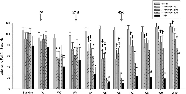
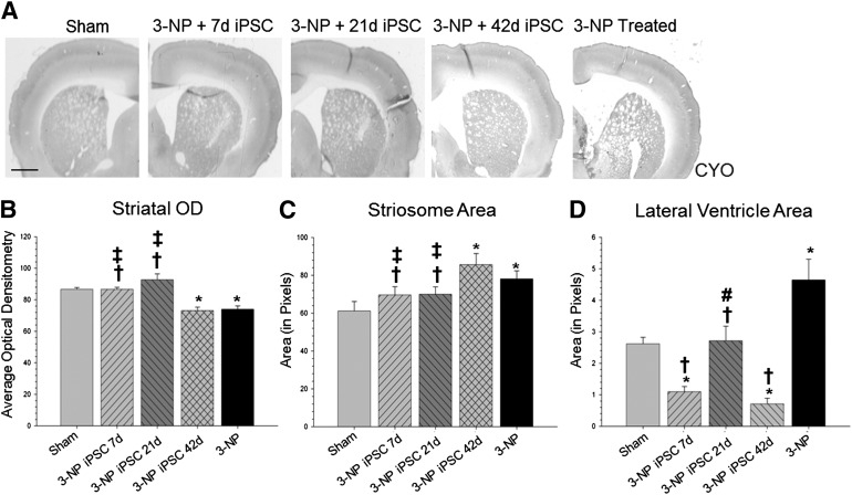
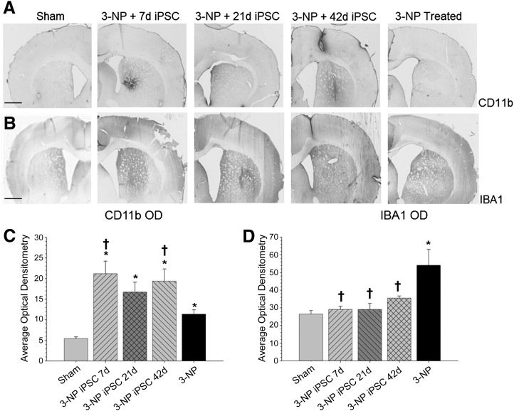
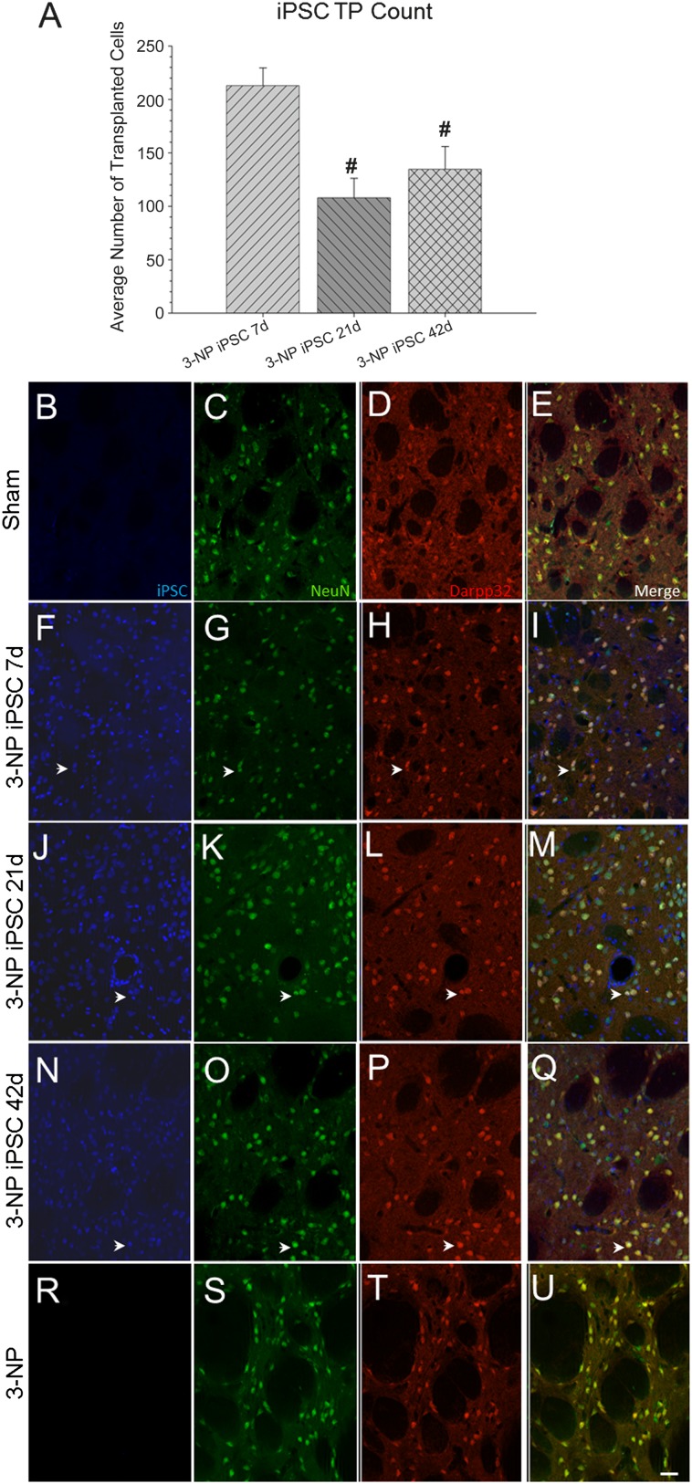
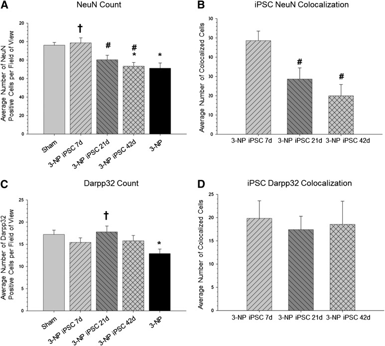
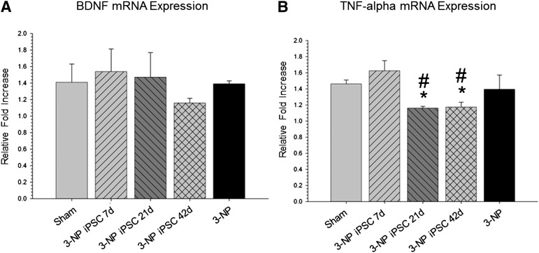
Induced pluripotent stem cells (iPSCs) show considerable promise for cell replacement therapies for Huntington's disease (HD). Our laboratory has demonstrated that tail-tip fibroblasts, reprogrammed into iPSCs via two adenoviruses, can survive and differentiate into neuronal lineages following transplantation into healthy adult rats. However, the ability of these cells to survive, differentiate, and restore function in a damaged brain is unknown. To this end, adult rats received a regimen of 3-nitropropionic acid (3-NP) to induce behavioral and neuropathological deficits that resemble HD. At 7, 21, and 42 days after the initiation of 3-NP or vehicle, the rats received intrastriatal bilateral transplantation of iPSCs. All rats that received 3-NP and vehicle treatment displayed significant motor impairment, whereas those that received iPSC transplantation after 3-NP treatment had preserved motor function. Histological analysis of the brains of these rats revealed significant decreases in optical densitometric measures in the striatum, lateral ventricle enlargement, as well as an increase in striosome size in all rats receiving 3-NP when compared with sham rats. The 3-NP-treated rats given transplants of iPSCs in the 7- or 21-day groups did not exhibit these deficits. Transplantation of iPSCs at the late-stage (42-day) time point did not protect against the 3-NP-induced neuropathology, despite preserving motor function. Transplanted iPSCs were found to survive and differentiate into region-specific neurons in the striatum of 3-NP rats, at all transplantation time points. Taken together, these results suggest that transplantation of adenovirus-generated iPSCs may provide a potential avenue for therapeutic treatment of HD.

Keywords: 3-Nitropropionic acid; Adenovirus; Huntington’s disease; Stem cell; Transplantation; iPSC.


Figures

Figure 1.
Figure 1.
Accelerod data from iPSC-transplanted 3-NP rats. Analysis of motor coordination revealed significant between-group differences for the latency to fall in the accelerod task. Starting at week 4, and continuing throughout the study, 3-NP rats that received iPSC transplantation at either 7 or 21 days had significantly higher latencies to fall than 3-NP rats that did not receive transplants. Also, 3-NP rats that received transplantation of iPSCs at 42 days showed significantly higher latencies to fall at week 9 than 3-NP rats that did not receive transplants. (Bar graph represents mean value; error bars represent SEM; arrows indicate the time of iPSC transplantation for each group; ∗, significantly different from sham rats, p < .05; †, significantly different from 3-NP rats, p < .05; #, significantly different from 3-NP iPSC 7-day rats, p < .05; ‡, significantly different from 3-NP iPSC 42-day rats, p < .05.) Abbreviations: 3-NP, 3-nitropropionic acid; iPSC, induced pluripotent stem cell.
Figure 2.
Figure 2.
Cytochrome oxidase labeling. (A): Analysis of the brain scans labeled with CYO revealed significant between-group differences in optical densitometry measures in the striatum (B). It was also revealed that rats receiving iPSC transplantation at 7 or 21 days had significantly higher optical densitometry measures in the striatum than 3-NP rats that did not receive transplants or 3-NP rats that received iPSC transplantation at 42 days. In addition, analysis of the striosome area revealed significant between-group differences (C). Rats receiving iPSC transplantation at 7 or 21 days had significantly smaller striosome area when compared with 3-NP rats that did not receive transplants or 3-NP rats that received iPSC transplantation at 42 days. Scans from the same sections also revealed significant between-group differences in the area of the lateral ventricles (D). All groups receiving iPSC transplantation had significantly smaller lateral ventricles when compared with 3-NP rats that did not receive transplants. (Bar graph represents mean value; error bars represent SEM; ∗, significantly different from sham rats, p < .05; †, significantly different from 3-NP rats without transplants, p < .05; #, significantly different from 3-NP iPSC 7-day rats, p < .05; ‡, significantly different from 3-NP iPSC 42-day rats, p < .05.) Abbreviations: 3-NP, 3-nitropropionic acid; iPSC, induced pluripotent stem cell; OD, optical density.
Figure 3.
Figure 3.
Measures of inflammation. Analysis of the immune response as measured by optical densitometry of CD11b (A) and IBA1 (B) to 3-NP and transplanted iPSCs. Analysis of expression levels revealed significant between-group differences in the average optical densitometry of CD11b, with all groups receiving 3-NP having higher optical densitometry measures for CD11b than sham rats (C). Significant between-group differences in the average optical densitometry of IBA1 (D) were also observed, revealing that 3-NP rats without transplants had significantly more macrophage infiltration than sham rats and all rats receiving iPSC transplantation. (Bar graph represents mean value; error bars represent SEM; ∗, significantly different from sham rats, p < .05; †, significantly different from 3-NP rats, p < .05.) Abbreviations: 3-NP, 3-nitropropionic acid; iPSC, induced pluripotent stem cell; OD, optical density.
Figure 4.
Figure 4.
Optical densitometry of GFAP from the transplant site. Astrocytes (GFAP; green) were observed around the transplanted iPSCs (blue), but no colocalization was observed between the two labels. A significant between-group difference in the average optical densitometry of GFAP (A) was observed. It was revealed that 3-NP rats receiving iPSC transplant at either 21 or 42 days displayed significantly higher GFAP labeling than sham rats and 3-NP rats that received transplantation of iPSCs at 7 days. The 3-NP rats receiving iPSC transplantation at 7 days displayed significantly more astrocyte activation than 3-NP rats that did not receive transplants. Sham rats are shown in (B–D); 7-day iPSC rats are shown in (E–G); 21-day iPSC rats are shown in (H–J); 42-day rats are shown in (K–M); 3-NP rats that did not receive transplants are shown in (N–P). (Scale bar = 50 μm; bar graph represents mean value; error bars represent SEM; ∗, significantly different from sham rats, p < .05; †, significantly different from 3-NP rats, p < .05; #, significantly different from 3-NP iPSC 7-day rats, p < .05.) Abbreviations: 3-NP, 3-nitropropionic acid; GFAP, glial fibrillary acidic protein; iPSC, induced pluripotent stem cell; OD, optical density.
Figure 5.
Figure 5.
Immunohistochemical analysis of transplanted iPSCs (blue), mature neurons (NeuN; green), and medium spiny neurons (Darpp32; red). All transplanted rats displayed surviving iPSCs (blue) around the injection site at the conclusion of the study. However, a significant between-group effect was observed for the average number of surviving iPSCs (A). It was revealed that the 7-day transplantation time point had significantly more surviving iPSCs than the 21- or 42-day transplantation group. Sham rats are shown in (B–E); 7-day iPSC rats are shown in (F–I); 21-day iPSC rats are shown in (J–M); 42-day rats are shown in (N–Q); 3-NP rats that did not receive transplants are shown in (R–U). (Scale bar = 50 μm; colocalization is shown by white arrowheads; bar graph represents mean value; error bars represent SEM; #, significantly different from 3-NP iPSC 7-day rats, p < .05.) Abbreviations: 3-NP, 3-nitropropionic acid; iPSC, induced pluripotent stem cell.
Figure 6.
Figure 6.
Cell counts of mature neurons, medium spiny neurons, and colocalization with transplanted iPSCs. A significant between-group difference in the number of NeuN-positive cells in the area around the transplant site (A) was observed. It was revealed that 3-NP rats that received iPSC transplantation at 7 days had a significantly greater amount of NeuN-positive cells than the 3-NP rats that did receive transplants or the 3-NP rats receiving transplantation at either 21 or 42 days. A significant between-group difference was found in the number of transplanted iPSC cells colocalizing with NeuN (B). It was found that the 7-day transplantation group had more iPSCs colocalized with NeuN than either the 21- or 42-day group. A significant between-group difference was observed in the number of DARPP32-positive cells in the area around the transplant site (C). It was found that 3-NP rats receiving iPSC transplantation at 21 days had significantly more DARPP32-positive cells than 3-NP control rats. However, no between-group differences were observed in the number of iPSCs colocalizing with DARPP32 (D). (∗, Significantly different from sham rats, p < .05; †, significantly different from 3-NP rats, p < .05; #, significantly different from 3-NP iPSC 7-day rats, p < .05.) Abbreviations: 3-NP, 3-nitropropionic acid; iPSC, induced pluripotent stem cell; NeuN, neuronal nuclei.
Figure 7.
Figure 7.
Quantitative real-time polymerase chain reaction (RT-PCR) of striatal tissue for mRNA expression of BDNF and TNF-α. Quantitative RT-PCR of striatal tissue revealed no significant differences in the mRNA expression of BDNF between all groups (A), but did reveal a significant between-group difference in the mRNA expression of TNF-α (B). No reduction in BDNF expression was observed in the 3-NP rats without transplantation, indicating that behavioral deficits are not the result of depletion of this trophic factor. The downregulation of TNF-α in the rats that received transplantation of iPSCs at 21 and 42 days is indicative of a general deactivation of the immune response to the transplanted cells. (Bar graph represents mean value; error bars represent SEM; ∗, significantly different from sham rat, p < .05; #, significantly different from 3-NP iPSC 7-day rats, p < .05.) Abbreviations: 3-NP, 3-nitropropionic acid; BDNF, brain-derived neurotrophic factor; iPSC, induced pluripotent stem cell; TNF, tumor necrosis factor.

Similar articles

Cited by

References

    1. The Huntington’s Disease Collaborative Research Group A novel gene containing a trinucleotide repeat that is expanded and unstable on Huntington’s disease chromosomes. Cell. 1993;72:971–983. - PubMed
    1. Estrada Sánchez AM, Mejía-Toiber J, Massieu L. Excitotoxic neuronal death and the pathogenesis of Huntington’s disease. Arch Med Res. 2008;39:265–276. - PubMed
    1. Southwell AL, Ko J, Patterson PH. Intrabody gene therapy ameliorates motor, cognitive, and neuropathological symptoms in multiple mouse models of Huntington’s disease. J Neurosci. 2009;29:13589–13602. - PMC - PubMed
    1. Bachoud-Lévi A-C. Neural grafts in Huntington’s disease: Viability after 10 years. Lancet Neurol. 2009;8:979–981. - PubMed
    1. Cicchetti F, Saporta S, Hauser RA, et al. Neural transplants in patients with Huntington’s disease undergo disease-like neuronal degeneration. Proc Natl Acad Sci USA. 2009;106:12483–12488. - PMC - PubMed

Publication types

MeSH terms