Intraoral zygoma reduction using L-shaped osteotomy
- PMID: 24657982
- PMCID: PMC4025629
- DOI: 10.1097/SCS.0000000000000759
Intraoral zygoma reduction using L-shaped osteotomy
Abstract
Background: Because of the various defects of malarplasty, including a large incision, much bleeding, visible scars after the operation, and so on, caused by the conventional coronal incision or the temporal incision with the intraoral incision approach, the malarplasty by simple intraoral approach is an innovative development.
Methods: Through the intraoral approach and subperiosteal dissection, we can reach the osteotomy point on the zygomatic body directly and arrive at the osteotomy point at the zygomatic arch end along the medial side of the zygoma. A new L osteotomy is applied with the reciprocating saw. In addition, the osteotomy was performed on the zygomatic arch from the inside out with an angle of 20 degrees horizontally.
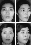
Results: From 1997 to 2010, we were satisfied with the results of 114 cases of malarplasty with the intraoral approach and L osteotomy as the observed objects. There are 103 cases for women and 11 for men. Ages ranged from 16 to 48 years. The mean operation time is approximately 1 hour. We just had a few complications: 3 nonunion at the osteotomy line and needed a second surgery to repair as well as 2 slight cheek drooping during the initial period and required face lifting.
Conclusions: The method of intraoral approach and L-shaped osteotomy for zygoma reduction can reduce prominent zygoma while maintaining the natural curves of the zygomatic body and arch. Because of the simple procedures, fewer complications, and excellent results, this method will be considered a relatively desirable way.
Level of evidence: Therapeutic, III.
Conflict of interest statement
The authors report no conflicts of interest.
Figures


References
-
- Jung H, Lee Y, Doo JH, et al. Prevention of complication and management of unfavorable results in reduction malarplasty. J Korean Soc Plast Reconstr Surg 2008; 35: 465– 470
-
- Onizuka T, Watanabe K, Takasu K, et al. Reduction malarplasty. Aesthetic Plast Surg 1983; 7: 121– 125 - PubMed
-
- Hahm JW, Baek RM, Oh KS, et al. 10 year experience on reduction malarplasty. J Korean Soc Plast Reconstr Surg 1997; 24: 1478– 1489
-
- Baek SM, Chung YD, Kim SS Reduction malarplasty. Plast Reconstr Surg 1991; 88: 53– 61 - PubMed
-
- Wang T, Gui L, Tang X, et al. Reduction malarplasty with a new L-shaped osteotomy through an intraoral approach: retrospective study of 418 cases. Plast Reconstr Surg 2009; 124: 1245– 1253 - PubMed
Publication types
MeSH terms
LinkOut - more resources
Full Text Sources
Other Literature Sources
Medical
Research Materials

