. 2014 Mar 21;9(3):e92309.
doi: 10.1371/journal.pone.0092309. eCollection 2014.

EphA4 activation of c-Abl mediates synaptic loss and LTP blockade caused by amyloid-β oligomers

Affiliations

EphA4 activation of c-Abl mediates synaptic loss and LTP blockade caused by amyloid-β oligomers

Lina M Vargas et al. PLoS One. .

Abstract

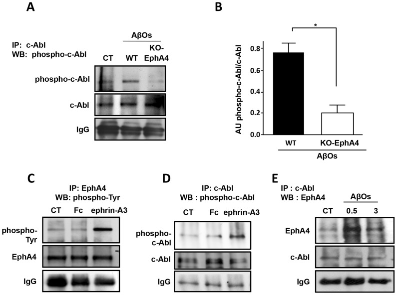
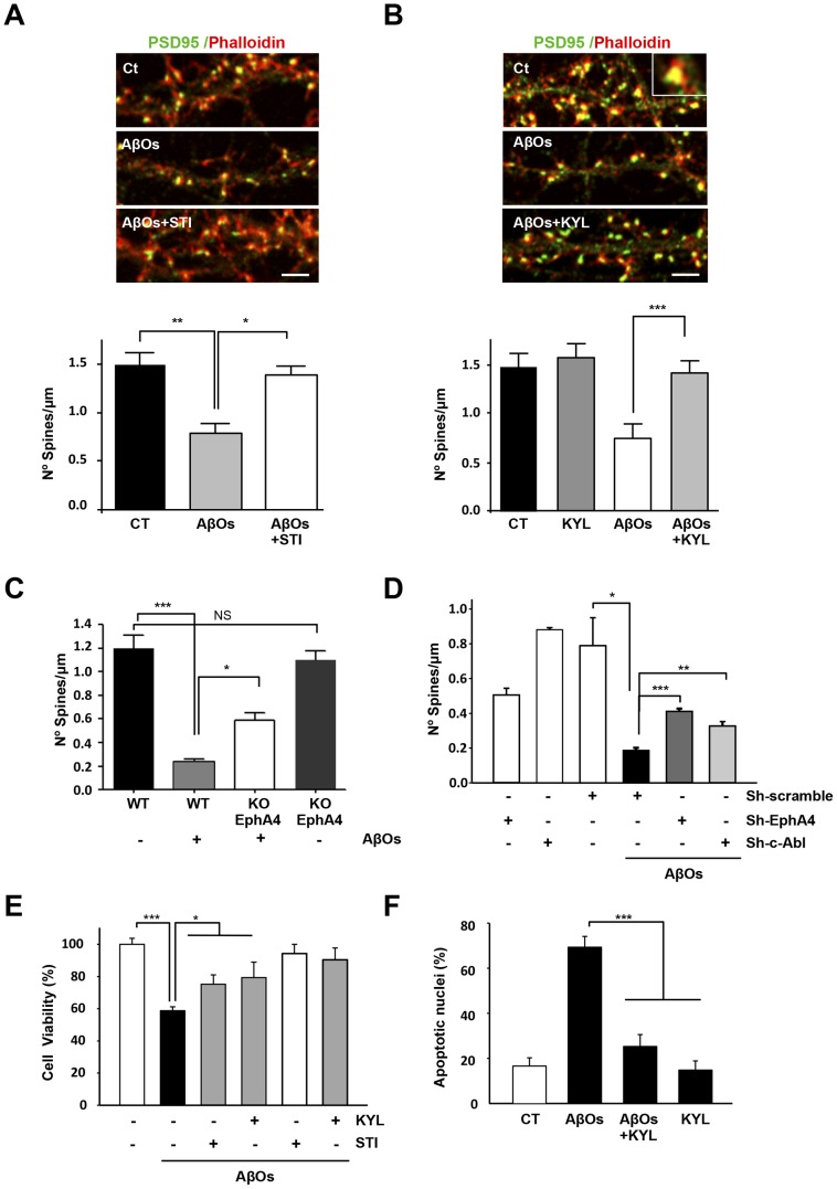
The early stages of Alzheimer's disease are characterised by impaired synaptic plasticity and synapse loss. Here, we show that amyloid-β oligomers (AβOs) activate the c-Abl kinase in dendritic spines of cultured hippocampal neurons and that c-Abl kinase activity is required for AβOs-induced synaptic loss. We also show that the EphA4 receptor tyrosine kinase is upstream of c-Abl activation by AβOs. EphA4 tyrosine phosphorylation (activation) is increased in cultured neurons and synaptoneurosomes exposed to AβOs, and in Alzheimer-transgenic mice brain. We do not detect c-Abl activation in EphA4-knockout neurons exposed to AβOs. More interestingly, we demonstrate EphA4/c-Abl activation is a key-signalling event that mediates the synaptic damage induced by AβOs. According to this results, the EphA4 antagonistic peptide KYL and c-Abl inhibitor STI prevented i) dendritic spine reduction, ii) the blocking of LTP induction and iii) neuronal apoptosis caused by AβOs. Moreover, EphA4-/- neurons or sh-EphA4-transfected neurons showed reduced synaptotoxicity by AβOs. Our results are consistent with EphA4 being a novel receptor that mediates synaptic damage induced by AβOs. EphA4/c-Abl signalling could be a relevant pathway involved in the early cognitive decline observed in Alzheimer's disease patients.


Conflict of interest statement

Competing Interests: The authors have declared that no competing interests exist.

Figures

Figure 1
Figure 1. AβOs induce the c-Abl activation in hippocampal neurons.
(A) c-Abl phosphorylated on tyrosine 412 (phospho-c-Abl; red) and β-tubulin (green). Hippocampal neurons (15 DIV) treated with 3 μM AβOs for 3 hours showed an increase in phospho-c-Abl immunoreactivity in the cell body and dendritic processes. Scale bar, 10 μm. (B) c-Abl was immunoprecipitated (IP) and then analyzed by western blotting (WB) for phospho-c-Abl and total c-Abl. IgG indicates the heavy chain of the antibodies used for immunoprecipitation. (C) The histogram shows densitometric analysis of phosphorylated c-Abl normalized to total immunoprecipitated c-Abl (average values ± standard error; ***p<0.001 by one-way ANOVA; n = 3). (D) Immunofluorescence labeling for c-Abl (gray) of 21 DIV hippocampal neurons treated with 3 μM AβOs 6 hours and postsynaptic protein PSD95 (green). Scale bar, 5 μm. (E) Cultured hippocampal neurons (21 DIV) were treated with 5 μM AβOs-FITC for 60 minutes and labeled for c-Abl phosphorylated on tyrosine 412 (phospho-c-Abl; red). Scale bar, 10 μm. Images of dendrites treated with AβOs-FITC for 30, 60 and 90 minutes labeled for phospho-c-Abl. (F) Histogram showing Manders overlap coefficient for AβOs-FITC and phospho-c-Abl at different times of treatment (**p<0.01 by one-way ANOVA; n = 15–20 neurites of 3 independent experiments). (G) Electron microscopy images of synatoneurosomes. The black arrows indicates vesicles accumulated in the active zone of presynaptic regions, asterisks indicate mitochondria, and white arrows indicate electron-dense regions corresponding to postsynaptic densities. (H) Immunoblot (phospho-c-Abl, total c-Abl and synapsin 1 (Syn)) of synaptoneurosomes exposed in vitro to 3 μM AβOs for 0, 15 and 60 minutes. (I) Densitometric analysis of phospho-c-Abl levels normalized to total c-Abl (AβOs for 15 minutes: **p<0.01 and for 60 minutes *p<0.05, by one-way ANOVA; n = 3).
Figure 2
Figure 2. AβOs colocalize with the EphA4 receptor and induce changes in its subcellular localization.
(A) Cultured hippocampal neurons (21 DIV) were treated with 5 μM AβOs-FITC (green) for 0.5–1 hours and immunolabeled for EphA4 (red). Scale bars, 10 μm in the left panel and 5 μm in the right panels. (B) Hippocampal neurons (21 DIV) were treated with 3 μM AβOs or 9.5 μg/ml ephrin A3 Fc as a soluble EphA4 ligand for 1 hour or left untreated as a control. The neurons were then immunolabeled with an EphA4 antibody (green; B) or double-labeled with an EphA4 antibody and phalloidin-TRITC (red; C).
Figure 3
Figure 3. EphA4 is activated in hippocampal neurons treated with AβOs and in Alzheimer's disease transgenic mice.
(A) Cultured hippocampal neurons (15 DIV) were treated with 3 μM AβOs for 0.5 and 1 hour EphA4 was immunoprecipitated and then analyzed by immunoblotting with an anti-phosphotyrosine antibody or (B) anti-phospho-EphA4 Tyr 602, shows that the quick response of EphA4 (15 minutes). The histogram shows densitometric analysis of phosphotyrosine levels normalized to total EphA4 levels (mean ± standard error; ***p<0.001 by one-way ANOVA; n = 3). (C) Synaptoneurosomes were treated with 3 μM AβOs for 0.5 and 1 hour. EphA4 was immunoprecipitated and then analyzed by immunoblotting using an anti-phosphotyrosine antibody. (D) The histogram shows densitometric analysis of phosphotyrosine levels at 0,5 and 1 hour normalized to total EphA4 levels (AβOs 0.5 hour **p<0.01, AβOs 1 hour *p<0.05 by Student's t-tests; n = 3). (E) Immunofluorescence showing phospho-EphA4 (p-EphA4) (red) and thioflavine-S (ThS) staining (green) in APPswe/PSEN1ΔE9 transgenic and wild-type (WT) hippocampi. Empty arrowheads show phospho-EphA4-positive neurons and white arrowheads show amyloid deposits stained with thioflavine-S (ThS). (F) Western blot of either wild-type or APPswe/PSEN1ΔE9 transgenic mouse hippocampus homogenates blotted against phospho-EphA4 Tyr 602 and total EphA4. Wild-type mouse lines 1–2 and APPswe/PSEN1ΔE9 transgenic mice lines 3–5.
Figure 4
Figure 4. EphA4 induces c-Abl activation in response to AβOs.
(A) Primary cultures of wild-type (WT) and EphA4 knockout (KO) hippocampal neurons were treated with AβOs for 60 minutes. c-Abl immunoprecipitates were probed by immunoblotting with anti-phospho c-Abl Tyr-412 antibody. (B) The histogram shows densitometric analysis of phospho-c-Abl levels normalized to total c-Abl in EphA4 WT and KO neurons treated with AβOs (mean ± standard error, *p<0.05 by one-way ANOVA; n = 3). C) Hippocampal slices from adult mice were treated for 90 minutes with 9.5 μg/ml ephrin-A3 ligand Fc or Fc as a control. EphA4 was immunoprecipitated, and the immunoprecipitates were probed by immunoblotting with an anti-phosphotyrosine antibody and reprobed for EphA4. (D) The hippocampal slices were stimulated as in C. c-Abl was immunoprecipitated, and immunoblotting was performed to detect anti-phospho c-Abl Tyr-412 antibody and reprobed for c-Abl. (E) Cultured hippocampal neurons (15 DIV) were treated with 3 μM AβOs for 0.5 or 3 hours, and c-Abl was then immunoprecipitated (IP). Immunoblotting was performed to detect EphA4 and c-Abl and showed that AβOs promote EphA4 association with c-Abl.
Figure 5
Figure 5. Inhibition of the EphA4-c-Abl pathway decreases the loss of dendritic spines and apoptosis induced by AβOs.
(A) Cultured hippocampal neurons (21 DIV) exposed for 5 hours to 3 μM AβOs with and without STI (5 μM) and (B) KYL (60 μM). The confocal images show F-actin labeled with phalloidin (red) and immunofluorescence labeling for PSD95 (green). Scale bar, 5 μm. Densitometric analysis (A. **p<0.01 for control versus AβOs; *p<0.05 for AβOs versus AβOs + STI. B. ***p<0.001 AβOs versus AβOs + KYL. By one-way ANOVA; n = 15–20 neurites of 3 independent experiments). All graphics show the average values ± standard error. (C) Quantification of dendritic spines in cultures of wild-type (EphA4+/+, WT) or EphA4 knockout (EphA4-/-, KO) hippocampal neurons (15 DIV) exposed to AβOs for 5 hours (*p<0.05, ***p<0.001, no significant (NS) by one-way ANOVA; n = 15–20 neurites of 3 independent experiments). (D) Quantification of dendritic spines in neurons transfected with pGFP, sh-EphA4, sh-c-Abl, scramble RNA EphA4 and c-Abl (SC) and treated with AβOs for 5 hours (*p<0.05 for SC versus pGFP+ AβOs; ***p<0.001 for pGFP+ AβOs verus Sh-EphA4+ AβOs or sh-c-Abl+ AβOs **p<0,01). (E) Cell viability assay in primary cultures of hippocampal neurons treated with AβOs and/or 15 or 30 μM KYL peptide for 24 hours (AβOs versus AβOs+ STI and AβOs+ KYL *p<0,05; n = 3). (F) Quantification of apoptotic nuclei in primary cultures of hippocampal neurons treated with AβOs and/or 30 μM KYL for 24 hours.
Figure 6
Figure 6. STI and KYL block the AβO effect in vitro, allowing for the induction of LTP of the hippocampal slices from wild-type mice.
(A) Hippocampal slices were exposed to ACSF (red) or AβOs (1 μM, white); arrow indicates the time of TBS and the plot show the fEPSP slope at different times. (B) Hippocampal slices were exposed to AβOs and STI (5 μM, black) or KYL (30 μM, gray), arrow indicates the time of TBS and the plot show the fEPSP slope at different times. (C) Inhibitors of c-Abl and EphA4 does not alter amplitude of basal fEPSP. Hippocampal slices were exposed to ACSF (white), STI (5 μM, black) or KYL (30 μM, gray) in basal conditions without stimulation. (D) Histogram showing Plot of fEPSP electrical recordings.

References

    1. Nelson PT, Alafuzoff I, Bigio EH, Bouras C, Braak H, et al. (2012) Correlation of Alzheimer disease neuropathologic changes with cognitive status: a review of the literature. J Neuropathol Exp Neurol 71: 362–381. - PMC - PubMed
    1. Walsh DM, Klyubin I, Fadeeva JV, Cullen WK, Anwyl R, et al. (2004) Deciphering the molecular basis of memory failure in Alzheimer's disease. Neuron 44: 181–193. - PubMed
    1. Klein WL (2013) Synaptotoxic, Amyloid-β Oligomers: A Molecular Basis for the Cause, Diagnosis, and Treatment of Alzheimer's Disease? J Alzheimers Dis 33 Suppl 1S49–S65. - PubMed
    1. Estrada LD, Zanlungo SM, Alvarez AR (2011) c-Abl tyrosine kinase signaling: a new player in AD tau pathology. Curr Alzheimer Res 6: 643–651. - PubMed
    1. Bradley WD, Koleske AJ (2009) Regulation of cell migration and morphogenesis by Abl-family kinases: emerging mechanisms and physiological contexts. J Cell Sci 1: 3441–3454. - PMC - PubMed

Publication types

MeSH terms