. 2014 May;13(5):515-23.
doi: 10.1038/nmat3912. Epub 2014 Mar 23.

Synthesis and patterning of tunable multiscale materials with engineered cells

Affiliations

Synthesis and patterning of tunable multiscale materials with engineered cells

Allen Y Chen et al. Nat Mater. 2014 May.

Abstract

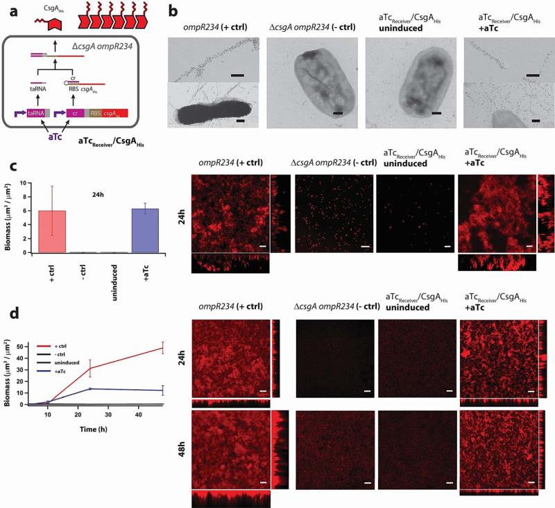
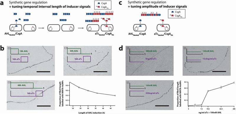
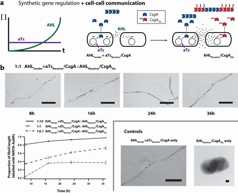
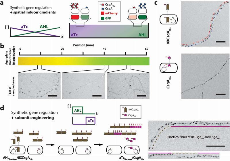
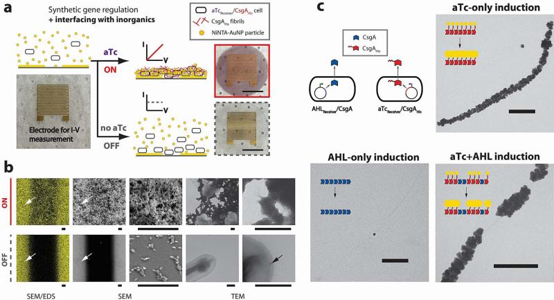
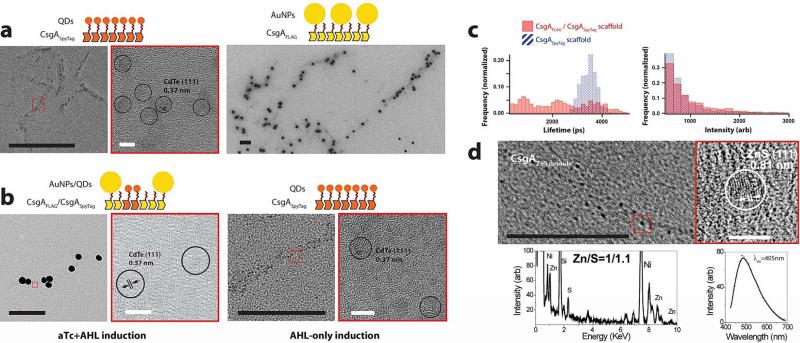
Many natural biological systems--such as biofilms, shells and skeletal tissues--are able to assemble multifunctional and environmentally responsive multiscale assemblies of living and non-living components. Here, by using inducible genetic circuits and cellular communication circuits to regulate Escherichia coli curli amyloid production, we show that E. coli cells can organize self-assembling amyloid fibrils across multiple length scales, producing amyloid-based materials that are either externally controllable or undergo autonomous patterning. We also interfaced curli fibrils with inorganic materials, such as gold nanoparticles (AuNPs) and quantum dots (QDs), and used these capabilities to create an environmentally responsive biofilm-based electrical switch, produce gold nanowires and nanorods, co-localize AuNPs with CdTe/CdS QDs to modulate QD fluorescence lifetimes, and nucleate the formation of fluorescent ZnS QDs. This work lays a foundation for synthesizing, patterning, and controlling functional composite materials with engineered cells.


Figures

Figure 1
Figure 1. Inducible production of engineered curli fibrils and biofilms
a, Riboregulator circuits tightly regulate expression of curli subunits, such as CsgAHis. Production of CsgAHis requires the expression of trans-activating RNA (taRNA). The taRNA prevents the cis-repressive (cr) sequence from blocking the ribosome-binding sequence (RBS) controlling translation of the mRNA transcript. In the absence of inducer, mRNA and taRNA levels are low, thus leading to significant repression of gene expression. The addition of aTc induces transcription of both csgAHis mRNA and taRNA, thus enabling CsgAHis production. Tight regulation of curli expression is useful for controlling patterning (Supplementary Fig. 19). b, Immuno-labelling of curli fibrils with rabbit anti-CsgA antibodies and gold-conjugated goat anti-rabbit antibodies. Positive-control (“+ ctrl”) MG1655 ompR234 cells (“ompR234”, see Supplementary Table 3), which have an intact endogenous csgA gene, produce curli fibrils that were labelled by anti-CsgA antibodies and are attached to cells. However, negative-control (“− ctrl”) cells with the csgA gene knocked out and no csgA-expressing circuits (“ΔcsgA ompR234”, see Supplementary Table 3), as well as aTcReceiver/CsgAHis cells in the absence of aTc, did not produce curli fibrils. Inducing aTcReceiver/CsgAHis cells with aTc enabled the synthesis of curli fibrils that were labelled by anti-CsgA antibodies and attached to cells. Scale bars are 200nm. c, Confocal microscopy and biomass quantification revealed that under static culture conditions, E. coli ompR234 cells formed thick adherent biofilms. However, E. coli ΔcsgA ompR234 cells, as well as aTcReceiver/CsgAHis cells in the absence of aTc, did not form biofilms. Inducing aTcReceiver/CsgAHis cells with aTc led to the formation of thick adherent biofilms. d, Confocal microscopy and biomass quantification revealed similar biofilm-forming capabilities by E. coli ompR234 and induced aTcReceiver/CsgAHis cells when grown in flow cells. To enable visualization, we transformed a constitutive mCherry-expressing plasmid into all strains (see Supplementary Methods). Cells were grown in liquid M63 media with glucose; the corresponding experiments for other media conditions are shown in Supplementary Figure 1 and 2. Scale bars in c) and d) are 50μm, and orthogonal XZ and YZ views are maximum-intensity projections.
Figure 2
Figure 2. Conversion of timing and amplitude of chemical inducer signals into material structure and composition
a, Inducible synthetic gene circuits couple curli subunit secretion to external chemical inducers. Engineered cells containing these circuits can translate induction pulse length into nanoscale structure and composition of block co-fibrils. b, We first used AHL to induce secretion of CsgA from AHLReceiver/CsgA and then used aTc to induce secretion of CsgAHis from aTcReceiver/CsgAHis. We tuned the relative block lengths and proportions of CsgA and CsgAHis (plot of the proportion of fibril length labelled by NiNTA-AuNP, solid grey line) by changing the relative lengths of AHL versus aTc induction times. Scale bars are 200nm. c, Synthetic genetic regulatory circuits that couple curli subunit secretion to external inducer signals can translate inducer concentration into nanoscale structure and composition of block cofibrils. d, Engineered cells AHL induced secretion of CsgA from AHLReceiver/CsgA, while at the same time, aTc induced secretion of CsgAHis from aTcReceiver/CsgAHis. We tuned the relative block lengths and proportions of CsgA and CsgAHis by changing the relative concentrations of AHL and aTc inducers applied simultaneously. The solid grey line indicates the proportion of fibril length labelled by NiNTA-AuNP with varying concentrations of aTc and constant 100nM AHL. Detailed histograms can be found in Supplementary Figure 6. Scale bars are 200nm.
Figure 3
Figure 3. Synthetic cellular communication for dynamic, autonomous material production and patterning
a, Synthetic gene circuits that couple curli subunit secretion to external inducer signals, when combined with synthetic cellular communication circuits, allow for the production of materials whose structure and composition changes autonomously with time. AHLSender+aTcReceiver/CsgA secreted both CsgA and AHL. As AHL signal accumulated, AHLReceiver/CsgAHis secreted increasing levels of CsgAHis. b, Using the autonomous cellular communication system, the length of CsgAHis blocks and the proportion of CsgAHis increased with time (plot of the proportion of fibril length labelled by NiNTA-AuNP, grey lines). This behaviour could be tuned by the ratio of the seeding density of AHLSender+aTcReceiver/CsgA cells to AHLReceiver/CsgAHis cells. When only AHLSender+aTcReceiver/CsgA cells were present, the resulting fibrils were almost uniformly unlabelled; when only AHLReceiver/CsgAHis cells were present, no curli fibrils were formed (Controls). Detailed histograms can be found in Supplementary Fig. 10. Scale bars are 200nm.
Figure 4
Figure 4. Multiscale patterning with cellular consortia via synthetic gene regulation combined with inducer gradients and subunit engineering
a, Synthetic gene circuits that couple curli subunit secretion to external inducer signals, when combined with a spatial inducer gradient, enable patterning across multiple length scales. We used an agar plate with opposing concentration gradients of AHL and aTc to achieve control at the macroscale (Supplementary Fig. 12). This was combined with regulation of nanoscale patterning to achieve multiscale patterning. Embedded in top agar were equal numbers of AHLReceiver/CsgA, aTcReceiver/CsgAHis, AHLReceiver/GFP, and aTcReceiver/mCherry cells. b, By combining synthetic gene regulation with spatial inducer gradients, we created a change in the nanoscale structure of fibrils across a distance of millimetres. This nanoscale and macroscale patterning was shown by changes in segment lengths of unlabelled and NiNTA-AuNP-labelled fibril segments at different locations across the agar plate. Inducer concentration gradients were demonstrated by overlaid GFP and mCherry fluorescence images of embedded AHLReceiver/GFP and and aTcReceiver/mCherry reporter cells. Scale bars are 200nm. c, We also achieved patterning at the nanoscale by protein engineering of curli subunits. Concatenating eight tandem repeats of CsgA and adding one histidine tag to the C-terminus (8XCsgAHis) resulted in fibrils that were labelled by a syncopated pattern of NiNTA-AuNPs, with clusters of particles separated by 33.3±27.1 (s.e.m.) nm. Scale bars are 100nm. d, Synthetic gene circuits that couple curli subunit secretion to external inducer signals, when combined with subunit engineering, enable patterning across multiple length scales (nanometres to micrometres). We used AHL to induce production of 8XCsgAHis from AHLReceiver/8XCsgAHis and then used aTc to induce production of CsgAHis from aTcReceiver/CsgAHis. In the TEM images, dashed brown lines refer to syncopated 8XCsgAHis segments while the solid amethyst lines indicate CsgAHis segments. Detailed histograms for data shown here can be found in Supplementary Figure 11. Scale bars are 100nm.
Figure 5
Figure 5. Environmentally switchable conductive biofilms and cell-based synthesis of curli-templated nanowires and nanorods
a, We used aTcReceiver/CsgAHis cells to form amyloid fibrils composed of CsgAHis in response to aTc. When combined with NiNTA-AuNPs, we created conductive biofilms that can be externally controlled as electrical switches. When aTc was added to aTcReceiver/CsgAHis cells grown in the presence of NiNTA-AuNPs, it triggered the formation of conductive biofilms on electrodes, with embedded 5nm gold particles giving biofilms a red colour (‘ON’, solid red box). However, in the absence of aTc, few cells adhered to the electrodes (‘OFF’, dashed grey box). Scale bars are 5mm. b, SEM/EDS elemental mapping of the aTc-induced ‘ON’ state for aTcReceiver/CsgAHis biofilms showed that networks of gold in the biofilms connected the electrodes (white arrows). SEM imaging showed that the biofilms bridged electrodes. TEM imaging showed networks of aggregated gold particles. In contrast, SEM/EDS mapping of the ‘OFF’ state showed no gold networks, SEM imaging showed only scattered cells in the gap between electrodes, and TEM imaging showed only scattered and isolated gold particles (black arrow). Scale bars of scanning electron micrographs are 20μm and scale bars of transmission electron micrographs are 200nm. c, A mixed population of aTcReceiver/CsgAHis and AHLReceiver/CsgA cells produced curli templates for organizing either gold nanowires or gold nanorods when they were induced with aTc only or both aTc and AHL, respectively. NiNTA-AuNPs were patterned on CsgAHis subunits within curli fibrils and then gold enhanced. Scale bars are 200nm.
Figure 6
Figure 6. Assembly and tuning of functional AuNP-QD heterostructures and nucleation of fluorescent ZnS QDs on cell-synthesized curli fibrils
a, CsgASpyTag fibrils specifically bind CdTe/CdS QDs conjugated to the SpyCatcher protein; the CdTe cores of QDs are seen under HRTEM. CsgAFLAG fibrils are specifically bound by anti-FLAG antibodies which are in turn bound by 40nm AuNPs conjugated to secondary antibodies. CsgA fibrils do not bind either CdTe/CdS QDs conjugated to SpyCatcher or 40nm AuNPs conjugated to antibodies (Supplementary Fig. 23a, b). b, A mixed population of aTcReceiver/CsgAFLAG and AHLReceiver/CsgASpyTag cells produced curli templates for either AuNP-QD heterostructures (cofibrils of CsgAFLAG and CsgASpyTag) or QD-only assemblies (CsgASpyTag fibrils) depending on whether they were induced by both aTc and AHL, or AHL only, respectively. c, Cell-patterned curli fibrils enable the tuning of stimuli-responsive inorganic-organic materials. AuNP-QD assemblies patterned on CsgAFLAG/CsgASpyTag scaffolds (solid red bars) exhibited different fluorescence lifetime and intensity properties than QD-only assemblies patterned on CsgASpyTag scaffolds (hashed blue bars). d, CsgAZnS peptide fibrils nucleated ~5nm nanoparticles with a cubic zinc blende ZnS (111) structure and approximately 1:1 ratio of zinc and sulphur. The particles were fluorescent, with an emission peak at 490nm when excited at 405nm. Control CsgA fibrils nucleated few such particles (Supplementary Fig. 23c). In a), b), and d) black scale bars are 200nm and white scale bars are 5nm; the images outlined by red boxes are zoomed-in versions of the inset red boxes.

References

    1. Fratzl P, Weinkamer R. Nature's hierarchical materials. Progress in Materials Science. 2007;52:1263–1334. doi: http://dx.doi.org/10.1016/j.pmatsci.2007.06.001. - DOI
    1. Kollmannsberger P, Bidan CM, Dunlop JWC, Fratzl P. The physics of tissue patterning and extracellular matrix organisation: how cells join forces. Soft Matter. 2011;7:9549–9560. doi:10.1039/c1sm05588g.
    1. Stevens MM, George JH. Exploring and engineering the cell surface interface. Science. 2005;310:1135–1138. doi:10.1126/science.1106587. - PubMed
    1. O'Toole G, Kaplan HB, Kolter R. Biofilm formation as microbial development. Annu Rev Microbiol. 2000;54:49–79. doi:10.1146/annurev.micro.54.1.4954/1/49 [pii] - PubMed
    1. Epstein AK, Pokroy B, Seminara A, Aizenberg J. Bacterial biofilm shows persistent resistance to liquid wetting and gas penetration. Proc Natl Acad Sci U S A. 2011;108:995–1000. doi:1011033108 [pii]10.1073/pnas.1011033108. - PMC - PubMed

Publication types

MeSH terms