

Glaucoma progression detection using structural retinal nerve fiber layer measurements and functional visual field points
- PMID: 24658239
- PMCID: PMC4248722
- DOI: 10.1109/TBME.2013.2295605
Glaucoma progression detection using structural retinal nerve fiber layer measurements and functional visual field points
Abstract
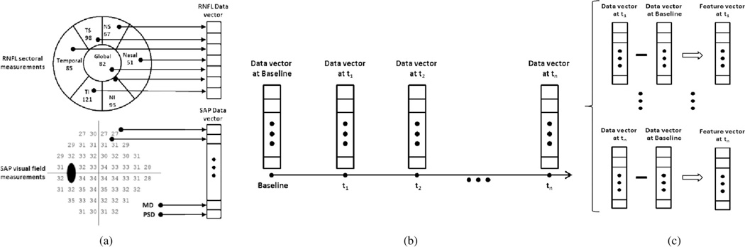
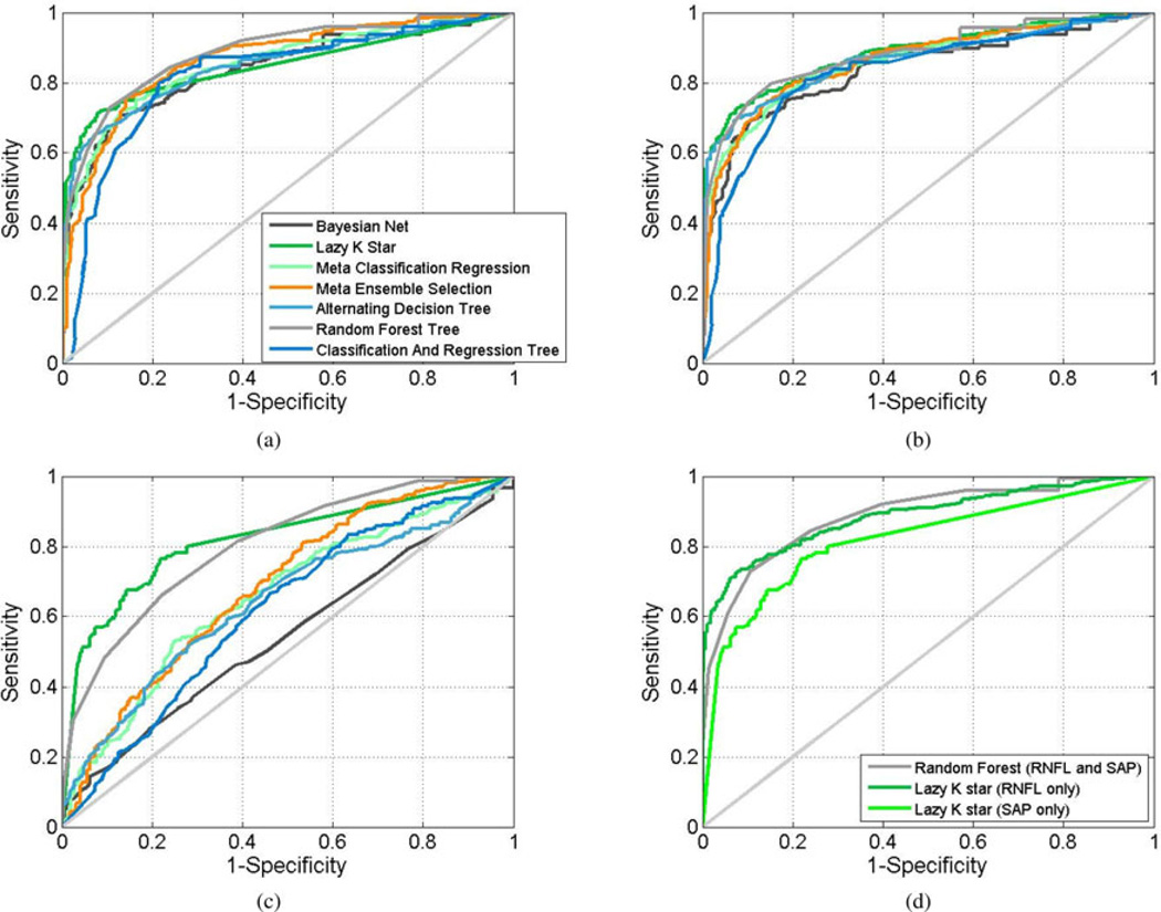
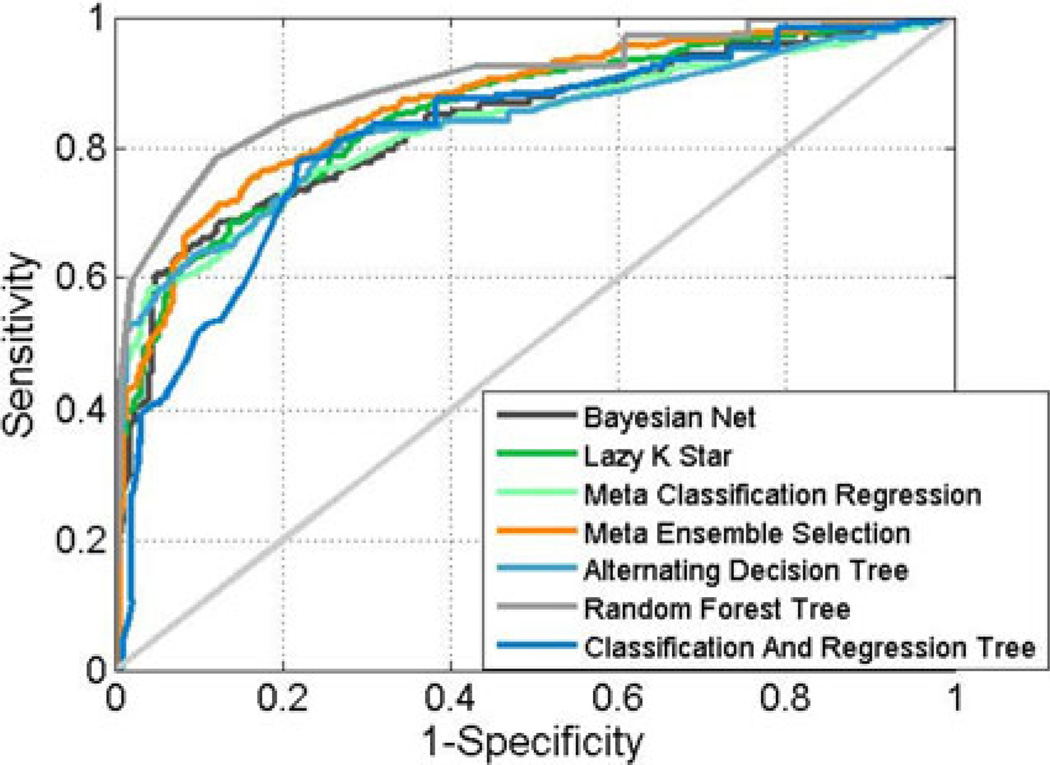
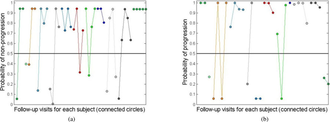
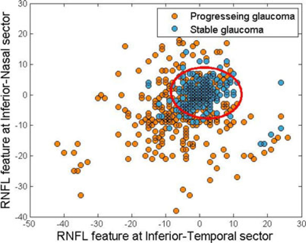
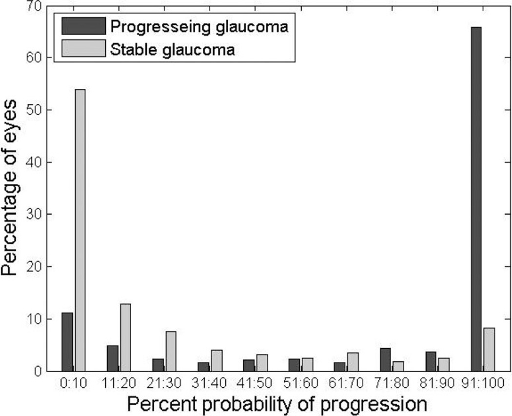
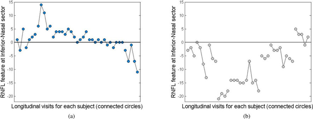
Machine learning classifiers were employed to detect glaucomatous progression using longitudinal series of structural data extracted from retinal nerve fiber layer thickness measurements and visual functional data recorded from standard automated perimetry tests. Using the collected data, a longitudinal feature vector was created for each patient's eye by computing the norm 1 difference vector of the data at the baseline and at each follow-up visit. The longitudinal features from each patient's eye were then fed to the machine learning classifier to classify each eye as stable or progressed over time. This study was performed using several machine learning classifiers including Bayesian, Lazy, Meta, and Tree, composing different families. Combinations of structural and functional features were selected and ranked to determine the relative effectiveness of each feature. Finally, the outcomes of the classifiers were assessed by several performance metrics and the effectiveness of structural and functional features were analyzed.
Figures
References
-
- Weinreb RN, Khaw PT. Primary open-angle glaucoma. Lancet. 2004 May 22;363:1711–1720. - PubMed
-
- Jonas JB, Dichtl A. Evaluation of the retinal nerve fiber layer. Surv. Ophthalmol. 1996 Mar-Apr;40:369–378. - PubMed
-
- Bowd C, Weinreb RN, Zangwill LM. Evaluating the optic disc and retinal nerve fiber layer in glaucoma—Part I: Clinical examination and photographic methods. Semin. Ophthalmol. 2000 Dec.15:194–205. - PubMed
Publication types
MeSH terms
Grants and funding
- R01EY008208/EY/NEI NIH HHS/United States
- K99EY020518/EY/NEI NIH HHS/United States
- R01 EY011008/EY/NEI NIH HHS/United States
- P30 EY022589/EY/NEI NIH HHS/United States
- R01 EY019869/EY/NEI NIH HHS/United States
- R00 EY020518/EY/NEI NIH HHS/United States
- R01 EY022039/EY/NEI NIH HHS/United States
- P30EY022589/EY/NEI NIH HHS/United States
- R01EY022039/EY/NEI NIH HHS/United States
- R01EY019869/EY/NEI NIH HHS/United States
- R01 EY021818/EY/NEI NIH HHS/United States
- R01 EY008208/EY/NEI NIH HHS/United States
- K99 EY020518/EY/NEI NIH HHS/United States
- R01EY011008/EY/NEI NIH HHS/United States
