. 2014 Apr;21(4):383-8.
doi: 10.1038/nsmb.2797. Epub 2014 Mar 23.

Structural basis for pure antagonism of integrin αVβ3 by a high-affinity form of fibronectin

Affiliations

Structural basis for pure antagonism of integrin αVβ3 by a high-affinity form of fibronectin

Johannes F Van Agthoven et al. Nat Struct Mol Biol. 2014 Apr.

Abstract

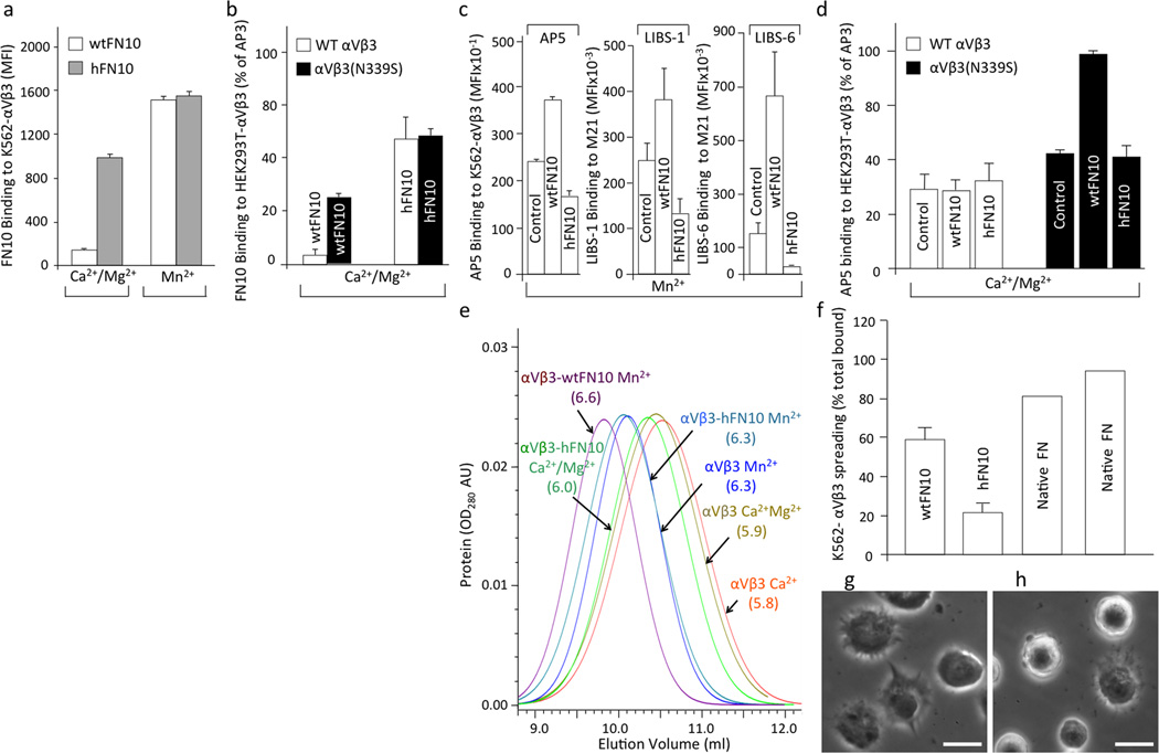
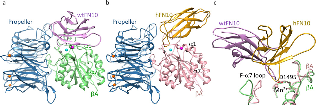
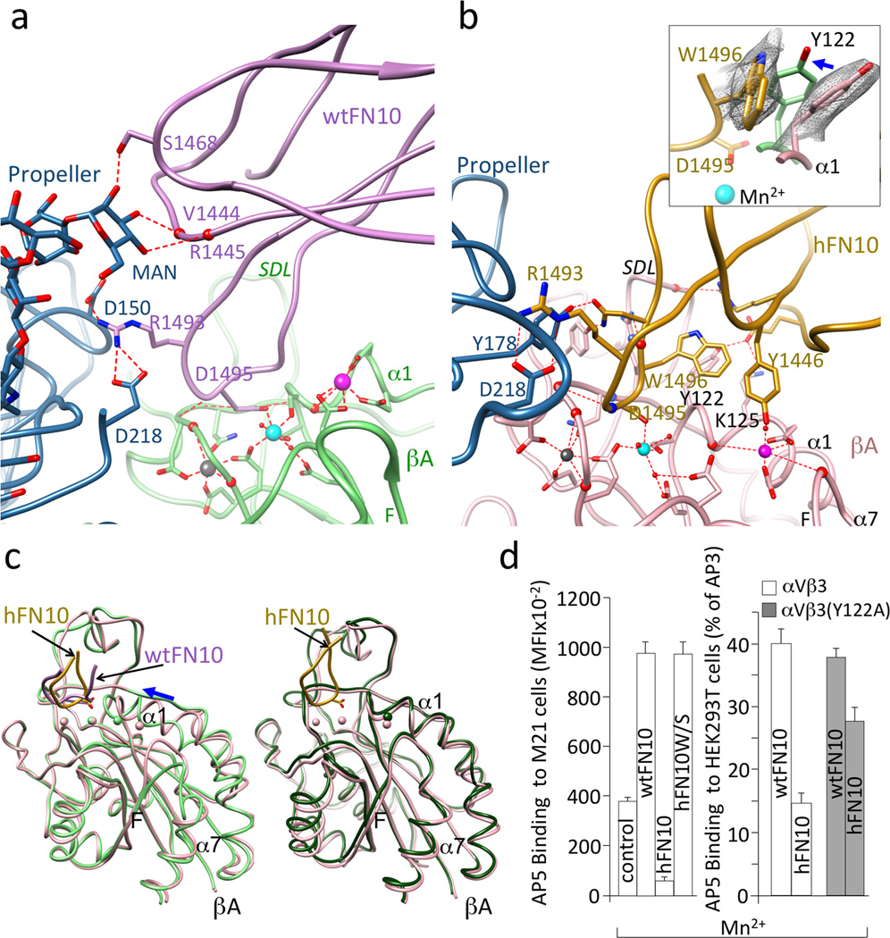
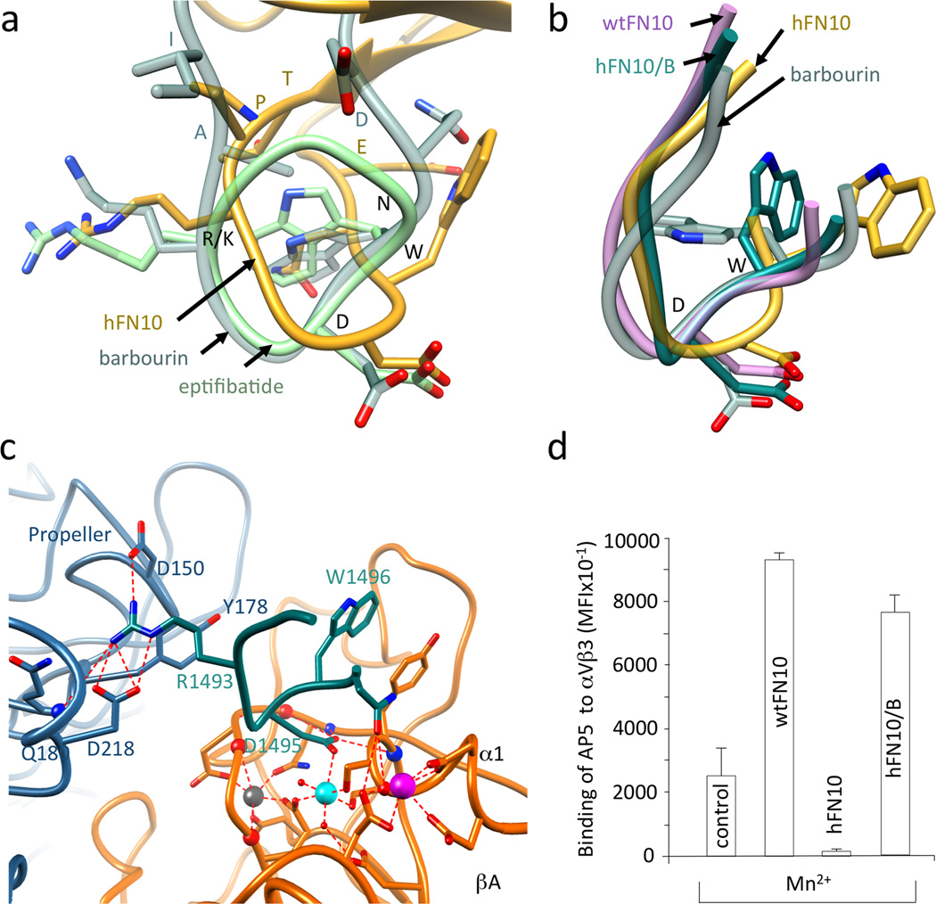
Integrins are important therapeutic targets. However, current RGD-based anti-integrin drugs are also partial agonists, inducing conformational changes that trigger potentially fatal immune reactions and paradoxical cell adhesion. Here we describe the first crystal structure of αVβ3 bound to a physiologic ligand, the tenth type III RGD domain of wild-type fibronectin (wtFN10), or to a high-affinity mutant (hFN10) shown here to act as a pure antagonist. Comparison of these structures revealed a central π-π interaction between Trp1496 in the RGD-containing loop of hFN10 and Tyr122 of the β3 subunit that blocked conformational changes triggered by wtFN10 and trapped hFN10-bound αVβ3 in an inactive conformation. Removing the Trp1496 or Tyr122 side chains or reorienting Trp1496 away from Tyr122 converted hFN10 into a partial agonist. These findings offer new insights into the mechanism of integrin activation and a basis for the design of RGD-based pure antagonists.


Figures

Figure 1
Figure 1. Binding properties of hFN10 and wtFN10 to αVβ3
Binding of fluoresceinated wtFN10 (a) and hFN10 (b) or LIBS mAbs (alone (c) or in presence of wtFN10 and hFN10 (d) to αVβ3+ cells. In (c), mAb-binding was assessed using K562-αVβ3 and M21 cells. MFI, mean fluorescence intensity. Histograms in a-d represent mean+SD, n=3 independent experiments. (e) Hydrodynamic analyses of unliganded αVβ3 and αVβ3-FN10 complexes in presence of Ca2+, Ca2+-Mg2+ or Mn2+. Stokes radii (in nm) are shown in parentheses. AU: absorbance unit. (f–h) Mn2+-induced spreading of K562-αVβ3 on wtFN10 (f), hFN10 (g) (mean+SD, n=3 independent experiments), or on full-length FN (f) (two independent experiments are shown). Spreading under all conditions was eliminated by mAb LM609 against αVβ3 (not shown). (g,h) Representative phase contrast images of K562-αVβ3 spreading on wtFN10 (g) and hFN10 (h). Bar = 20µM.
Figure 2
Figure 2. Structures of αVβ3 bound to FN10
Ribbon diagrams of αVβ3 head bound to wtFN10 (a) or hFN10 (b). Orientation of the integrin head in (a) and (b) is identical. Mn2+ ions at LIMBS (gray), MIDAS (cyan) and ADMIDAS (magenta) are shown as spheres (also in Figs. 3a–b, 4c). (c) Orientation of bound FN10 relative to the superimposed βA domains (chain colors as in a, b). Mn2+ at MIDAS the ligand Asp (D1495) and the F-α7 loop are shown.
Figure 3
Figure 3. αVβ3-FN10 interfaces, conformational changes and structure validation
Ribbon diagrams showing key electrostatic and H-bond interactions and metal ion coordinations in αVβ3-wtFN10 (a) and αVβ3-hFN10 (b) structures. Chain colors are as in Fig. 2. Inset in b, enlarged view of σA weighted 2Fo-Fc map contoured at 1.0σ of Trp1496 and Tyr122 side-chains in αVβ3-hFN10 complex. Inward movement (blue arrow) of Tyr122 (in light green) in wtFN10-bound βA would clash with Trp1496 side chain. (c) Left panel, βA domain from αVβ3-hFN10 (in pink) superimposed on that of αVβ3-wtFN10 (in light green) and on βA domain (in dark green) from unliganded αVβ3 (pdb 3ije) (right panel). Blue arrow in left panel in (c) indicates direction of wtFN10-induced inward movement of α1 helix (and ADMIDAS ion) towards MIDAS. Spheres representing the three metal ions bear the color of respective βA. The major tertiary change observed in the F-α7 loop of wtFN10-bound βA (c, left panel) was not translated into a one-turn displacement of α7, possibly the result of crystal contacts when the complete ectodomain is used in crystallization. Except for ligand-occupancy and resulting changes in SDL loop, structures of unliganded- and hFN10-bound βA domains are identical (c, right panel) (LIMBS and MIDAS are not occupied by metal in unliganded βA). (d) Left panel, binding of fluoresceinated AP5 to M21 cells in absence (control) or presence of unlabeled wtFN10, hFN10 or hFN10W/S. Right panel, binding of fluoresceinated AP5 to αVβ3+ or or αVβ3(Y122A) + HEK293T in presence of unlabeled wt- or hFN10. Histograms represent mean±SD, n=3 independent experiments.
Figure 4
Figure 4. RGD-containing loop structures in wild type and modified FN10
(a) Superimposed R/KGD-containing loops of hFN10, eptifibatide (pdb id 2vdn) and barbourin (pdb id 1q7j, model 2). Residues R/KGDWN common to hFN10 and barbourin are labeled in black and the three flanking residues are in the respective loop color. (b) Superimposed structures of RGD-containing loops of αVβ3-hFN10, αVβ3-wtFN10, barbourin and αVβ3-hFN10/B. The position the Cα and Cβ of Trp1496 in the αVβ3-hFN10/B complex is as that in barbourin or eptifibatide. (c) Main ionic interactions at the αVβ3-hFN10/B interface involving the RGD-containing loop (in dark cyan). (d) Binding (mean±SD, n=3 independent experiments) of fluoresceinated AP5 mAb to M21 cells in absence (control) and presence of wtFN10, hFN10 or hFN10/B.

References

    1. Hynes RO. Integrins: bidirectional, allosteric signaling machines. Cell. 2002;110:673–687. - PubMed
    1. Xiong JP, et al. Crystal structure of the extracellular segment of integrin alpha Vbeta3. Science. 2001;294:339–345. - PMC - PubMed
    1. Arnaout MA, Goodman SL, Xiong JP. Structure and mechanics of integrin-based cell adhesion. Curr Opin Cell Biol. 2007;19:495–507. - PMC - PubMed
    1. Xiong JP, et al. Crystal structure of the complete integrin alphaVbeta3 ectodomain plus an alpha/beta transmembrane fragment. The Journal of cell biology. 2009;186:589–600. - PMC - PubMed
    1. Honda S, et al. Topography of ligand-induced binding sites, including a novel cation-sensitive epitope (AP5) at the amino terminus, of the human integrin beta 3 subunit. The Journal of biological chemistry. 1995;270:11947–11954. - PubMed

Publication types