

Emerging endoscopic imaging technologies for bladder cancer detection
- PMID: 24658832
- PMCID: PMC4043447
- DOI: 10.1007/s11934-014-0406-5
Emerging endoscopic imaging technologies for bladder cancer detection
Abstract
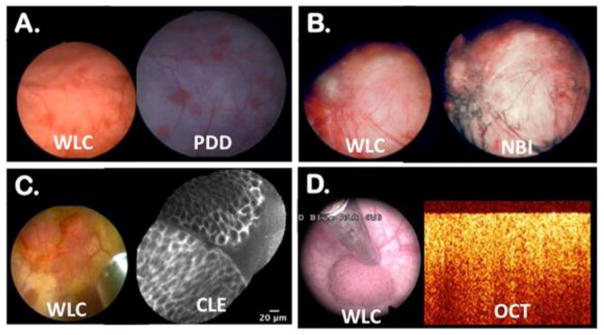
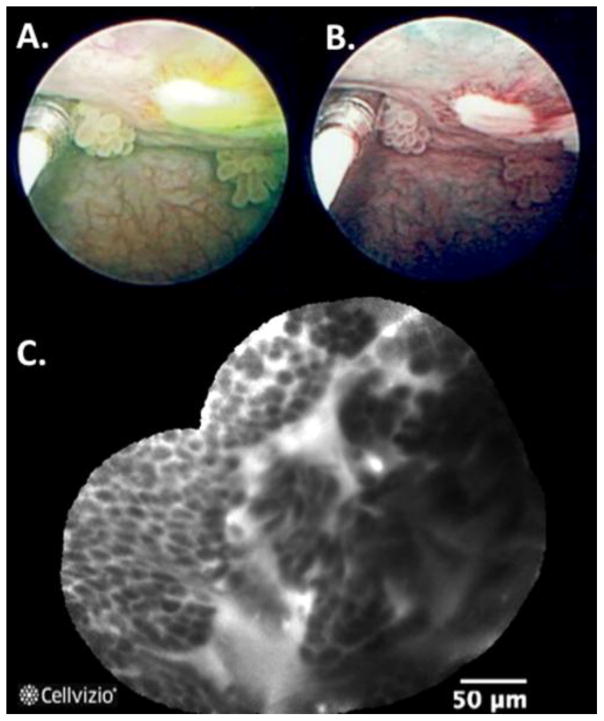
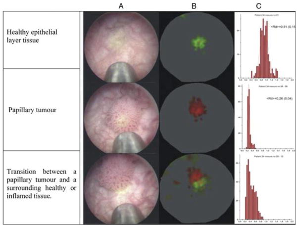
Modern urologic endoscopy is the result of continuous innovations since the early nineteenth century. White-light cystoscopy is the primary strategy for identification, resection, and local staging of bladder cancer. While highly effective, white light cystoscopy has several well-recognized shortcomings. Recent advances in optical imaging technologies and device miniaturization hold the potential to improve bladder cancer diagnosis and resection. Photodynamic diagnosis and narrow band imaging are the first to enter the clinical arena. Confocal laser endomicroscopy, optical coherence tomography, Raman spectroscopy, UV autofluorescence, and others have shown promising clinical and pre-clinical feasibility. We review their mechanisms of action, highlight their respective advantages, and propose future directions.
Conflict of interest statement
Dr. Aristeo Lopez declares no potential conflicts of interest relevant to this article.
Dr. Joseph C. Liao received a research grant from NIH. Dr. Liao received travel support from Mauna Kea Technologies including expenses covered or reimbursed, and payment for the development of educational presentations from Storz.
Figures
Similar articles
-
New optical imaging technologies for bladder cancer: considerations and perspectives.J Urol. 2012 Aug;188(2):361-8. doi: 10.1016/j.juro.2012.03.127. Epub 2012 Jun 13. J Urol. 2012. PMID: 22698620 Free PMC article. Review.
-
New imaging techniques for nonmuscle invasive bladder cancer.Curr Opin Urol. 2014 Sep;24(5):532-9. doi: 10.1097/MOU.0000000000000093. Curr Opin Urol. 2014. PMID: 25051025 Review.
-
[Supplementary optical techniques for the detection of nonmuscle invasive bladder cancer].Urologe A. 2018 Feb;57(2):139-147. doi: 10.1007/s00120-017-0539-5. Urologe A. 2018. PMID: 29110046 Review. German.
-
Recent advances in optical imaging technologies for the detection of bladder cancer.Photodiagnosis Photodyn Ther. 2018 Dec;24:192-197. doi: 10.1016/j.pdpdt.2018.10.009. Epub 2018 Oct 10. Photodiagnosis Photodyn Ther. 2018. PMID: 30315954 Review.
-
High magnification cystoscopy in the primary diagnosis of bladder tumors.Curr Opin Urol. 2011 Sep;21(5):398-402. doi: 10.1097/MOU.0b013e32834956ad. Curr Opin Urol. 2011. PMID: 21730856 Review.
Cited by
-
Probe-Based Confocal Laser Endomicroscopy During Transurethral Resection of Bladder Tumors Improves the Diagnostic Accuracy and Therapeutic Efficacy.Ann Surg Oncol. 2019 Apr;26(4):1158-1165. doi: 10.1245/s10434-019-07200-6. Epub 2019 Feb 4. Ann Surg Oncol. 2019. PMID: 30719635 Free PMC article.
-
Fluorescence-Based Microendoscopic Sensing System for Minimally Invasive In Vivo Bladder Cancer Diagnosis.Biosensors (Basel). 2022 Aug 11;12(8):631. doi: 10.3390/bios12080631. Biosensors (Basel). 2022. PMID: 36005027 Free PMC article.
-
Bioinformatics in urology - molecular characterization of pathophysiology and response to treatment.Nat Rev Urol. 2024 Apr;21(4):214-242. doi: 10.1038/s41585-023-00805-3. Epub 2023 Aug 21. Nat Rev Urol. 2024. PMID: 37604982 Review.
-
Confocal laser endomicroscopy of bladder and upper tract urothelial carcinoma: a new era of optical diagnosis?Curr Urol Rep. 2014 Sep;15(9):437. doi: 10.1007/s11934-014-0437-y. Curr Urol Rep. 2014. PMID: 25002073 Free PMC article. Review.
-
Nanotechnology in bladder cancer: current state of development and clinical practice.Nanomedicine (Lond). 2015;10(7):1189-201. doi: 10.2217/nnm.14.212. Nanomedicine (Lond). 2015. PMID: 25929573 Free PMC article. Review.
References
-
- Herr H. Early History of Endoscopic Treatment of Bladder Tumors From Grunfeld’s Polypenkneipe to the Stern-McCarthy Resectoscope. J Endourol. 2006;20(2):85–91. This article highlights the most noteworthy innovations that occurred in urologic endoscopy providing an overview of the history of endoscopy and the endoscopic management of bladder tumors. - PubMed
-
- Natalin Ra, Landman J. Where next for the endoscope? Nat Rev Urol. 2009 Nov;6(11):622–8. - PubMed
-
- Samplaski MK, Jones JS. Two centuries of cystoscopy: the development of imaging, instrumentation and synergistic technologies. BJU Int. 2009 Jan;103(2):154–8. - PubMed
-
- Witjes Ja. Bladder Carcinoma in Situ in 2003: State of the Art. Eur Urol. 2004 Feb;45(2):142–146. - PubMed
-
- Cheng L, Neumann RM, Weaver aL, Cheville JC, Leibovich BC, Ramnani DM, Scherer BG, Nehra a, Zincke H, Bostwick DG. Grading and staging of bladder carcinoma in transurethral resection specimens. Correlation with 105 matched cystectomy specimens. Am J Clin Pathol. 2000 Feb;113(2):275–9. - PubMed
Publication types
MeSH terms
Grants and funding
LinkOut - more resources
Full Text Sources
Other Literature Sources
Medical
