Review
. 2014 Nov;34(6):1168-216.
doi: 10.1002/med.21315. Epub 2014 Mar 21.

Recent advances on plasmin inhibitors for the treatment of fibrinolysis-related disorders

Affiliations
Review

Recent advances on plasmin inhibitors for the treatment of fibrinolysis-related disorders

Rami A Al-Horani et al. Med Res Rev. 2014 Nov.

Abstract

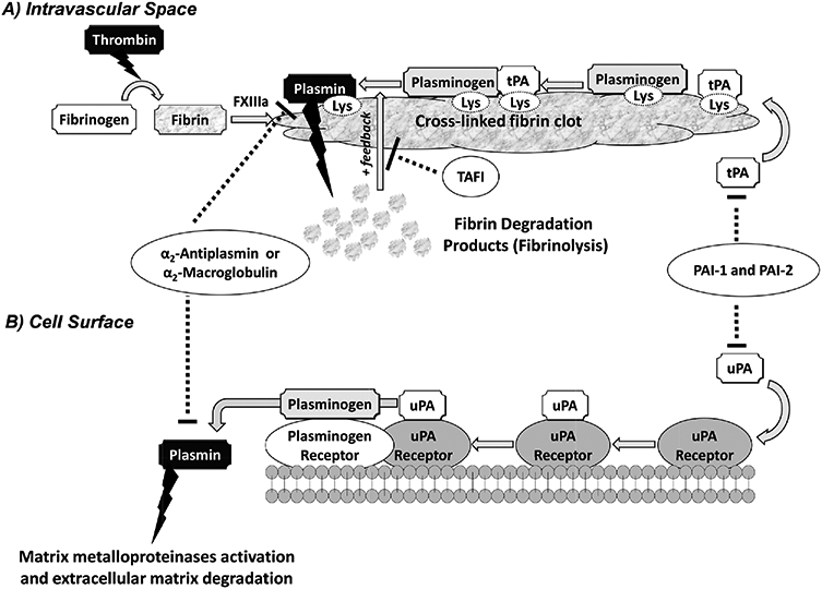
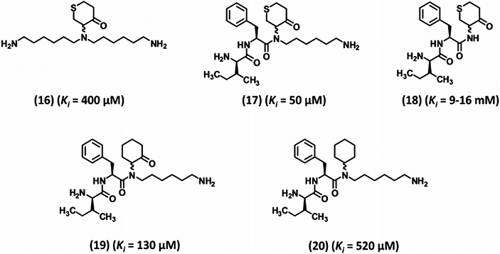
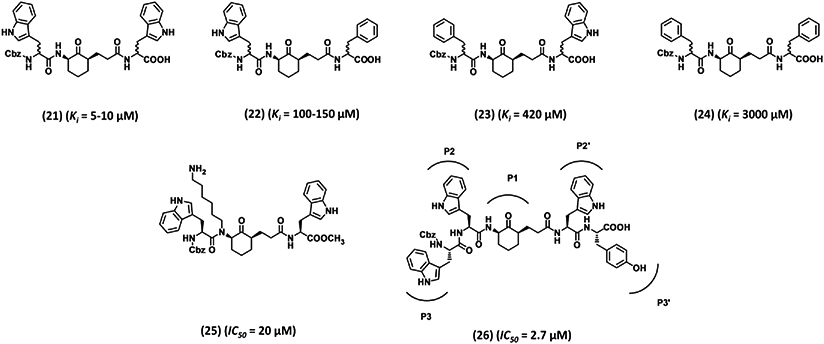
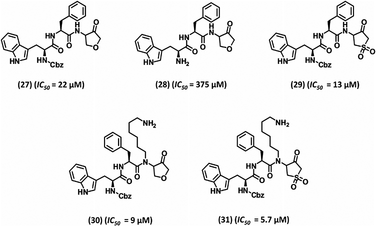
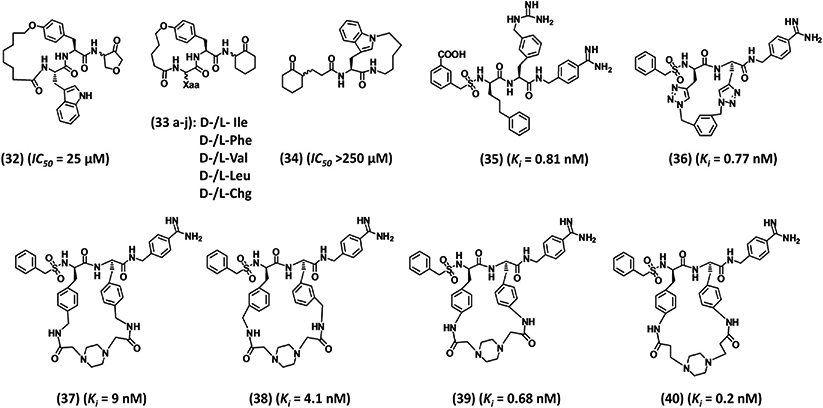
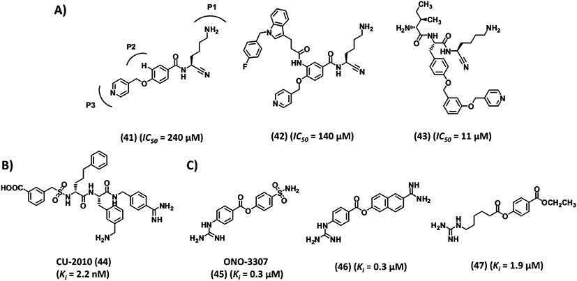
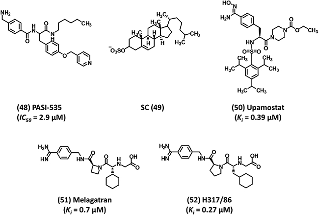
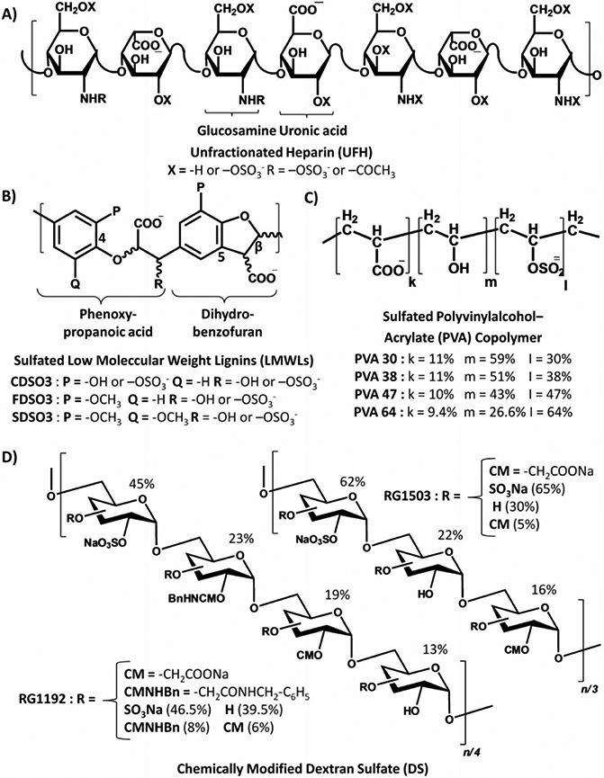
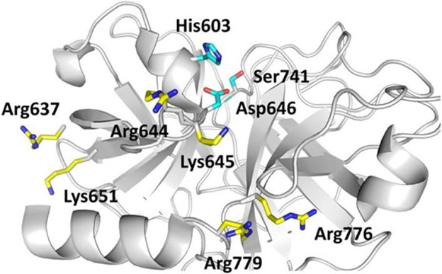
Growing evidence suggests that plasmin is involved in a number of physiological processes in addition to its key role in fibrin cleavage. Plasmin inhibition is critical in preventing adverse consequences arising from plasmin overactivity, e.g., blood loss that may follow cardiac surgery. Aprotinin was widely used as an antifibrinolytic drug before its discontinuation in 2008. Tranexamic acid and ε-aminocaproic acid, two small molecule plasmin inhibitors, are currently used in the clinic. Several molecules have been designed utilizing covalent, but reversible, chemistry relying on reactive cyclohexanones, nitrile warheads, and reactive aldehyde peptidomimetics. Other major classes of plasmin inhibitors include the cyclic peptidomimetics and polypeptides of the Kunitz and Kazal-type. Allosteric inhibitors of plasmin have also been designed including small molecule lysine analogs that bind to plasmin's kringle domain(s) and sulfated glycosaminoglycan mimetics that bind to plasmin's catalytic domain. Plasmin inhibitors have also been explored for resolving other disease states including cell metastasis, cell proliferation, angiogenesis, and embryo implantation. This review highlights functional and structural aspects of plasmin inhibitors with the goal of advancing their design.

Keywords: allosteric inhibition; aprotinin; cyclic peptidomimetics; glycosaminoglycan mimetics; plasmin(ogen); serine proteases antifibrinolytics; tranexamic acid.


Conflict of interest statement

CONFLICT OF INTEREST

Authors declare no competing financial conflict of interest.

Figures

Figure 1.
Figure 1.
A simplified schematic representation of the plasminogen-plasmin system. Plasminogen is activated in the intravascular space by tissue plasminogen activator (tPA) (A) or at the cell surface by urokinase plasminogen activator (uPA) (B). (A) During coagulation, thrombin converts fibrinogen into soluble fibrin monomers, which cross-link by the action of factor XIIIa resulting in formation of insoluble cross-linked fibrin clot. If the fibrinolysis is to be initiated, plasminogen and tPA bind to fibrin through their lysine-binding sites (LBSs) present on the kringle domains. Formation of such ternary complex activates plasminogen and releases plasmin, which hydrolyzes fibrin. In a positive feedback mechanism, plasmin promotes its own formation by exposing more C-terminal lysine residues of fibrin. Four physiologic inhibitors regulate fibrinolysis including plasminogen-activator inhibitor −1 and −2 (PAI-1 and PAI-2), which inhibit tPA and uPA, and α2-antiplasmin and α2-macroglobulin, which inactivate any unbound plasmin. In addition, activated thrombin-activatable fibrinolysis inhibitor, which upon activation by thrombin removes the C-terminal lysine residues of fibrin, also prevents plasmin generation. (B) Activator uPA binds to its receptor at the cell surface and activates plasminogen that is bound to its receptor nearby. This releases plasmin into the extracellular matrix. Plasmin generated at the cell surface is primarily regulated by the action of α2-antiplasmin, PAI-1, and -2. Plasmin generated at the cell surface plays key roles in MMP activation and ECM degradation.
Figure 2.
Figure 2.
A schematic depiction of the plasminogen structure. Plasminogen possesses an N-terminal plasmin-apple-nematode (PAN) domain (1–77), five kringle domains K1–K5 (residues 162–542), and a catalytic domain (562–791). K77-K78 and R561-V562 are two cleavage sites. Cleavage at the R561-V562 scissile bond by tPA and other activators produces full-length active plasmin. Cleavage at the K77-K78 bond can produce a shorter zymogen called Lys-plasminogen. Ligand- or substrate-binding sites are also shown (see legend). Competitive inhibitors such as the polypeptides aprotinin, DX-1000, KDI-L17R, and the cyclic peptidomimetics 39 and 40 bind to the active site, whereas lysine analogs such asTXAand EACA bind to the kringle domains. GAG mimetics bind to the putative heparin-binding site on the catalytic domain.
Figure 3.
Figure 3.
Structures of lysine analogs 1–6. EACA (1) and TXA (2) are the most widely used antifibrinolytic agents. These agents are noncompetitive inhibitors of plasmin binding to kringle domains except inhibitor (3), which is a competitive inhibitor. Inhibitor 6 was recently identified by computational chemistry and exhibited fourfold better potency than TXA (2). Ki is the inhibition constant for plasmin.
Figure 4.
Figure 4.
Structures of trans-4-aminomethylcyclohexanecarbonyl-conjugated inhibitors 7–15. These inhibitors are active site inhibitors. The development of inhibitor 15 was accomplished through two stages of P2/P1′ optimization followed by P1/P1′ optimization. IC50 refers to inhibition of plasmin amidolysis.
Figure 5.
Figure 5.
Early development of cyclohexanone-based peptidomimetic inhibitors 16–20. These inhibitors are reversible, covalent inhibitors (except inhibitor 20) targeting the active site of human plasmin. Inhibition involves formation of hemiketal tetrahedral complex with the Ser residue of the catalytic triad. Ki is the inhibition constant for plasmin.
Figure 6.
Figure 6.
Advanced cyclohexanone-based peptidomimetic inhibitors 21–26. These inhibitors were developed by combinatorial chemistry approach to reversibly inhibit plasmin. Inhibitor 26 has six domains spaning plasmin active site from subsite S3-S3′. Ki is the inhibition constant for plasmin. IC50 refers to inhibition of plasmin amidolysis.
Figure 7.
Figure 7.
Development of five membered heterocycle-based peptidomimtic inhibitors 27–31. Inhibitors were developed by divalent classical bioisosterism replacement of position 4 methylene unit in previously designed inhibitors with either ether or sulfone moiety. IC50 refers to inhibition of plasmin amidolysis.
Figure 8.
Figure 8.
Structures of various macrocyclic inhibitors 32–40. Inhibitors 39 and 40 represent the most potent and selective small peptidomimetics reported in the literature. Ki is the inhibition constant for plasmin. IC50 refers to inhibition of plasmin amidolysis.
Figure 9.
Figure 9.
Structures of diverse group of human plasmin inhibitors 41–49. (A) Examples of nitrile warhead-based covalent inhibitors. (B) CU-2010 is amidinobenzyl-based peptidomimetic inhibitor that showed high promise, but its development was recently stopped. (C) Nonspecific inhibitors that are commonly used in complex pathologies such as DIC and pancreatitis. Ki is the inhibition constant for plasmin. IC50 refers to inhibition of plasmin amidolysis.
Figure 10.
Figure 10.
Structures of plasmin inhibitors that show activities other than antifibrinolytic activity. Inhibitor 48 exhibits antitumor potential; SC 49 has been implicated in embryo implantation; Upamostat 50 is in pancreatic cancer Phase II studies; melagatran 51 was approved as a direct thrombin inhibitor; and H317/86 52, a benzamidine derivative related to upamostat, also inhibits plasmin. IC50 refers to inhibition of plasmin fibrinolysis.
Figure 11.
Figure 11.
Structures of glycosaminoglycan (GAG) mimetics. (A) Heparin is a polysaccharide that consists of variably sulfated glucosamine and uronic acid units. (B) Sulfated LMWLs are liginin-based polymers that have variable level of sulfation and hydrophobicity. (C) PVA copolymers. (D) Chemically modified dextran sulfates.
Figure 12.
Figure 12.
A snapshot of the active site of human microplasmin (PDB ID: 3UIR) and the adjacent putative heparin-binding site. The pose shows basic amino acids that are likely to be targeted by GAGs mimetics of the sulfated LMWLs type. The putative heparin-binding site includes Arg637, Arg644, Lys645, Lys651, Arg776, and Arg779 in addition to other hydrophobic amino acid (see text). The catalytic triad of His603, Asp646, and Ser741 (His57, Asp102, and Ser195 in chymotrypsinogen numbering) is also shown. Colors used are gray for the protein backbone, cyan for carbons of the catalytic triad, yellow for the carbons of the putative heparin-binding site, red for oxygen, and blue for nitrogen atoms. The figure was generated in PyMOL (http://www.pymol.org/).

References

    1. Levy JH. Pharmacologic methods to reduce perioperative bleeding. Transfusion 2008;48:31S–38S. - PubMed
    1. Dhir A Antifibrinolytics in cardiac surgery. Ann Card Anaesth 2013;16:117–125. - PubMed
    1. Spahn DR, Goodnough LT. Alternatives to blood transfusion. Lancet 2013;381:1855–1865. - PMC - PubMed
    1. Shander A, Hofmann A, Ozawa S, Theusinger OM, Gombotz H, Spahn DR. Activity-based costs of blood transfusions in surgical patients at four hospitals. Transfusion 2010;50:753–765. - PubMed
    1. Trudell J, McMurdy N. Current antifibrinolytic therapy for coronary artery revascularization. AANA 2008;76:121–124. - PubMed

Publication types

MeSH terms