

doi: 10.1002/adhm.201400065.
Epub 2014 Mar 24.
Assessing cellular response to functionalized α-helical peptide hydrogels
Affiliations
- PMID: 24659615
- PMCID: PMC4276410
- DOI: 10.1002/adhm.201400065
Item in Clipboard
Assessing cellular response to functionalized α-helical peptide hydrogels
Adv Healthc Mater.
2014 Sep.
Abstract
α-Helical peptide hydrogels are decorated with a cell-binding peptide motif (RGDS), which is shown to promote adhesion, proliferation, and differentiation of PC12 cells. Gel structure and integrity are maintained after functionalization. This opens possibilities for the bottom-up design and engineering of complex functional scaffolds for 2D and 3D cell cultures.
Keywords: 2D cell culture; PC12 cells; peptide hydrogels; self-assembled fibers; tissue engineering.
© 2014 The Authors. Published by WILEY-VCH Verlag GmbH & Co. KGaA, Weinheim.
Figures
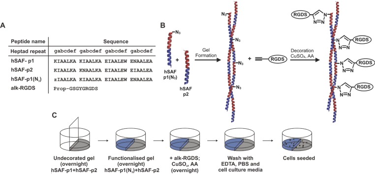
Peptide sequences, a schematic of the click reaction, and the half-moon model. A) Peptide
sequences used for this study. Key: z, azido norleucine; Prop, propiolate. B) The gel was formed
using an N-terminally azido-modified hSAF-p1. Decoration was achieved by performing
a click reaction with alk-RGDS on azide-containing gels catalyzed by CuSO4 with ascorbic
acid (AA). C) Side-by-side gel formation in 24-well cell-culture plates allowed a direct comparison
of cellular behavior on undecorated hSAF- and RGDS-decorated hSAF gels. Key: undecorated hSAF gel,
gray; and RGDS-decorated hSAF gel, blue.
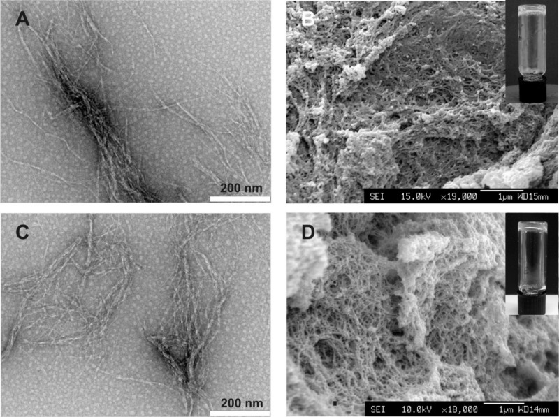
Fiber morphology and gel structure. Transmission electron images for the A) undecorated hSAF and
C) RGDS-decorated hSAF fibers. Average fiber diameters were 13 ± 5 nm for hSAF-undecorated
fibers and 17 ± 4 nm for RGDS-decorated hSAF fibers. B,D) Scanning electron images showing
interconnected fibers forming porous hydrogels of similar morphology D) with and B) without
alk-RGDS. The gels are self-supporting (insets). Scale bars on (A,C) equal 200 nm while scale bars
on (B,D) equal 1 μm.
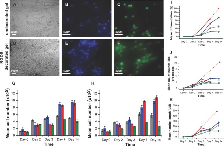
Response of PC12 cells to hydrogels. A,D) Light microscopy images showing PC12 attachment, and
elongated cell morphology, to undecorated hSAF- and RGDS-decorated hSAF gels after 14 d. B,E)
Representative fluorescent images for DAPI-stained cells on undecorated hSAF- and RGDS-decorated
hSAF gels. C,F) Viable cells on undecorated hSAF- and RGDS-decorated hSAF gels indicated by
calcein-AM staining. G) Proliferation of PC12 cells on gels and TCP over 14 d as judged by MTT
assays. H) DNA quantification using Hoechst dye for PC12 cells on the gels and TCP over 14 d. I)
PC12 differentiation, J) number of neurite-like processes, and K) lengths of processes as a function
of time. Due to a high proliferation rate, individual cell processes were difficult to identify at
day 14 on Matrigel. Dashed lines represent the projections for Matrigel assuming that the underlying
trend from the early time points continues. Key: undecorated hSAF gel, gray; RGDS-decorated hSAF
gel, blue; Matrigel, red; and TCP, green.
References
-
- Dhandayuthapani B, Yoshida Y, Maekawa T, Kumar DS. Int. J. Polym. Sci. 2011;2011:1.
-
- Jeon O, Song SJ, Lee KJ, Park MH, Lee SH, Hahn SK, Kim S, Kim BS. Carbohyr. Polym. 2007;70:251.
-
- Hou QP, Grijpma DW, Feijen J. Biomaterials. 2003;24:1937. - PubMed
Publication types
MeSH terms
Substances
Grants and funding
LinkOut - more resources
Full Text Sources
Other Literature Sources
