. 2014 May 1;192(9):4284-93.
doi: 10.4049/jimmunol.1303238. Epub 2014 Mar 21.

Integrin α4β1 is necessary for CD4+ T cell-mediated protection against genital Chlamydia trachomatis infection

Affiliations

Integrin α4β1 is necessary for CD4+ T cell-mediated protection against genital Chlamydia trachomatis infection

Sergio J Davila et al. J Immunol. .

Abstract

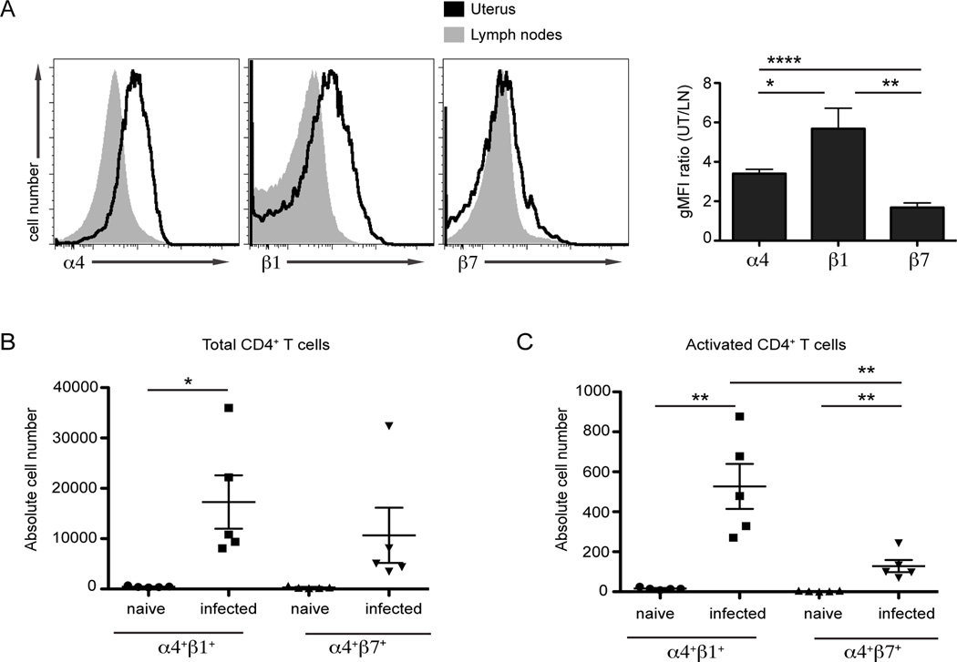
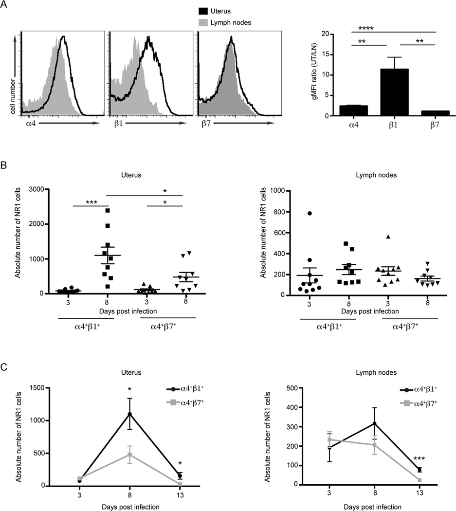
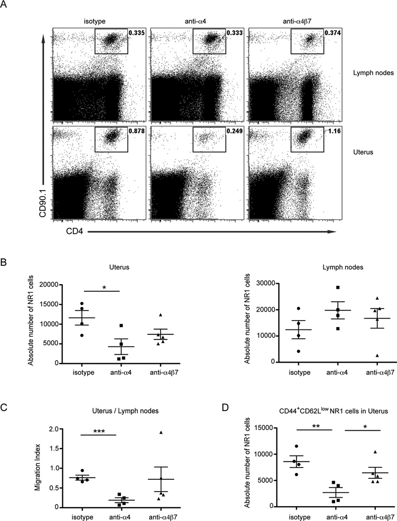
Chlamydia trachomatis infection is the most common sexually transmitted bacterial infection in the United States and a significant health burden worldwide. Protection from Chlamydia infection in the genital mucosa is dependent on IFN-γ derived from CD4(+) Th1 cells. These CD4(+) T cells must home successfully to the genital tract to exert their effector function and decrease C. trachomatis burden. Although adhesion receptors expressed by CD4(+) T cells in the genital tract have been characterized, the integrin receptor required for Chlamydia-specific CD4(+) T cell-mediated protection has not been explored. In this study, we demonstrate that C. trachomatis infection of the upper genital tract results in recruitment of Chlamydia-specific CD4(+) T cells robustly expressing the integrin α4β1. Interfering with α4β1, but not α4β7, function resulted in defective CD4(+) T cell trafficking to the uterus and high bacterial load. We conclude that integrin α4β1 is necessary for CD4(+) T cell-mediated protection against C. trachomatis infection in the genital mucosa. By identifying homing molecules required for successful CD4(+) T cell trafficking to C. trachomatis-infected tissues, we will be better equipped to design vaccines that elicit sterilizing, long-lasting immunity without inducing immune pathologies in the upper genital tract.


Conflict of interest statement

Disclosures

The authors declare no financial conflicts of interest.

Figures

FIGURE 1
FIGURE 1
C. trachomatis infection leads to robust integrin α4β1 surface expression on polyclonal CD4+ T cells in the genital tract. C57BL/6 mice were transcervically infected with 106 IFU of C. trachomatis. Seven days following infection the indicated tissues were harvested and prepared for flow cytometry. After gating on live cells that were CD3+CD4+ the surface expression of α4, β1 and β7 was quantified. (A) The integrin surface expression was analyzed by comparing the geometric mean fluorescence intensity (gMFI) ratio of CD4+ T cells localized in the genital mucosa to those in the draining lymph node. (B) The absolute number of α4+β1+ and α4+β7+ CD4+ T cells were quantified in the genital tract of naïve or mice infected with C. trachomatis for seven days. (C) The absolute number of activated α4+β1+ and α4+β7+ CD4+ T cells was quantified in the genital tract of naïve or infected mice; activation was determined by CD44+ staining. Shown are representative results from one of two independent experiments. * p < 0.05, ** p < 0.01, and **** p < 0.0001.
FIGURE 2
FIGURE 2
C. trachomatis infection leads to robust α4β1 surface expression on C. trachomatis-specific CD4+ T cells responding to the genital tract. One million CD90.1+ NR1 cells were transferred intravenously into CD90.2+ recipient mice. The following day mice were infected transcervically with 106 IFU of C. trachomatis. The uterus and draining lymph nodes were harvested at the indicated time points following infection and prepared for flow cytometry. For integrin surface staining analysis, cells were pre-gated as live Vα2+CD4+CD90.1+ cells and then examined for the surface expression of α4, β1, and β7. (A) Eight days after C. trachomatis infection, the integrin surface expression on NR1 cells was analyzed by comparing the gMFI ratio of NR1 cells in uterus to those localized in the draining lymph nodes. (B) The absolute numbers of α4+β1+ and α4+β7+ NR1 cells was quantified 3 and 8 days after transcervical infection in the uterus (left) and the draining lymph nodes (right). (C) Quantification of the trafficking kinetics of α4+β1+ and α4+β7+ NR1 cells in the uterus (left) and draining lymph nodes (right) at the indicated time points. Shown are representative results from one of three independent experiments. * p < 0.05, ** p < 0.01, *** p < 0.001, and **** p < 0.0001.
FIGURE 3
FIGURE 3
Antibody blockade of α4 but not α4β7 exacerbates C. trachomatis burden in the genital mucosa. NR1 cells were skewed in vitro to a Th1 phenotype for 5 days. NR1 cells were pretreated with the indicated antibodies and then transferred intravenously into naïve recipients. The following day, mice were infected transcervically with 5×10⁶ IFU of C. trachomatis. Groups were injected with the respective integrin or isotype control antibody 1 and 3 days following infection. Five days after infection, the genital tract was isolated and genomic DNA was purified. The levels of Chlamydia 16S DNA relative to the levels of host GADPH were quantified using qPCR. Shown are representative results from one of two independent experiments. * p < 0.05, and ** p < 0.01.
FIGURE 4
FIGURE 4
Blockade of α4 but not α4β7 impairs C. trachomatis-specific CD4+ T cell trafficking to the genital tract following infection. NR1 cells were skewed in vitro to a Th1 phenotype for 5 days. NR1 cells were pretreated with the indicated antibodies and then 106 CD90.1+ NR1 cells were transferred intravenously into CD90.2+ host mice. The following day mice were infected transcervically with 5×10⁶ IFU of C. trachomatis. Groups were injected with the respective integrin or control antibody 1 and 3 days following infection. The uterus and draining lymph nodes were harvested 5 days following infection and prepared for flow cytometry. For quantification of NR1 cell trafficking, we gated on live Vα2+CD4+CD90.1+ cells. (A) Representative flow cytometry plots indicate the percentage of NR1 cells following the indicated antibody treatment in the draining lymph nodes (top) and uterus (bottom) (B) The absolute number of NR1 cells were quantified in the uterus (left) and draining lymph nodes (right). (C) A migration index for each antibody treatment was calculated by comparing the absolute number of live NR1 cells in the uterus directly to the number of NR1 cells in draining lymph nodes within each animal. A lower migration index ratio indicates decreased NR1 cell recruitment to the uterus. (D) The absolute number of effector NR1 cells was determined by examining the CD44+CD62Llow gated population in the genital mucosa. Shown are representative results from one of two independent experiments. * p < 0.05, ** p < 0.01, and *** p < 0.001.
FIGURE 5
FIGURE 5
Chlamydia-specific CD4+ T cells deficient in integrin β1 are unable to protect mice from C. trachomatis infection. The indicated NR1 genotypes were skewed in vitro to a Th1 phenotype for 5 days. (A) The expansion of NR1 cells was compared for the three genotypes. (B) The activation of live NR1 cells was assessed by gating for CD25+CD44+ cells. (C) TNF-α and IFN-γ production was examined using intracellular cytokine staining. (D) After 5 days of in vitro stimulation, 105 NR1 cells were transferred intravenously into naïve hosts. The following day, mice were transcervically infected with 5x10⁶ IFU of C. trachomatis. Five days after infection, the genital tract was harvested and genomic DNA was purified. The levels of Chlamydia 16S DNA relative to the levels of host GADPH were quantified using qPCR. Shown are representative results from one of three independent experiments. * p < 0.05 and ** p < 0.01.
FIGURE 6
FIGURE 6
β1 deficient Chlamydia-specific CD4+ T cells are unable to traffic efficiently to the genital tract following infection. An equivalent number of CD45.2/CD90.1 integrin wildtype NR1 cells and CD45.2/CD90.2 wildtype, β1−/−, or β7−/− NR1 cells were transferred intravenously into CD45.1/CD90.2 host mice. The next day, mice were infected transcervically with 106 IFU of C. trachomatis. The genital tract, draining lymph nodes and spleen were harvested 7 days after infection and prepared for flow cytometry. We examined the recruitment of NR1 cells by pre-gating on live Vα2+CD4+CD45.2+ cells. We then differentiated the competing NR1 populations by examining the number of CD90.1+ or CD90.2+ cells. (A) Shown are representative plots of integrin sufficient and deficient NR1 cells in the draining lymph node (top) and genital mucosa (bottom). (B) The migration index within each group was calculated by comparing the percentage of CD90.2+ to CD90.1+ NR1 cells in the uterus to the CD90.2+ to CD90.1+ NR1 cells in the draining lymph nodes (left) or spleen (right). A lower migration index indicates less efficient trafficking of integrin-deficient NR1 cells specifically to the uterus relative to the circulation. Shown are representative results from one of three independent experiments. ** p < 0.01, *** p < 0.001, and **** p < 0.0001.

References

    1. Brunham R, Rey-Ladino J. Immunology of Chlamydia infection: implications for a Chlamydia trachomatis vaccine. Nature reviews. Immunology. 2005;5:149–161. - PubMed
    1. Cohen C, Brunham R. Pathogenesis of Chlamydia induced pelvic inflammatory disease. Sexually transmitted infections. 1999;75:21–24. - PMC - PubMed
    1. Roan N, Starnbach M. Immune-mediated control of Chlamydia infection. Cellular microbiology. 2008;10:9–19. - PubMed
    1. Perry L, Su H, Feilzer K, Messer R, Hughes S, Whitmire W, Caldwell H. Differential sensitivity of distinct Chlamydia trachomatis isolates to IFN-gamma-mediated inhibition. Journal of immunology (Baltimore, Md. : 1950) 1999;162:3541–3548. - PubMed
    1. Thomas S, Garrity L, Brandt C, Schobert C, Feng G, Taylor M, Carlin J, Byrne G. IFN-gamma-mediated antimicrobial response. Indoleamine 2,3-dioxygenase-deficient mutant host cells no longer inhibit intracellular Chlamydia spp. or Toxoplasma growth. Journal of immunology (Baltimore, Md. : 1950) 1993;150:5529–5534. - PubMed

Publication types

Substances