. 2014 Jun;196(11):1980-91.
doi: 10.1128/JB.01573-14. Epub 2014 Mar 21.

Intracellular hydrogen peroxide and superoxide poison 3-deoxy-D-arabinoheptulosonate 7-phosphate synthase, the first committed enzyme in the aromatic biosynthetic pathway of Escherichia coli

Affiliations

Intracellular hydrogen peroxide and superoxide poison 3-deoxy-D-arabinoheptulosonate 7-phosphate synthase, the first committed enzyme in the aromatic biosynthetic pathway of Escherichia coli

Jason M Sobota et al. J Bacteriol. 2014 Jun.

Abstract

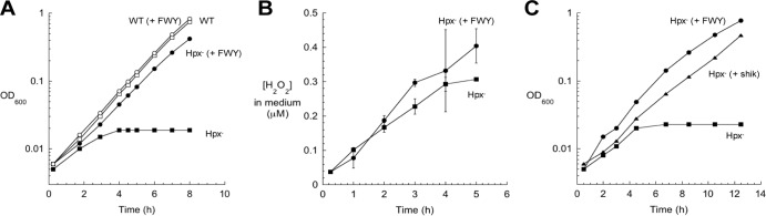
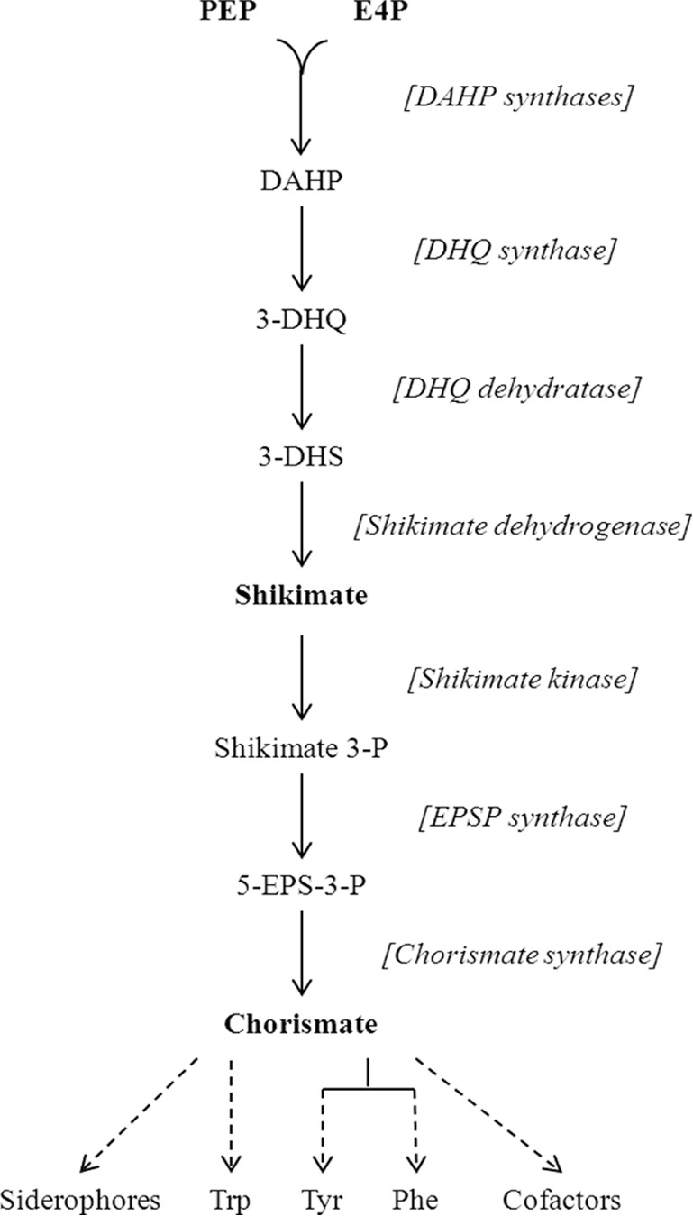
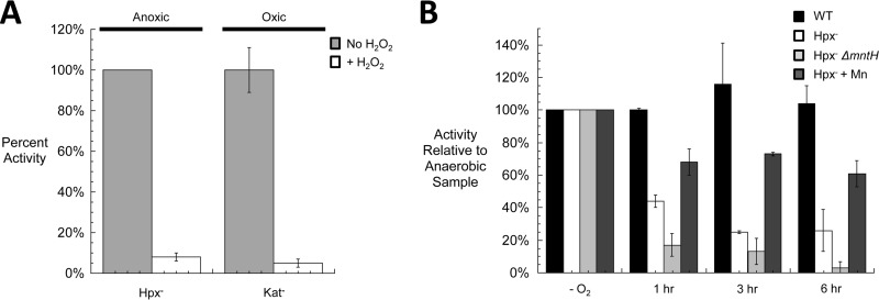
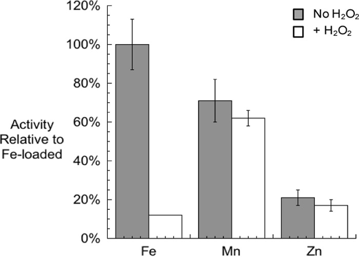
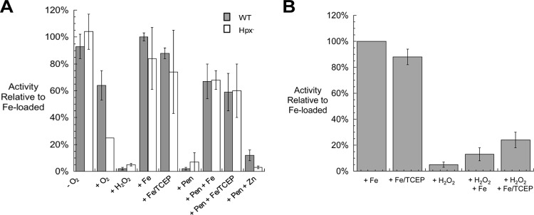
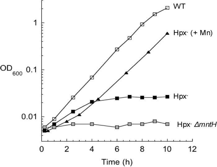
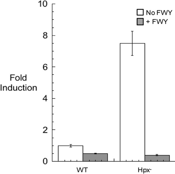
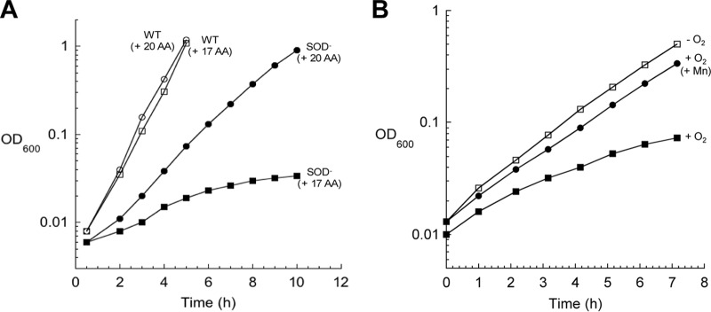
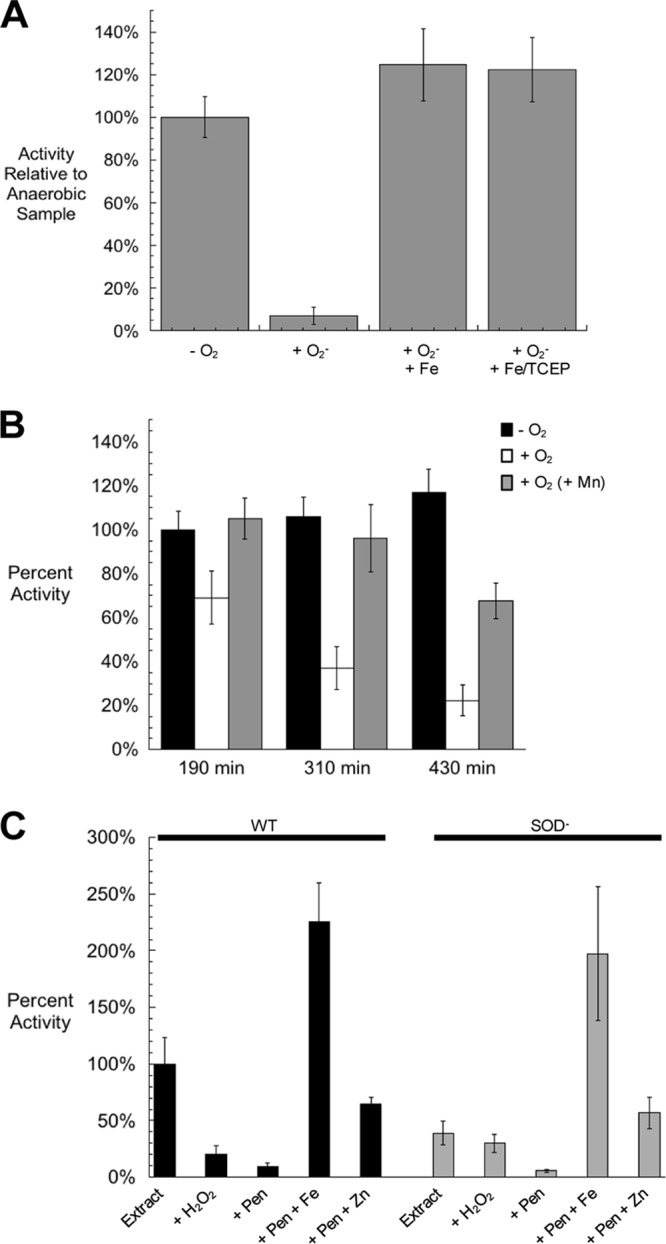
In Escherichia coli, aromatic compound biosynthesis is the process that has shown the greatest sensitivity to hydrogen peroxide stress. This pathway has long been recognized to be sensitive to superoxide as well, but the molecular target was unknown. Feeding experiments indicated that the bottleneck lies early in the pathway, and the suppressive effects of fur mutations and manganese supplementation suggested the involvement of a metalloprotein. The 3-deoxy-D-arabinoheptulosonate 7-phosphate synthase (DAHP synthase) activity catalyzes the first step in the pathway, and it is provided by three isozymes known to rely upon a divalent metal. This activity progressively declined when cells were stressed with either oxidant. The purified enzyme was activated more strongly by ferrous iron than by other metals, and only this metalloform could be inactivated by hydrogen peroxide or superoxide. We infer that iron is the prosthetic metal in vivo. Both oxidants displace the iron atom from the enzyme. In peroxide-stressed cells, the enzyme accumulated as an apoprotein, potentially with an oxidized cysteine residue. In superoxide-stressed cells, the enzyme acquired a nonactivating zinc ion in its active site, an apparent consequence of the repeated ejection of iron. Manganese supplementation protected the activity in both cases, which matches the ability of manganese to metallate the enzyme and to provide substantial oxidant-resistant activity. DAHP synthase thus belongs to a family of mononuclear iron-containing enzymes that are disabled by oxidative stress. To date, all the intracellular injuries caused by physiological doses of these reactive oxygen species have arisen from the oxidation of reduced iron centers.


Figures

FIG 1
FIG 1
Submicromolar intracellular H2O2 disrupts the initial stage of aromatic amino acid synthesis. (A) At time zero, wild-type (WT) and mutant cultures growing exponentially in anoxic medium containing glucose (glucose medium) were diluted into aerobic medium. Where indicated, three aromatic amino acids (phenylalanine, tryptophan, and tyrosine) were added to the medium [(+ FWY)]. OD600, optical density at 600 nm. (B) Accumulation of H2O2 in the medium of Hpx mutants. The H2O2 equilibrates across the membrane sufficiently quickly that the extracellular and intracellular H2O2 concentrations are effectively the same (17). Error bars represent the standard deviations from the means of three independent replicate cultures. (C) Cultures growing exponentially in anoxic glucose medium were diluted at time zero into aerobic medium. Where indicated, 50 μM shikimate (shik) was added to the medium.
FIG 2
FIG 2
Enzymes and intermediates of the shikimate pathway. The solid black arrows represent reactions, dashed arrows represent multiple reactions, chemical intermediates are named, and enzymes are denoted in brackets next to the reactions that they catalyze. Abbreviations: PEP, phosphoenolpyruvate; E4P, erythrose 4-phosphate; DAHP, 3-deoxy-d-arabino-heptulosonate 7-phosphate; DHQ, dehydroquinate; DHS, dehydroshikimate; Shikimate 3-P, Shikimate 3-phosphate; 5-EPS-3-P, 5-enolpyruvoylshikimate-3-phosphate; Trp, tryptophan; Tyr, tyrosine; Phe, phenylalanine.
FIG 3
FIG 3
DAHP synthase enzymes are inactivated by H2O2 in vitro and in vivo. (A) Cultures were grown exponentially to 0.2 OD in anoxic medium (Hpx cells) or oxic medium (catalase-deficient cells). Extracts were prepared anoxically. Where indicated, extracts were exposed to 100 μM H2O2 for 5 min prior to the assay of DAHP synthase activity. (B) Cultures growing exponentially in anoxic medium were diluted at time zero into oxic medium. At various intervals (time shown in hours), aliquots were shifted to the anaerobic chamber, and extracts were prepared and assayed. Where indicated, 1 μM MnCl2 was added to the medium. For both panels, the error bars represent the standard deviations from the means of three independent experiments.
FIG 4
FIG 4
Activities of pure DAHP synthase activated with various metals. Purified apo-DAHP synthase was treated with 50 μM TCEP and 500 μM each metal. The enzyme activities were then determined with or without exposure to 100 μM H2O2. Activities are given relative to Fe-loaded DAHP synthase without H2O2 challenge. Error bars represent the standard deviations from the means of three independent samples.
FIG 5
FIG 5
Reactivation of DAHP synthase that has been inactivated by H2O2 in vivo and in vitro. (A) Cultures were grown exponentially in anoxic glucose medium. An anaerobic sample was harvested, and the remaining cells were diluted to 0.05 OD into oxic medium and cultured to 0.15 OD before harvesting. Where indicated, samples were treated with 100 μM H2O2 for 5 min; 500 μM Fe, Zn, or both Fe and TCEP (Fe/TCEP) for 15 min; or 5 mM penicillamine (Pen) for 20 min followed by Zn, Fe, or Fe/TCEP treatments, prior to the assay. (B) Cell extracts were prepared from anoxically grown cells. Where indicated, samples were treated in vitro with 100 μM H2O2 for 5 min and then with either 500 μM Fe or Fe/TCEP for 15 min prior to the assay. For both panels, the error bars represent the standard deviations from the means of three independent cultures.
FIG 6
FIG 6
Manganese supplements suppress the aromatic biosynthetic defect of Hpx cells. Exponentially growing Hpx cells in anoxic glucose medium were diluted at time zero into oxic medium. Where indicated, 50 μM Mn was added to the medium.
FIG 7
FIG 7
aroF transcription increases in response to H2O2 stress. Hpx strain JS299 containing an aroF::lacZ transcriptional fusion in the lambda attachment site was subcultured into oxic glucose medium and grown for 4 h. Extracts were prepared, and β-galactosidase activity was determined. Error bars represent the standard deviations from the means of three independent cultures.
FIG 8
FIG 8
SOD mutants are aromatic amino acid auxotrophs. (A) Cultures were grown anaerobically overnight in glucose medium supplemented with 17 amino acids [(+ 17 AA)] (lacking Phe, Tyr, and Trp). At time zero, cells were diluted into oxic medium with or without aromatic amino acids. (B) Cultures were treated as in panel A. Where indicated, 25 μM MnCl2 was added to the medium.
FIG 9
FIG 9
O2 damages purified DAHP synthase in vitro and in vivo. (A) Purified Fe-loaded DAHP synthase was exposed to O2 generated by xanthine and xanthine oxidase. Catalase was included in all samples. Superoxide stress was terminated by the addition of SOD. All superoxide-treated samples were returned to anoxic conditions and, where indicated, treated with ferrous iron with or without TCEP prior to the assay. Error bars represent the standard deviations from the means of three independent samples. (B) Exponentially growing cultures were diluted from anoxic to oxic glucose medium containing 17 amino acids (lacking Phe, Tyr, and Trp) at time zero. Where indicated, 25 μM MnCl2 was added to the medium. At three different time points, DAHP synthase activity was assayed. (C) Extracts were prepared from wild-type and SOD cultures after growth to 0.2 OD in oxic medium. Where indicated, samples were treated with 100 μM H2O2 for 5 min, 5 mM penicillamine for 20 min, and 750 μM Fe or Zn for 15 min, prior to the assay. For all panels, the error bars represent the standard deviations from the means of three independent experiments.

References

    1. Loew O. 1900. A new enzyme of general occurrence in organisms. Science 11:701–702. 10.1126/science.11.279.701 - DOI - PubMed
    1. McCord JM, Fridovich I. 1969. Superoxide dismutase. An enzymic function for erythrocuprein (hemocuprein). J. Biol. Chem. 244:6049–6055 - PubMed
    1. Imlay JA. 2013. The molecular mechanisms and physiological consequences of oxidative stress: lessons from a model bacterium. Nat. Rev. Microbiol. 11:443–454. 10.1038/nrmicro3032 - DOI - PMC - PubMed
    1. Imlay JA, Chin SM, Linn S. 1988. Toxic DNA damage by hydrogen peroxide through the Fenton reaction in vivo and in vitro. Science 240:640–642. 10.1126/science.2834821 - DOI - PubMed
    1. Winterbourn CC, Metodiewa D. 1999. Reactivity of biologically important thiol compounds with superoxide and hydrogen peroxide. Free Radic. Biol. Med. 27:322–328. 10.1016/S0891-5849(99)00051-9 - DOI - PubMed

Publication types

MeSH terms