. 2014 May 9;289(19):13142-54.
doi: 10.1074/jbc.M113.530311. Epub 2014 Mar 21.

Essential roles of neutral ceramidase and sphingosine in mitochondrial dysfunction due to traumatic brain injury

Affiliations

Essential roles of neutral ceramidase and sphingosine in mitochondrial dysfunction due to traumatic brain injury

Sergei A Novgorodov et al. J Biol Chem. .

Abstract

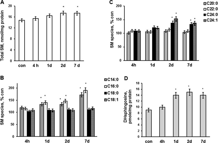
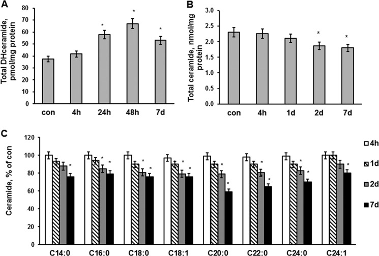
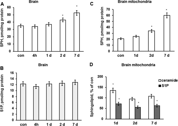
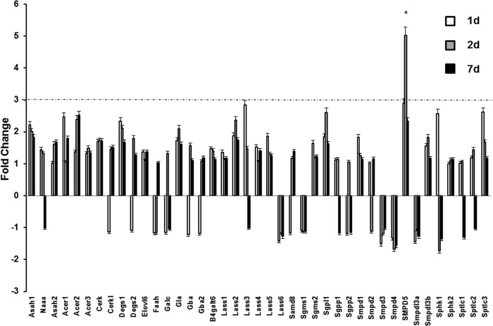
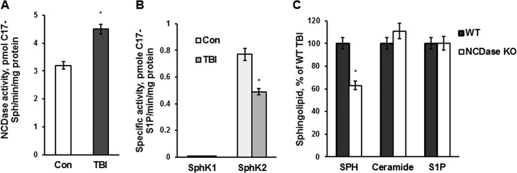
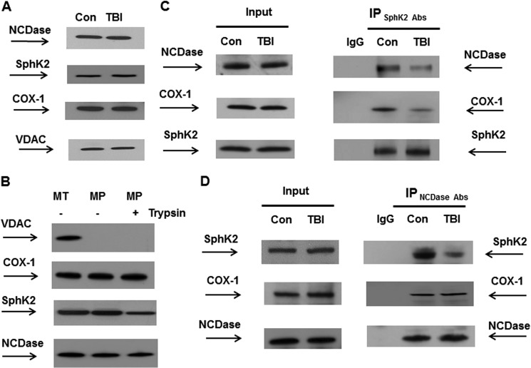
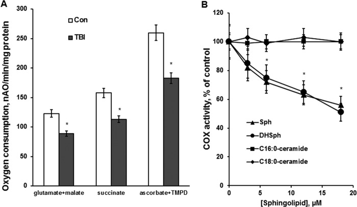
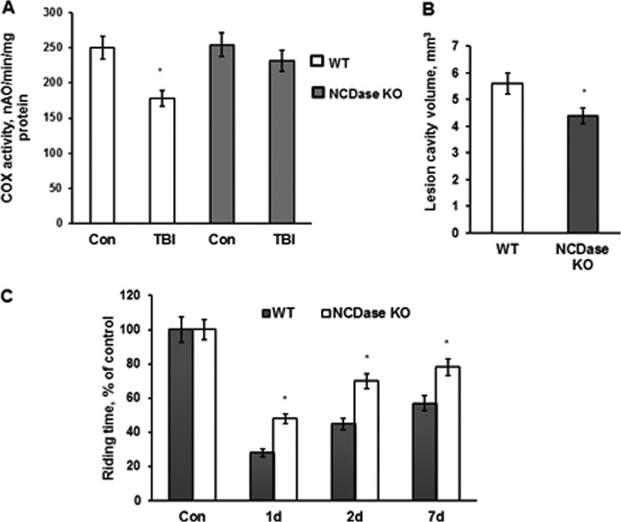
In addition to immediate brain damage, traumatic brain injury (TBI) initiates a cascade of pathophysiological events producing secondary injury. The biochemical and cellular mechanisms that comprise secondary injury are not entirely understood. Herein, we report a substantial deregulation of cerebral sphingolipid metabolism in a mouse model of TBI. Sphingolipid profile analysis demonstrated increases in sphingomyelin species and sphingosine concurrently with up-regulation of intermediates of de novo sphingolipid biosynthesis in the brain. Investigation of intracellular sites of sphingosine accumulation revealed an elevation of sphingosine in mitochondria due to the activation of neutral ceramidase (NCDase) and the reduced activity of sphingosine kinase 2 (SphK2). The lack of change in gene expression suggested that post-translational mechanisms are responsible for the shift in the activities of both enzymes. Immunoprecipitation studies revealed that SphK2 is complexed with NCDase and cytochrome oxidase (COX) subunit 1 in mitochondria and that brain injury hindered SphK2 association with the complex. Functional studies showed that sphingosine accumulation resulted in a decreased activity of COX, a rate-limiting enzyme of the mitochondrial electron transport chain. Knocking down NCDase reduced sphingosine accumulation in mitochondria and preserved COX activity after the brain injury. Also, NCDase knockdown improved brain function recovery and lessened brain contusion volume after trauma. These studies highlight a novel mechanism of secondary TBI involving a disturbance of sphingolipid-metabolizing enzymes in mitochondria and suggest a critical role for mitochondrial sphingosine in promoting brain injury after trauma.

Keywords: Brain; Cell Death; Mitochondria; Respiratory Chain; Sphingolipid.


Figures

FIGURE 1.
FIGURE 1.
SM and dihydrosphingosine changes in brain tissue after TBI. Sphingolipids were analyzed in brain tissue lysate made from the brain hemisphere ipsilateral to the injury imposed by a control cortical impact device as described under “Experimental Procedures.” Sham-injured animal brain was used as a control (con). A, total SM content was increased in the injured brain tissue. B and C, SM species changes in the injured tissue. D, dihydrosphingosine (DHsphingosine) was increased in the injured brain. Data are means ± S.E. (error bars); *, p < 0.05, n = 16. Each sample was normalized to its respective total protein levels.
FIGURE 2.
FIGURE 2.
Dihydroceramide and ceramide changes in brain tissue after TBI. Brain tissue samples were prepared from the mouse brain hemisphere ipsilateral to the damage. Sham-injured animal brain was used as a control (con). Dihydroceramide (DHceramide) (A), total ceramide (B), and ceramide species (C) were measured in the injured brain tissue after TBI as described under “Experimental Procedures.” Data are means ± S.E. (error bars); *, p < 0.05, n = 16. Each sample was normalized to its respective total protein levels.
FIGURE 3.
FIGURE 3.
Sphingosine and S1P changes in brain tissue and mitochondria after TBI. Brain tissue samples and mitochondria were prepared from the mouse brain hemisphere ipsilateral to the damage. Sham-injured animal brain was used as a control (con). Sphingosine content was up-regulated (A), and S1P content was unchanged (B) in the brain during the first week post-TBI. Data are means ± S.E. (error bars); *, p < 0.05; n = 16. Each sample was normalized to its respective total protein levels. C, sphingosine was gradually elevated in mitochondria after TBI. D, S1P was reduced in brain mitochondria after TBI, whereas ceramide was increased at 1 day, returning to control levels at 2 days post-TBI. The ceramide content of control mitochondria was 1318 ± 94.7 pmol/mg of protein, whereas S1P content was 8.7 ± 0.9 pmol/mg of protein. Data are means ± S.E.; *, p < 0.05, n = 12. Each sample was normalized to its respective total protein levels.
FIGURE 4.
FIGURE 4.
Sphingolipid-metabolizing enzyme gene expression after TBI. Brain samples were prepared from the ipsilateral hemisphere after TBI and sham-injured brain (control). RNA was extracted and converted to cDNA for analysis by real-time PCR using a PCR array as described under “Experimental Procedures.” The data are presented as -fold change TBI/sham injury and are means of three independent experiments. The dashed line represents a 3-fold change. *, p < 0.05; n = 6. Error bars, S.E.
FIGURE 5.
FIGURE 5.
TBI-induced mitochondrial sphingosine accumulation is due to an activation of NCDase and could be partially rescued by knocking down NCDase. Mitochondria were purified from the injured hemisphere of a mouse brain (TBI) and sham-injured mouse brain (Con) at 7 days post-TBI. A, specific NCDase activity was measured with C17-C18-ceramide as a substrate (42), yielding C17-sphingosine (C17-Sph) as described under “Experimental Procedures.” Data are means ± S.E. (error bars); *, p < 0.05; n = 8. B, specific SphK2 and SphK1 activities were measured with C17-sphingosine as a substrate, yielding C17-S1P, as described under “Experimental Procedures.” The specific activities of SphK1 and SphK2 in brain homogenate were 1.10 ± 0.12 and 0.68 ± 0.14 pmol of C17-S1P/min/mg of protein, respectively. Data are means ± S.E.; *, p < 0.05; n = 8. C, cerebral mitochondria were purified from WT and NCDase KO mice at day 7 post-TBI. Sphingolipid content was measured by tandem MS. Data are means ± S.E.; *, p < 0.05; n = 12. Each sample was normalized to its respective total protein levels.
FIGURE 6.
FIGURE 6.
TBI hindered SphK2 association with NCDase and COX. Mitochondria were purified from the injured hemisphere of mouse brain and sham-injured brain (con) at 7 days post-TBI. A and B, mitochondrial lysate was loaded into the lane (30 μg/lane) (A). To investigate submitochondrial localization of NCDase and SphK2, equal volume samples of untreated mitochondria (MT), mitoplasts (MP), or mitoplasts treated with trypsin (MPT) were loaded into the lane (B). Blots were analyzed using specific anti-NCDase (Bethyl Laboratories), anti-SphK2 (Abcam), anti-COX-1 (Santa Cruz Biotechnology), and anti-VDAC (Cell Signaling) antibodies. C and D, SphK2 association with NCDase and COX-1 was detected in reciprocal immunoprecipitation (IP) experiments. Mitochondria were immunoprecipitated using anti-SphK2 (Abcam) antibodies, and the blots were probed with anti-NCDase (Bethyl Laboratories), anti-COX-1 (Santa Cruz Biotechnology), or anti-SphK2 (Santa Cruz Biotechnology) antibodies. Input load was 30 mg/lane (C). Mitochondria were immunoprecipitated with anti-NCDase antibody (Bethyl Laboratories) and probed using anti-SphK2 (Santa Cruz Biotechnology), anti-COX-1 (Santa Cruz Biotechnology), or anti-NCDase (Santa Cruz Biotechnology) antibodies. Input load was 30 μg/lane (D). As a control, the same immunoprecipitation procedure was performed except for primary antibody application (IgG).
FIGURE 7.
FIGURE 7.
Respiratory chain activity was reduced at the level of COX after TBI and sphingosine inhibited the COX activity in baseline mitochondria. A, mitochondria were purified from the ipsilateral hemisphere of WT mouse brain at day 7 post-TBI. Sham-injured animal brain was used as a control (Con). Mitochondrial respiration was measured by recording oxygen consumption with a Clark-type oxygen electrode in the presence of Complex I substrate 5 mm glutamate plus 5 mm malate (glutamate + malate), Complex II substrate 10 mm succinate (succinate), and Complex IV (COX) substrate 1 mm ascorbate plus 250 μm TMPD (ascorbate + TMPD) in the presence of 100 μm ADP (state 3). Data are means ± S.E. (error bars); *, p < 0.05, n = 8. B, mitochondrial COX activity was measured by recording oxygen consumption of baseline mitochondria in the presence of COX substrate (1 mm ascorbate plus 250 μm TMPD), 1 μm antimycin, and 50 μm 2,4-DNP. Sphingosine (Sph) and dihydrosphingosine (DHSph) were delivered in ethanol. C16:0-ceramide and C18:0-ceramide were delivered in ethanol/dodecane (98:2, v/v). Data are means ± S.E.; *, p < 0.05, n = 8.
FIGURE 8.
FIGURE 8.
NCDase knockdown rescued COX activity defect, attenuated brain damage, and improved sensorimotor deficit recovery after TBI. A, mitochondria were purified from the ipsilateral hemisphere of WT and NCDase KO mouse brain at day 7 post-TBI. Sham-injured animal brain was used as a control (Con). Mitochondrial COX activity was measured by recording oxygen consumption in the presence of COX substrate (1 mm ascorbate plus 250 μm TMPD), 1 μm antimycin, and 50 μm 2,4-DNP (state 3u). Data are means ± S.E. (error bars); *, p < 0.05; n = 8. B, lesion cavity volume was measured at day 28 post-TBI by staining of brain sections with 0.1% cresyl violet and image analysis as described under “Experimental Procedures.” Data are means ± S.E.; *, p < 0.05; n = 12. C, sensorimotor deficits were assessed using a standard rotarod test. Each day for 3 days prior to injury, animals were trained on the rotarod at a speed of 18 rpm in the acceleration mode (0–18 rpm/90 s). Animals were tested with the rotarod apparatus using three trials in session, with a minimum of 5 min resting between trials. Data are means ± S.E.; *, p < 0.05; n = 16.

Similar articles

Cited by

References

    1. Spaethling J. M., Geddes-Klein D. M., Miller W. J., von Reyn C. R., Singh P., Mesfin M., Bernstein S. J., Meaney D. F. (2007) Linking impact to cellular and molecular sequelae of CNS injury: modeling in vivo complexity with in vitro simplicity. Prog. Brain Res. 161, 27–39 - PubMed
    1. Robertson C. L., Scafidi S., McKenna M. C., Fiskum G. (2009) Mitochondrial mechanisms of cell death and neuroprotection in pediatric ischemic and traumatic brain injury. Exp. Neurol. 218, 371–380 - PMC - PubMed
    1. Sullivan P. G., Keller J. N., Mattson M. P., Scheff S. W. (1998) Traumatic brain injury alters synaptic homeostasis: implications for impaired mitochondrial and transport function. J. Neurotrauma 15, 789–798 - PubMed
    1. Gilmer L. K., Roberts K. N., Joy K., Sullivan P. G., Scheff S. W. (2009) Early mitochondrial dysfunction after cortical contusion injury. J. Neurotrauma 26, 1271–1280 - PMC - PubMed
    1. Singh I. N., Sullivan P. G., Hall E. D. (2007) Peroxynitrite-mediated oxidative damage to brain mitochondria: protective effects of peroxynitrite scavengers. J. Neurosci. Res. 85, 2216–2223 - PubMed

Publication types

MeSH terms